File:Keibel1897 plate01.jpg
Original file (1,504 × 2,000 pixels, file size: 365 KB, MIME type: image/jpeg)
Plate 1.
Fig. 1
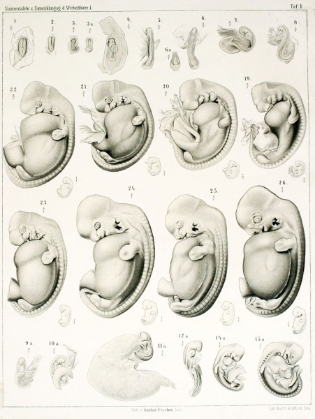
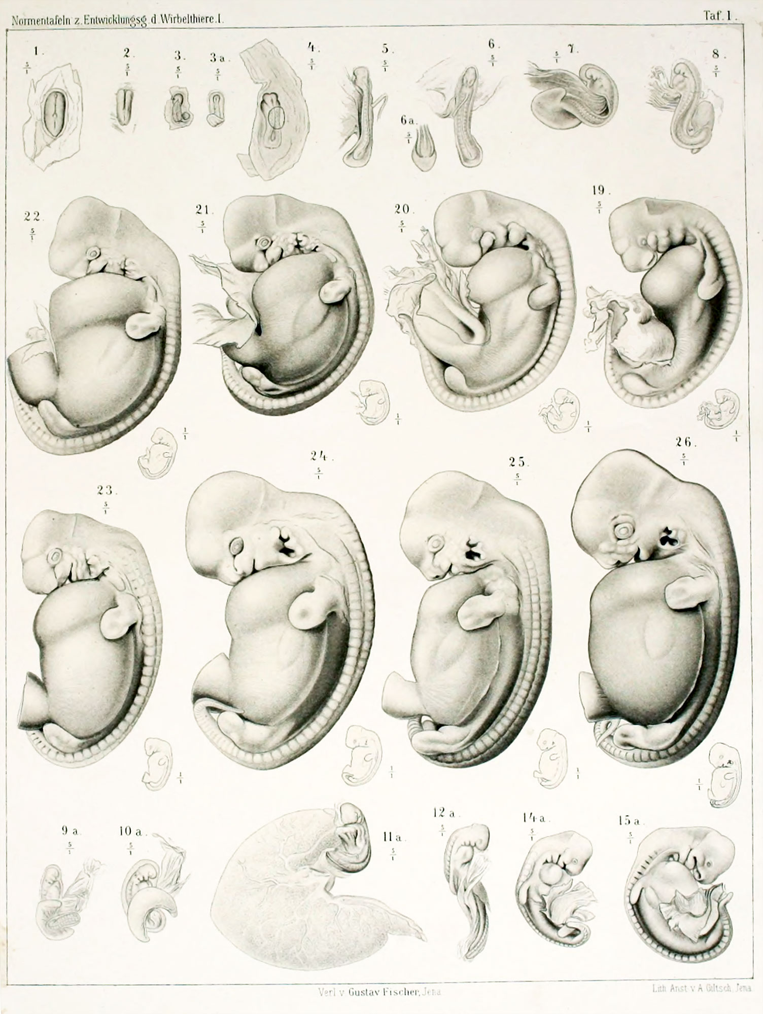
The embryonic disc p.7 b. 93 is from a 14-Days 10 hours before the slaughter sow occupied. After the embryonic disc was removed in the usual way with chrome acetic uterus, it was fixes to sublimate on. FIG. 1 illustrates the embryonic disc is how they presented itself to me after this treatment in 70 "Alcohol. M., and M, are a draft after ZiKGi.ER'schen model, which was produced with a basis Plattenreconstruction. We see, as the primitive streak has already been limited to the rear half of the embryonic disc. Before the primitive streak region we see the first installation of the medullary and the medullary. The medullary groove extends up close to the front end of the embryonic disc, rear end already on the point, the front end to embrace the primitive streak. The area in front of the front end of Primitiv.streifens is easy aufgewulstet, and this bead forms the base of the rear part, the medullary groove. At the front end of the embryonic disc a little bump already at surface observation stands right in front of the medullary groove clearly emerges. As can be seen from the average series, called these bumps, the front end of the head extension resp. the notochord, it corresponds to the front end of the embryo at all. The small protrusion that is noticeable at the rear end of the embryonic disc is caused by Mesodermwucherung and the mesodermal Allantoisanlage. It should be emphasized that in this embryonic disc, the posterior intestinal bay is already well trained. The occurrence of the amnion was already mentioned in the previous embryonic disc, the development of the amnion has made progress, we find, as in the previous stage, evenly developed in circumference around the embryonic disc.
| Historic Disclaimer - information about historic embryology pages |
|---|
 |
Reference
Franz Keibel, Normentafeln zur Entwicklungsgeschichte der Wirbelthiere (Normal plates of the development of vertebrates) Volume Hft.1 (1897) Jena, G. Fischer, Germany.
Cite this page: Hill, M.A. (2026, October 15) Embryology Keibel1897 plate01.jpg. Retrieved from https://embryology.med.unsw.edu.au/embryology/index.php/File:Keibel1897_plate01.jpg
- © Dr Mark Hill 2026, UNSW Embryology ISBN: 978 0 7334 2609 4 - UNSW CRICOS Provider Code No. 00098G
File history
Yi efo/eka'e gwa ebo wo le nyangagi wuncin ye kamina wunga tinya nan
| Gwalagizhi | Nyangagi | Dimensions | User | Comment | |
|---|---|---|---|---|---|
| current | 10:49, 20 November 2013 | | 1,504 × 2,000 (365 KB) | Z8600021 (talk | contribs) |
You cannot overwrite this file.
File usage
The following 3 pages use this file: