File:Intestine histology 001.jpg: Difference between revisions
From Embryology
No edit summary |
No edit summary |
||
| Line 5: | Line 5: | ||
* cross-section of villi extending into the lumen | * cross-section of villi extending into the lumen | ||
* lacteal - surrounded by a layer of flattened endothelial, no blood cells in lumen | * lacteal - surrounded by a layer of flattened endothelial, no blood cells in lumen | ||
:Links: [[:File:Intestine histology 002.jpg|Unlabeled version]] | |||
{{Blue Histology}} | {{Blue Histology}} | ||
Revision as of 15:51, 23 February 2012
Jejunum
- Baboon, Stain H&E
- jejunum
- cross-section of villi extending into the lumen
- lacteal - surrounded by a layer of flattened endothelial, no blood cells in lumen
- Links: Unlabeled version
Links: Histology | Histology Stains | Blue Histology images copyright Lutz Slomianka 1998-2009. The literary and artistic works on the original Blue Histology website may be reproduced, adapted, published and distributed for non-commercial purposes. See also the page Histology Stains.
Cite this page: Hill, M.A. (2024, June 1) Embryology Intestine histology 001.jpg. Retrieved from https://embryology.med.unsw.edu.au/embryology/index.php/File:Intestine_histology_001.jpg
- © Dr Mark Hill 2024, UNSW Embryology ISBN: 978 0 7334 2609 4 - UNSW CRICOS Provider Code No. 00098G
Jej20he.jpg
File history
Click on a date/time to view the file as it appeared at that time.
| Date/Time | Thumbnail | Dimensions | User | Comment | |
|---|---|---|---|---|---|
| current | 15:42, 23 February 2012 | 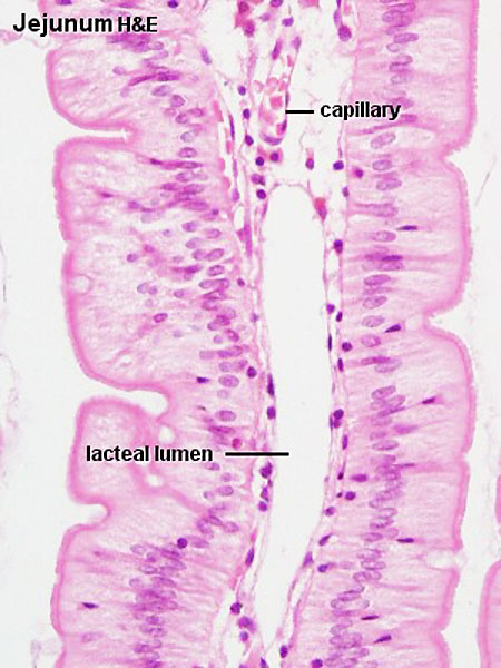 | 450 × 600 (65 KB) | Z8600021 (talk | contribs) | Jej20he.jpg |
You cannot overwrite this file.