User:Z3290270: Difference between revisions
| Line 13: | Line 13: | ||
--[[User:Z3290270|Maeda Sadeghpour]] 12:29, 15 September 2011 (EST) | --[[User:Z3290270|Maeda Sadeghpour]] 12:29, 15 September 2011 (EST) | ||
--[[User:Z3290270|Maeda Sadeghpour]] 11:34, 22 September 2011 (EST) | |||
== '''Lab 1 Assignment:''' == | == '''Lab 1 Assignment:''' == | ||
Revision as of 11:34, 22 September 2011
Lab attendence:
--Z3290270 12:56, 28 July 2011 (EST)
--Maeda Sadeghpour 11:48, 4 August 2011 (EST)
--z3290270 11:47, 11 August 2011 (EST)
--Maeda Sadeghpour 11:09, 18 August 2011 (EST)
--Maeda Sadeghpour 11:06, 25 August 2011 (EST)
--Maeda Sadeghpour 12:03, 1 September 2011 (EST)
--Maeda Sadeghpour 12:29, 15 September 2011 (EST)
--Maeda Sadeghpour 11:34, 22 September 2011 (EST)
Lab 1 Assignment:
1. Identify the origin of In Vitro Fertilization and the 2010 nobel prize winner associated with this technique. The history of IVF dates back to early as 1890s with physicians conducting experiemnts on animal embryo transplantation. With increased research into egg maturation cycle and optimal conditions of fertilization in the womb, the first IVF animal was born in 1959. Robert Edwards was awarded The Nobel Prize in Physiology or Medicine 2010 for the "for the development of in vitro fertilization".
2. Identify a recent paper on fertilisation and describe its key findings.
Carson C, Kelly Y, Kurinczuk J, Sacker A, Redshaw M, Quigley M. (2011). Effect of pregnancy planning and fertility treatment on cognitive outcomes in children at ages 3 and 5: longitudinal cohort study. BMJ, 343(d4473). The UK based cohort study recorded cognitive outcomes in children born after planned and unplanned pregnancy. The study found that there is no evidence that pregnancy planning, subfertility, or assisted fertility had a direct affect on the subject’s cognitive development at age 3 or 5. Children conceived through unplanned pregnancies score poorly in comparison to those born as a result of planned pregnancy but this was linked to socioeconomic factors and inequalities.
3. Identify 2 congenital anomalies.
Hirschsprung’s disease, Spina bifida
--Mark Hill 09:49, 3 August 2011 (EST) These answers are fine, you seem to be able to work online for this course. I will also be showing you how the reference can be linked to the internet database.
Lab 2 Assignment:
1. Identify the ZP protein that spermatozoa binds and how is this changed (altered) after fertilisation.
ZP3 – Zona pellucida glycoprotein 3, which acts as a receptor for spermatozoa. ZP3 stimulates the acrosome reaction, allowing enzymes to be released from the acrosome of the sperm which subsequently digest a path through the zona pellucida. This causes intracellular calcium levels in the oocyte to increase, causing the activation of cortical granules. An enzyme of the cortical granules clips the terminal sugar residues of ZP3 carbohydrate chains which makes the egg impenetrable by another sperm.
2. Identify a review and a research article related to your group topic. (Paste on both group discussion page with signature and on your own page)
Review: Padmanabhan, R. (2006). Etiology, pathogenesis and prevention of neural tube defects. Congenital Anomalies, 46(2), 55-67.
Research: Joó, J. G., Beke, A., Papp, C., Tóth-Pál, E., Csaba, A., Szigeti, Z., Papp, Z. (2007). Neural tube defects in the sample of genetic counselling. Prenatal Diagnosis, 27(10), 912-21.
Lab 3 Assignment:
What is the maternal dietary requirement for late neural development?
Iodine, 250μg/day. Iodine is critical in the synthesis of thyroid hormones which have a significant role in fetal development.
Shah, D., & Sachdev, H. P. (2004). Maternal micronutrients and fetal outcome. Indian J Pediatr, 71(11), 985-90.
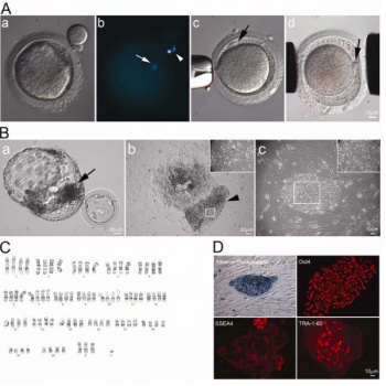
Establishment of HD hybrid cell line
(A) First polar body of mature rhesus macaque oocyte was removed by gentle squeezing through a slit of zona pellucida (A-a). Staining of 1st polar body DNA (arrowhead) and oocyte DNA (arrow) (A-b). HD monkey skin cell was placed under the zona pellucida (black arrow) (A-c). Reconstructed oocyte with HD monkey skin cell (A-d; yellow arrow) was placed between two electrodes for electrofusion (A-d). (B) Day 12 hatching blastocyst derived from HD monkey hybrid embryo (B-a; arrow indicated ICM). HD monkey hybrid blastocyst outgrowth at six days after attached onto feeder cells (B-b). High magnification of selected region (inset) of the ICM outgrowth (arrowhead). HD monkey hybrid cell line (TrES1) at passage 10 (B-c). (C) G-banding analysis of TrES1. Cytogenetic analysis of TrES1 demonstrated tetraploid chromosome (84; XXXY). (D) Expression of ES-cell specific markers: Alkaline phosphatase, Oct4, SSEA4 and TRA-1-60.
http://www.ncbi.nlm.nih.gov/pmc/articles/PMC2833146/?tool=pmcentrez
This is an Open Access article distributed under the terms of the Creative Commons Attribution License (http://creativecommons.org/licenses/by/2.0), which permits unrestricted use, distribution, and reproduction in any medium, provided the original work is properly cited. --Maeda Sadeghpour 21:21, 17 August 2011 (EST)
Lab 4 Assignment:
1. The allantois, identified in the placental cord, is continuous with what anatomical structure?
Ventral outgrowth of the hindgut in developing embryo, expanding from the caudal wall of the yolk sac.
2. Identify the 3 vascular shunts, and their location, in the embryonic circulation.
1. Ductus Venosus - connecting portal and umbilical veins to the inferior vena cava.
2. Ductus Arteriosus - connects pulmonary trunk to aorta.
3. Foramen Ovale - connects right and left atrium.
3. Identify the group project sub-section that you will be researching.
- Pathogenesis
- Genetics --z3290270 23:31, 24 August 2011 (EST)
Lab 5 Assignment:
Which side (L/R) is most common for diaphragmatic hernia and why?
Diaphragmatic hernias are more common on the left side. It is most likely due to the earlier closure of the right pleuroperitoneal opening. This could be life threatening due to compression of the lungs and other organs.
--Maeda Sadeghpour 00:48, 1 September 2011 (EST)
Lab 6 Assignment:
1. What week of development do the palatal shelves fuse?
The palatal shelves fuse in week 9 of development. The fusion takes place between both secondary and primary palate which is shapes at approximately week 6.
2. What animal model helped elucidate the neural crest origin and migration of cells?
The quail-chick chimeras.
3. What abnormality results from neural crest not migrating into the cardiac outflow tract?
Tetralogy of fallot which results in failure of the pulmonary trunk and aorta to divide functionally.
--Maeda Sadeghpour 01:20, 15 September 2011 (EST)
Lab assignment 7:
1. Are satellite cells (a) necessary for muscle hypertrophy and (b) generally involved in hypertrophy?
Satelite cells are not necessary for hypertrophy but b) they are generally involved in hypertrophy.
2. Why does chronic low frequency stimulation cause a fast to slow fibre type shift?
Chronic low frequency stimualtion damages the muscle cells which in turn stimualtes satelite cell recruitment which differentiates the fast fibres to slow fibre types. This method can be used to increase aerobic capacity.
--z3290270 21:13, 21 September 2011 (EST)
Peer review:
- Growth curves for trisomy- Although the graph was easy to understand, there was no description that explained the significance of the results, and punctuation was not used correctly in the one sentence used to describe the image.
- References: The fact that the references had so many subheadings, specifically in the opening page, led me to believe that it was merely there to give a false impression of comprehensive research. Even though there were some aspects of the writings that were all-inclusive.
- Lack of a glossary – This page would be available to a broad range of readers, and it might be worthwhile and of value to invest some time in developing a glossary describing the terminology used. For example atrioventricualr canal in the abnormalities segment.
- Defnitions as research: In quite a few sections there wasn't much detail explaining the concepts but just definitions.For example the “Prevalence is measure of the proportion of a population that are disease cases at a point in time. Generally used to measure only relatively stable conditions, not suitable for acute disorders.” And “Aneuploidy is the term used to describe any abnormal number of chromosomes either an increase or decrease in total number.” These should all be moved to the glossary segment. The fact that the definitions are taking up almost 1/3rd of the space dedicated to the specific section shows lack of research and dedication.
- Associated congenital abnormalities – Some of these can well be represented by pictures – either hand drawn or photographs would make the page more pleasing to look at and would encourage readers to understand to read on. The detail given to explain the abnormalities is very poor. They have not shown any link between the defect and the abnormalities (even if they haven’t been well understood, you can still mention any current or past research trying to discover these links), nor have they explained what these abnormalities are. For example: "leukemia - Acute lymphocytic (lymphoblastic) leukemia (ALL) and Acute megakaryocytic leukemia (AML). AML occurs 200 to 400 times more frequently in Down syndrome." This is a very interesting point, but it there is nothing explaining what AML/ALL are or how they are linked to trisomy 21.
- Structure of writing:
Most of the writings were in dot-point format. This may be an efficient way to make a draft of what will be discussed, but inappropriate as the final copy. It looked very rushed and “last-minute”.
- Recent findings: This section was very interesting, but perhaps it would be best positioned towards the end of the website. It would be best suited in my opinion to introduce and explain the disease before moving onto current research.
--z3290270 21:33, 21 September 2011 (EST)

