Developmental Mechanism - Cell Junctions: Difference between revisions
mNo edit summary |
mNo edit summary |
||
| Line 38: | Line 38: | ||
Tight junction | Tight junction | ||
|} | |} | ||
==Adherens Junction== | |||
{| | |||
| | |||
* | |||
* actin microfilaments anchor the plaque that occurs under the membrane of each cell | |||
* plaques not as dense also occur as hemi-form | |||
* heart muscle, layers covering body organs, digestive tract | |||
* transmembrane proteins | |||
* Cadherin | |||
* clustered as focal adhesion | |||
Cadherin isoform naming was based upon the site of original identification: P-cadherin (placenta cadherin, [https://www.omim.org/entry/114021 cadherin 3]), E-cadherin (epithelial cadherin [https://www.omim.org/entry/192090 cadherin 1]), N-cadherin (neural cadherin, [https://www.omim.org/entry/114020 cadherin 2]). | |||
| [[File:Adherens Junction 01.jpg|300px]] | |||
Adherens Junction | |||
|} | |||
[[:File:Rat blood–testis barrier 02.jpg|image - blood–testis barrier]] | |||
==External Links== | ==External Links== | ||
Revision as of 09:58, 26 April 2020
Embryology - 23 Oct 2026    Expand to Translate Expand to Translate
|
|---|
| Google Translate - select your language from the list shown below (this will open a new external page) |
|
العربية | català | 中文 | 中國傳統的 | français | Deutsche | עִברִית | हिंदी | bahasa Indonesia | italiano | 日本語 | 한국어 | မြန်မာ | Pilipino | Polskie | português | ਪੰਜਾਬੀ ਦੇ | Română | русский | Español | Swahili | Svensk | ไทย | Türkçe | اردو | ייִדיש | Tiếng Việt These external translations are automated and may not be accurate. (More? About Translations) |
Introduction
Many developmental mechanisms require dynamic changes in cell adhesion that acts through different types of cell junctions.
Adhesion Cartoons

Tight junction
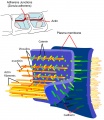
Adherens Junction
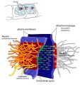
Desmosome
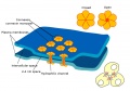
Gap Junction


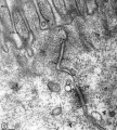
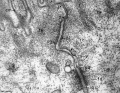
Adhesion EM Images
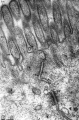
GIT epithelia EM1
GIT epithelia EM2
GIT epithelia EM3
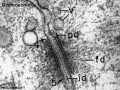
Desmosome EM


- Adhesion EM Images: GIT epithelia EM1 | GIT epithelia EM2 | GIT epithelia EM3 | Desmosome EM
- Adhesion Cartoons: Tight junction | Adherens Junction | Desmosome | Gap Junction
Tight Junction

Tight junction |
Adherens Junction
Cadherin isoform naming was based upon the site of original identification: P-cadherin (placenta cadherin, cadherin 3), E-cadherin (epithelial cadherin cadherin 1), N-cadherin (neural cadherin, cadherin 2). |

Adherens Junction |
External Links
External Links Notice - The dynamic nature of the internet may mean that some of these listed links may no longer function. If the link no longer works search the web with the link text or name. Links to any external commercial sites are provided for information purposes only and should never be considered an endorsement. UNSW Embryology is provided as an educational resource with no clinical information or commercial affiliation.
Glossary Links
- Glossary: A | B | C | D | E | F | G | H | I | J | K | L | M | N | O | P | Q | R | S | T | U | V | W | X | Y | Z | Numbers | Symbols | Term Link
Cite this page: Hill, M.A. (2026, October 23) Embryology Developmental Mechanism - Cell Junctions. Retrieved from https://embryology.med.unsw.edu.au/embryology/index.php/Developmental_Mechanism_-_Cell_Junctions
- © Dr Mark Hill 2026, UNSW Embryology ISBN: 978 0 7334 2609 4 - UNSW CRICOS Provider Code No. 00098G

