File:Gladstone1935 plate6.jpg: Difference between revisions
({{Ref-Gladstone1935}}) |
mNo edit summary |
||
| Line 1: | Line 1: | ||
== | ==Plate VI== | ||
Section of pineal cyst shown in [[:File:Gladstone1935 plate5.jpg|Plate V] C and E. The drawing also shows the relation of the dorsal diverticulum to the pineal gland, and chorioidal villi projecting into its lumen. Corpora arenacea are present in the tissue surrounding the pineal cyst and the diverticulum, and are also imbedded in their walls. | |||
Section of pineal tumour showing the lobulated arrangement of the tumour tissue, and tracts of degenerating glial tissue, containing thin-walled blood vessels. | |||
===Reference=== | |||
{{Ref-Gladstone1935}} | {{Ref-Gladstone1935}} | ||
Latest revision as of 09:07, 10 February 2020
Plate VI
Section of pineal cyst shown in [[:File:Gladstone1935 plate5.jpg|Plate V] C and E. The drawing also shows the relation of the dorsal diverticulum to the pineal gland, and chorioidal villi projecting into its lumen. Corpora arenacea are present in the tissue surrounding the pineal cyst and the diverticulum, and are also imbedded in their walls.
Section of pineal tumour showing the lobulated arrangement of the tumour tissue, and tracts of degenerating glial tissue, containing thin-walled blood vessels.
Reference
Gladstone RJ. and Wakeley CPG. Development and histogenesis of the human pineal organ. (1935) J Anat. 69: 427-454.11. PMID 17104550
File history
Yi efo/eka'e gwa ebo wo le nyangagi wuncin ye kamina wunga tinya nan
| Gwalagizhi | Nyangagi | Dimensions | User | Comment | |
|---|---|---|---|---|---|
| current | 09:03, 10 February 2020 | 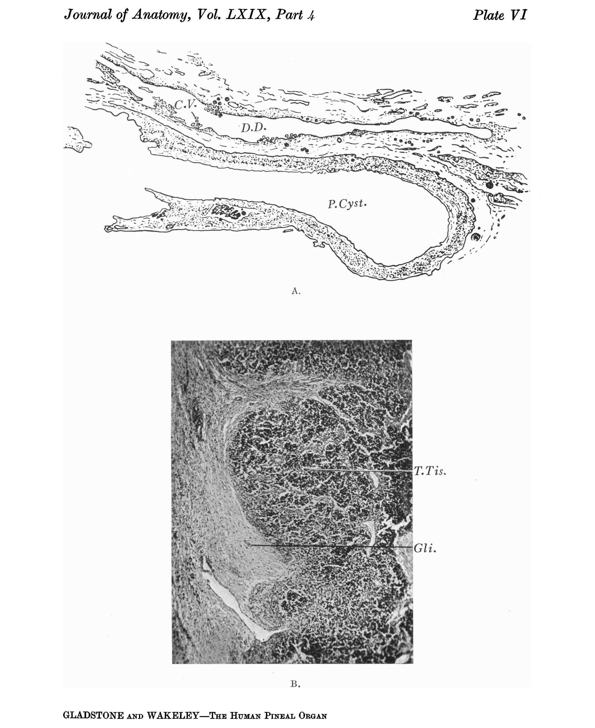 | 2,076 × 2,541 (728 KB) | Z8600021 (talk | contribs) | {{Ref-Gladstone1935}} |
You cannot overwrite this file.
File usage
The following page uses this file: