File:Ossification endochondral 01.jpg: Difference between revisions
From Embryology
(→Legend) |
|||
| Line 1: | Line 1: | ||
==Developing Vertebra - Endochondral Ossification== | ==Developing Vertebra - Endochondral Ossification== | ||
Histological image of a developing vertebra (neonatal {{rat}}), scale bar 160 microns | |||
{| | |||
| | |||
* vertebra - cartilage template and developing bony collar (centre of image) | * vertebra - cartilage template and developing bony collar (centre of image) | ||
* Note the dying cartilage cells (pale white regions) adjacent to the newly forming bony collar. | * Note the dying cartilage cells (pale white regions) adjacent to the newly forming bony collar. | ||
See also adjacent region image of [[:File:Ossification endochondral 1.jpg|Developing Intervertebral Disc]] | See also adjacent region image of [[:File:Ossification endochondral 1.jpg|Developing Intervertebral Disc]] | ||
| | |||
===Legend=== | ===Legend=== | ||
* blue - cartilage matrix | * blue - cartilage matrix | ||
* red - bone matrix | * red - bone matrix | ||
|} | |||
:'''Links:''' {{axial skeleton}} | [[:File:Ossification endochondral 1.jpg|Image - Intervertebral Disc]] | [[:File:Ossification_endochondral_01.jpg|Image - Vertebra]] | {{bone}} | [[Cartilage Histology]] | [[Bone Histology]] | |||
{{Footer}} | |||
[[Category:Histology]] [[Category:Musculoskeletal]] [[Category:Notochord]] [[Category:Cartilage]] [[Category:Bone]] [[Category:Axial Skeleton]] | [[Category:Histology]] [[Category:Musculoskeletal]] [[Category:Notochord]] [[Category:Cartilage]] [[Category:Bone]] [[Category:Axial Skeleton]] | ||
Revision as of 19:36, 2 May 2018
Developing Vertebra - Endochondral Ossification
Histological image of a developing vertebra (neonatal rat), scale bar 160 microns
See also adjacent region image of Developing Intervertebral Disc |
Legend
|
- Links: axial skeleton | Image - Intervertebral Disc | Image - Vertebra | bone | Cartilage Histology | Bone Histology
Cite this page: Hill, M.A. (2024, June 3) Embryology Ossification endochondral 01.jpg. Retrieved from https://embryology.med.unsw.edu.au/embryology/index.php/File:Ossification_endochondral_01.jpg
- © Dr Mark Hill 2024, UNSW Embryology ISBN: 978 0 7334 2609 4 - UNSW CRICOS Provider Code No. 00098G
File history
Click on a date/time to view the file as it appeared at that time.
| Date/Time | Thumbnail | Dimensions | User | Comment | |
|---|---|---|---|---|---|
| current | 12:33, 23 March 2012 | 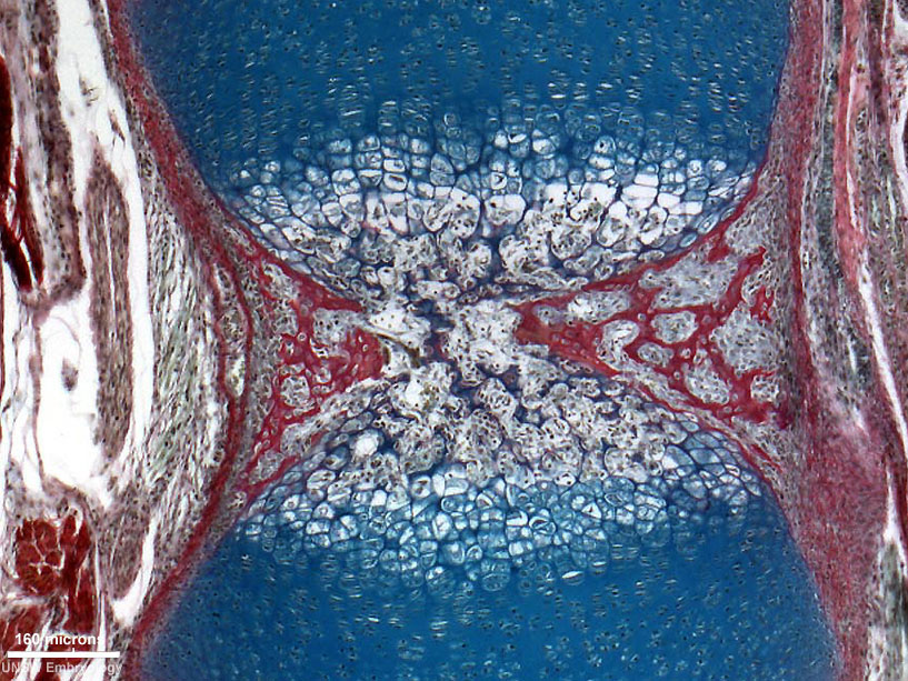 | 817 × 613 (198 KB) | Z8600021 (talk | contribs) | ==Developing Vertebra - Endochondral Ossification== * Histological image of a developing vertebra and intervertebral disc (neonatal rat). * intervertebral disc - nucleus pulposus and annular fibrocartilage (bottom of image) * vertebra - cartilage templa |
You cannot overwrite this file.
File usage
The following 2 pages use this file: