Talk:Draft 2016: Difference between revisions
From Embryology
mNo edit summary |
mNo edit summary |
||
| Line 1: | Line 1: | ||
== | ==Lymph Nodes== | ||
=== | ===Lymph Node Anatomy=== | ||
{| | {| | ||
! | ! Lymph Node Anatomy | ||
|- | |- | ||
| [[File:Thymus cartoon.jpg|400px]] | | [[File:Thymus cartoon.jpg|400px]] | ||
| Line 22: | Line 22: | ||
|} | |} | ||
=== | ===Lymph Node Functions=== | ||
{| class="wikitable mw-collapsible mw-collapsed" | {| class="wikitable mw-collapsible mw-collapsed" | ||
! colspan=2| | ! colspan=2|Lymph Node Functions | ||
|- | |- | ||
| valogn=top| | | valogn=top| | ||
| Line 55: | Line 55: | ||
|} | |} | ||
=== | ===Lymph Node Structure=== | ||
{| class="wikitable mw-collapsible mw-collapsed" | {| class="wikitable mw-collapsible mw-collapsed" | ||
! | ! Lymph Node Structure | ||
|- | |- | ||
| '''Structure Overview''' | | '''Structure Overview''' | ||
| Line 96: | Line 96: | ||
|} | |} | ||
=== | ===Lymph Node Histology=== | ||
{| class="wikitable mw-collapsible mw-collapsed" | {| class="wikitable mw-collapsible mw-collapsed" | ||
! | ! Lymph Node Histology | ||
|- | |- | ||
| | | | ||
| Line 133: | Line 133: | ||
{{Thymus Histology}} | {{Thymus Histology}} | ||
|} | |} | ||
==Lymph Nodes== | |||
[[File:Lymphatic-system-overview.jpg]] | |||
* Location throughout the entire body - Concentrated in axilla, groin, mesenteries | |||
* Encapsulated organ (1 mm - 2 cm) | |||
* Antigen transformed lymphocytes from the blood | |||
* In lymph vessel pathways “filter” | |||
* Afferent- towards node | |||
* Efferent- away from node | |||
Lymph flow | |||
* enters the node through '''afferent vessels''' | |||
* filters through the '''sinuses''' | |||
* leaves through '''efferent vessels''' | |||
===Lymph Node Structure=== | |||
[[File:Lymph_node_cartoon_01.jpg|Lymph_node_cartoon_01.jpg]] | |||
Connective Tissue | |||
* '''Capsule''' - dense connective tissue (irregular CT, some adipocytes)) | |||
* '''Trabeculae''' - dense connective tissue | |||
* '''Reticular Tissue''' - Reticular cells and fibers, supporting meshwork (collagen type III) | |||
** Reticular cell produces reticular fibers ('''collagen type III''') and surrounds the fibers with its cytoplasm | |||
** reticular fibbers can also be produced by fibroblasts | |||
<gallery> | |||
File:Lymph node cartoon 03.jpg|Cartoon with histology features | |||
File:Lymph node histology 01.jpg|Subcapsular Sinus (marginal sinus, continuation of trabecular sinus) | |||
File:Lymph node histology 02.jpg|Follicle | |||
File:Lymph node histology 03.jpg|Germinal Centre | |||
File:Lymph node histology 04.jpg|Medullary Cords and Sinuses | |||
File:Lymph node histology 05.jpg|High Endothelial Venules | |||
File:Lymph_node_histology_06.jpg|Macrophages | |||
</gallery> | |||
{| | |||
| | |||
===Lymphocyte (T and B) Traffic=== | |||
# Enter from high endothelial venules (HEVs also called post-capillary venules) | |||
# Spend 8 to 24 h in the lymph node interstitium. | |||
# Enter a network of medullary sinuses. | |||
# Drain from sinuses into efferent lymphatic vessels. | |||
'''Important -''' Please note that the lymphocytes exit from the node by efferent lymphatic vessels. | |||
[http://www.ncbi.nlm.nih.gov/books/NBK27092/figure/A47 See Figure] | |||
See also [[:File:Lymph node cartoon 02.jpg|Image - Cell Trafficking into and out of Lymph Nodes]]. | |||
| {{Lymph Node Movie 7}} | |||
T and B motility | |||
| | |||
{{Lymph Node Movie 8}} | |||
T and B interaction | |||
|[[File:Lymph node histology 05.jpg|200px]] | |||
High Endothelial Venules | |||
See also [[:File:Lymph node - high endothelial venule.jpg|Lymphocyte Migration at High Endothelial Venule Model]] | |||
|} | |||
'''Links:''' [http://www.ncbi.nlm.nih.gov/books/NBK27092/figure/A47 Immunobiology - Figure 1.8. Organization of a lymph node] | |||
Revision as of 17:42, 26 February 2016
Lymph Nodes
Lymph Node Anatomy
| Lymph Node Anatomy | |
|---|---|

|

|
| adult thymus - bilobed, superior mediastinum, anterior to heart | infant thymus - large |
| Thymus Involution | |
|---|---|

|
Overall Size Changes with age
|
Lymph Node Functions
| Lymph Node Functions | ||||
|---|---|---|---|---|
|

| |||

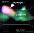
Macrophages phagocytosis of T cells
|

EM - T and B lymphocytes (look the same) | |||
Lymph Node Structure
| Lymph Node Structure | ||||||
|---|---|---|---|---|---|---|
Structure Overview

|
Lymph Node Histology
| Lymph Node Histology | |||||||||
|---|---|---|---|---|---|---|---|---|---|
Infant thymus
|


Lymph Nodes

- Location throughout the entire body - Concentrated in axilla, groin, mesenteries
- Encapsulated organ (1 mm - 2 cm)
- Antigen transformed lymphocytes from the blood
- In lymph vessel pathways “filter”
- Afferent- towards node
- Efferent- away from node
Lymph flow
- enters the node through afferent vessels
- filters through the sinuses
- leaves through efferent vessels
Lymph Node Structure

Connective Tissue
- Capsule - dense connective tissue (irregular CT, some adipocytes))
- Trabeculae - dense connective tissue
- Reticular Tissue - Reticular cells and fibers, supporting meshwork (collagen type III)
- Reticular cell produces reticular fibers (collagen type III) and surrounds the fibers with its cytoplasm
- reticular fibbers can also be produced by fibroblasts
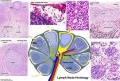
Cartoon with histology features
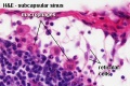
Subcapsular Sinus (marginal sinus, continuation of trabecular sinus)
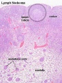
Follicle
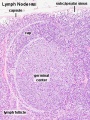
Germinal Centre
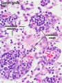
Medullary Cords and Sinuses
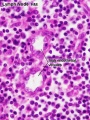
High Endothelial Venules
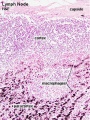
Macrophages


Lymphocyte (T and B) Traffic
Important - Please note that the lymphocytes exit from the node by efferent lymphatic vessels. See also Image - Cell Trafficking into and out of Lymph Nodes. |
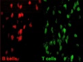
T and B motility |
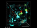
T and B interaction |

High Endothelial Venules See also Lymphocyte Migration at High Endothelial Venule Model |
Links: Immunobiology - Figure 1.8. Organization of a lymph node


