File:Adult mouse brain - prosomeric model.jpg: Difference between revisions
| Line 3: | Line 3: | ||
* <font color=crimson>'''Red'''</font> - hypothalamo-telencephalic prosomeres (hp1) | * <font color=crimson>'''Red'''</font> - hypothalamo-telencephalic prosomeres (hp1) | ||
* <font color=darkorange>'''Orange'''</font> - hypothalamo-telencephalic prosomeres (hp2) | * <font color=darkorange>'''Orange'''</font> - hypothalamo-telencephalic prosomeres (hp2) | ||
* <font color= | * <font color=gold>'''Yellow'''</font> - diencephalic prosomeres (p1–p3) | ||
* <font color=lawngreen>'''Green'''</font> - midbrain mesomeres (m1–m2) | * <font color=lawngreen>'''Green'''</font> - midbrain mesomeres (m1–m2) | ||
* <font color=deepskyblue>'''Blue'''</font> - Hindbrain rhombomeres and cryptorhombomeres (r0–r11) | * <font color=deepskyblue>'''Blue'''</font> - Hindbrain rhombomeres and cryptorhombomeres (r0–r11) | ||
Revision as of 11:30, 6 February 2016
Prosomeric Model of Adult Mouse Brain
- Red - hypothalamo-telencephalic prosomeres (hp1)
- Orange - hypothalamo-telencephalic prosomeres (hp2)
- Yellow - diencephalic prosomeres (p1–p3)
- Green - midbrain mesomeres (m1–m2)
- Blue - Hindbrain rhombomeres and cryptorhombomeres (r0–r11)
The roof, alar, basal and floor parts are not differentiated, for simplicity, but exist in every case (note the anterior commissure represents the rostralmost roof domain; the rostralmost floor corresponds to the mamillary area-M).
Abbreviations: ac, anterior commissure; cc, corpus callosum; VPall, LPall, DPall, MPall, ventral, lateral, dorsal and medial pallial sectors; PallSe, pallial septum; SPallSe, subpallial septum; OB, olfactory bulb; POA, preoptic area; THy, terminal hypothalamus; PHy, peduncular hypothalamus; PTh, prethalamus; Th, thalamus; PT, pretectum; M, mamillary body; APit, anterior pituitary; PPit, posterior pituitary; pc, posterior commissure; tc, tectal commissure; icc, intercollicular commissure.
Reference
<pubmed>25852489</pubmed>
http://dx.doi.org/10.3389/fnana.2015.00027
Copyright
Copyright © 2015 Puelles and Rubenstein.
This is an open-access article distributed under the terms of the Creative Commons Attribution License (CC BY). The use, distribution or reproduction in other forums is permitted, provided the original author(s) or licensor are credited and that the original publication in this journal is cited, in accordance with accepted academic practice. No use, distribution or reproduction is permitted which does not comply with these terms.
Original file name Figure 1. Fnana-09-00027-g001.jpg PMID added to figure.
File history
Yi efo/eka'e gwa ebo wo le nyangagi wuncin ye kamina wunga tinya nan
| Gwalagizhi | Nyangagi | Dimensions | User | Comment | |
|---|---|---|---|---|---|
| current | 10:59, 5 February 2016 | 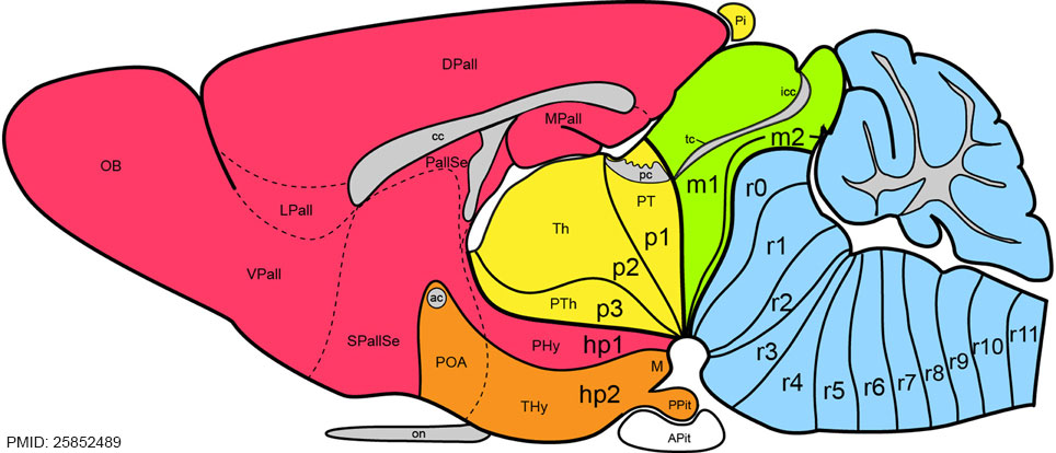 | 964 × 414 (91 KB) | Z8600021 (talk | contribs) | ==Prosomeric Model of Adult Mouse Brain== Hindbrain rhombomeres and cryptorhombomeres (r0–r11) are in blue, midbrain mesomeres (m1–m2) in green, diencephalic prosomeres (p1–p3) in yellow, and hypothalamo-telencephalic prosomeres (hp1–hp2) in r... |
You cannot overwrite this file.
File usage
The following 2 pages use this file: