File:Liver animated cartoon.gif: Difference between revisions
mNo edit summary |
mNo edit summary |
||
| Line 14: | Line 14: | ||
[[Category:Gastrointestinal Tract]] [[Category:Liver]] [[Category:Cartoon]] | [[Category:Gastrointestinal Tract]] [[Category:Liver]] [[Category:Cartoon]] | ||
[[Category:Animation]] | |||
Latest revision as of 17:02, 20 Ocak 2016
The Liver Lobule
An idealized representation of the "classical" liver lobule is a six-sided prism about 2 mm long and 1 mm in diameter. It is delimited by interlobular connective tissue (only little, if any, visible in humans; plentiful in e.g. pigs). In its corners we find the portal triads. In cross sections, the lobule is filled by cords of hepatic parenchymal cells, hepatocytes, which radiate from the central vein and are separated by vascular sinusoids.
There are other ways of dividing the parenchyma of the liver into units. Two common ways are divisions into portal lobules and liver acini.
- Portal lobules emphasize the afferent blood supply and bile drainage by the vessels of the portal triads. The secretory function of the liver is emphasized by the term liver acinus.
- Acini are smaller units than portal or "classical" liver lobules and relate structural units to terminal branches formed by the vessels of the portal triad but not the portal triad itself. Representations of portal lobules and liver acini vary in different textbooks.
Links: Histology | Histology Stains | Blue Histology images copyright Lutz Slomianka 1998-2009. The literary and artistic works on the original Blue Histology website may be reproduced, adapted, published and distributed for non-commercial purposes. See also the page Histology Stains.
Cite this page: Hill, M.A. (2026, October 3) Embryology Liver animated cartoon.gif. Retrieved from https://embryology.med.unsw.edu.au/embryology/index.php/File:Liver_animated_cartoon.gif
- © Dr Mark Hill 2026, UNSW Embryology ISBN: 978 0 7334 2609 4 - UNSW CRICOS Provider Code No. 00098G
File history
Yi efo/eka'e gwa ebo wo le nyangagi wuncin ye kamina wunga tinya nan
| Gwalagizhi | Nyangagi | Dimensions | User | Comment | |
|---|---|---|---|---|---|
| current | 15:45, 22 August 2010 | 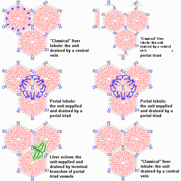 | 300 × 200 (239 KB) | S8600021 (talk | contribs) | ==The Liver Lobule== An idealized representation of the "classical" liver lobule is a six-sided prism about 2 mm long and 1 mm in diameter. It is delimited by interlobular connective tissue (only little, if any, visible in humans; plentiful in e.g. pigs) |
You cannot overwrite this file.


