Category:Electron Micrograph: Difference between revisions
From Embryology
mNo edit summary |
mNo edit summary |
||
| Line 1: | Line 1: | ||
This | This {{Embryology}} category shows pages and media related to the research imaging technique of scanning electron micrographs (SEM) in development. | ||
Note: | |||
* images in this category may also include some of the associated bright field images taken before SEM fixation and imaging. | |||
* there is a separate category [[:Category:Scanning EM|Scanning Electron Micrograph]]. | |||
:'''Links:''' [[Scanning Electron Microscopy]] | |||
Latest revision as of 11:09, 11 June 2015
This Embryology category shows pages and media related to the research imaging technique of scanning electron micrographs (SEM) in development. Note:
- images in this category may also include some of the associated bright field images taken before SEM fixation and imaging.
- there is a separate category Scanning Electron Micrograph.
- Links: Scanning Electron Microscopy
Pages in category 'Electron Micrograph'
The following 22 pages are in this category, out of 22 total.
H
P
- Paper - Cell-to-cell communication and ovulation - A study of the cumulus-oocyte complex
- Paper - Electron microscopy of the sperm tail - results obtained with a new fixative
- Paper - Fine structure of the human ovum in the pronuclear stage
- Paper - Studies on the fine structure of the mammalian testis 1
- Paper - Studies on the human oocyte and its follicle 1
- Template:Placenta EM links
R
Media in category 'Electron Micrograph'
The following 200 files are in this category, out of 201 total.
(previous page) (next page)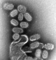 1918 influenza virus virions EM.jpg 700 × 743; 82 KB
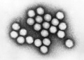
 2009 influenza virus virions EM.jpg 700 × 676; 126 KB
 Adenovirus.jpg 700 × 499; 45 KB
 Amphibian oocyte transcription.jpg 1,200 × 841; 475 KB
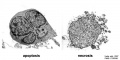 Apoptosis and necrosis.jpg 800 × 400; 60 KB
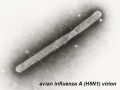 Avian influenza virion.jpg 320 × 240; 12 KB
 B lymphocyte EM08.jpg 1,000 × 730; 111 KB
 B lymphocyte EM09.jpg 673 × 1,000; 89 KB
 B lymphocyte EM10.jpg 671 × 1,000; 92 KB
 B lymphocytes EM08-10.jpg 677 × 996; 124 KB
 Blood capillary EM 01.jpg 1,107 × 714; 260 KB
 Blood capillary EM 02.jpg 600 × 600; 99 KB
 Blood capillary EM 03.jpg 1,560 × 1,230; 441 KB
 Blood capillary EM 04.jpg 1,560 × 1,230; 462 KB
 Blood capillary EM 05.jpg 1,015 × 800; 205 KB
 Blood capillary EM 06.jpg 1,015 × 800; 216 KB
 Blood-brain barrier EM01.jpg 1,656 × 810; 250 KB
 Blood-thymus barrier EM01.jpg 1,280 × 1,747; 375 KB
 BurgosFawcett1955 fig11.jpg 1,453 × 2,015; 528 KB
 BurgosFawcett1955 fig13.jpg 1,460 × 2,049; 501 KB
 BurgosFawcett1955 fig14.jpg 1,456 × 1,965; 381 KB
 Cajal body EM.jpg 600 × 487; 29 KB
 Cardiac muscle EM01.jpg 1,072 × 735; 231 KB
 Cardiac muscle EM02.jpg 1,072 × 735; 224 KB
 Cardiac muscle EM03.jpg 849 × 615; 135 KB
 Cardiac muscle EM04.jpg 1,000 × 680; 191 KB
 Cardiac Muscle EM05.jpg 992 × 733; 158 KB
 Cartilage em01.jpg 800 × 551; 176 KB
 Collagen EM01.jpg 3,839 × 2,979; 7.77 MB
 Coxsackie B4 virus.jpg 400 × 266; 14 KB
 Cytomegalovirus infected spermatozoa EM01.jpg 990 × 991; 204 KB
 Cytomegalovirus infected spermatozoa.jpg 1,000 × 1,260; 324 KB
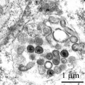
 Cytomegalovirus virions EM.jpg 911 × 987; 212 KB
 Cytoplasmic lattices in GV oocyte cytoplasm.jpg 1,048 × 846; 291 KB
 Cytoplasmic lattices in oocytes and two-cell embryos.jpg 753 × 1,000; 226 KB
 Desmosome 02.jpg 600 × 450; 74 KB
 Endotheliochorial placenta EM01.jpg 600 × 602; 134 KB
 Epithelial junctions EM01.jpg 658 × 1,000; 232 KB
 Epithelial junctions EM02.jpg 900 × 1,000; 288 KB
 Epithelial junctions EM03.jpg 1,291 × 1,000; 344 KB
 Erythrocyte and lymphocyte SEM01.jpg 800 × 522; 74 KB
 Erythrocyte and lymphocyte SEM02.jpg 800 × 522; 78 KB
 Erythrocyte and lymphocyte SEM03.jpg 800 × 522; 80 KB
 Fawcett1975 fig31.jpg 1,280 × 403; 128 KB
 Fawcett1975 fig34.jpg 1,280 × 1,746; 506 KB
 Haemomonochorial human placenta EM01.jpg 792 × 775; 79 KB
 Haemomonochorial placenta EM01.jpg 600 × 602; 111 KB
 Hepatitis A virus.jpg 700 × 537; 90 KB
 Hepatitis B virus.jpg 700 × 1,030; 123 KB
 Hepatitis E virus.jpg 700 × 464; 61 KB
 Herpes virus.jpg 320 × 240; 22 KB
 HertigAdams1967 fig01-4.jpg 1,691 × 2,353; 390 KB
 HertigAdams1967 fig01.jpg 531 × 547; 33 KB
 HertigAdams1967 fig02.jpg 531 × 547; 32 KB
 HertigAdams1967 fig03.jpg 518 × 545; 29 KB
 HertigAdams1967 fig04.jpg 1,628 × 1,714; 383 KB
 HertigAdams1967 fig25.jpg 1,637 × 1,452; 256 KB
 Human corpus luteum - light-and-electron-micrograph.jpg 936 × 711; 208 KB
 Human embryo skin 24 week EGA.jpg 596 × 939; 165 KB
 Human embryo skin 8-9 week EGA desmosomes.jpg 800 × 198; 40 KB
 Human embryo skin 8-9 week EGA.jpg 657 × 872; 188 KB
 Human embryo skin 9-11 week EGA.jpg 623 × 804; 176 KB
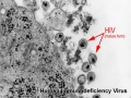 Human immunodeficiency virus.jpg 320 × 240; 25 KB
 Human lung inter-alveolar septum em01.jpg 785 × 589; 168 KB
 Human oocyte em01.jpg 600 × 589; 65 KB
 Human ovary follicle basement membrane EM01.jpg 558 × 697; 100 KB
 Human ovary follicles light and electron microscopy 01.jpg 586 × 1,080; 225 KB
 Human papilloma virus.jpg 961 × 721; 218 KB
 Human pronuclear stage EM02.jpg 639 × 1,000; 194 KB
 Human pronuclear stage EM022.jpg 1,100 × 705; 225 KB
 Human pronuclear stage EM03-05.jpg 982 × 439; 104 KB
 Human pronuclear stage EM06.jpg 907 × 1,000; 245 KB
 Human pronuclear stage EM07.jpg 357 × 509; 52 KB
 Human pronuclear stage EM08.jpg 359 × 513; 56 KB
 Human pronuclear stage EM09.jpg 361 × 506; 53 KB
 Human pronuclear stage EM10.jpg 630 × 781; 140 KB
 Human pronuclear stage EM11.jpg 625 × 768; 130 KB
 Human pronuclear stage EM12.jpg 998 × 777; 265 KB
 Human pronuclear stage EM13.jpg 998 × 771; 247 KB
 Human pronuclear stage EM14-16.jpg 1,013 × 459; 141 KB
 Human pronuclear stage EM17.jpg 1,104 × 504; 156 KB
 Human pronuclear stage EM18.jpg 477 × 541; 66 KB
 Human pronuclear stage EM19.jpg 478 × 534; 67 KB
 Human pronuclear stage EM20.jpg 1,114 × 762; 237 KB
 Human pronuclear stage EM21.jpg 366 × 587; 58 KB
 Human pronuclear stage EM22.jpg 366 × 581; 59 KB
 Human pronuclear stage EM25.jpg 1,013 × 782; 201 KB
 Human pronuclear stage EM26.jpg 1,010 × 784; 187 KB
 Human pronuclear stage EM27.jpg 997 × 777; 227 KB
 Human pronuclear stage EM28.jpg 993 × 774; 239 KB
 Human pronuclear stage EM29.jpg 967 × 763; 183 KB
 Human pronuclear stage EM30.jpg 955 × 853; 192 KB
 Human sperm pathologies EM01.jpg 761 × 759; 148 KB
 Human sperm pathology EM02.jpg 800 × 256; 22 KB
 Human spermatid electron micrograph.jpg 619 × 918; 206 KB
 Human spermatid EM01.jpg 1,000 × 762; 162 KB
 Human spermatid EM02.jpg 1,000 × 762; 186 KB
 Human spermatozoa nucleus EM01.jpg 600 × 476; 27 KB
 Human spermatozoa nucleus EM02.jpg 597 × 476; 52 KB
 Human spermatozoa nucleus EM03.jpg 600 × 475; 44 KB
 Human-spermatozoa EM01.jpg 1,000 × 204; 26 KB
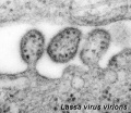 Lassa virus.jpg 320 × 278; 28 KB
 Leydig cell PMID13693345 EM02.jpg 1,359 × 957; 341 KB
 Leydig cell PMID13693345 EM03.jpg 1,359 × 957; 325 KB
 Listeria-bacterium.jpg 320 × 240; 15 KB
 Liver histology EM01.jpg 1,028 × 708; 141 KB
 Liver histology EM02.jpg 1,028 × 707; 154 KB
 Lutein cell glycogen granule em01.jpg 1,149 × 749; 169 KB
 Lutein cell lipid and glycogen em01.jpg 1,156 × 828; 149 KB
 Lutein cell lipid and glycogen em02.jpg 1,109 × 796; 227 KB
 Lymphocyte rosettes EM01-06.jpg 1,364 × 2,100; 334 KB
 Lymphocyte rosettes EM01.jpg 661 × 665; 58 KB
 Lymphocyte rosettes EM012.jpg 618 × 661; 59 KB
 Lymphocyte rosettes EM02.jpg 661 × 665; 62 KB
 Lymphocyte rosettes EM03.jpg 661 × 665; 51 KB
 Lymphocyte rosettes EM04.jpg 661 × 665; 53 KB
 Lymphocyte rosettes EM05.jpg 661 × 665; 55 KB
 Malaria and red blood cell em.jpg 500 × 536; 82 KB
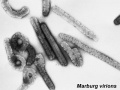 Marburg virus.jpg 320 × 240; 15 KB
 Measles virus.jpg 700 × 535; 91 KB
 Merkel cell EM 01.jpg 984 × 738; 209 KB
 Merkel cell EM 02.jpg 984 × 685; 166 KB
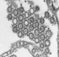
 MERS-CoV EM1.jpg 537 × 537; 68 KB
 Mitochondria EM01.jpg 640 × 480; 96 KB
 Model capacitation-induced acrosome docking to sperm membrane.jpg 600 × 489; 73 KB
 Monocyte EM01.jpg 923 × 1,000; 221 KB
 Mouse antral follicle 01.jpg 932 × 1,095; 374 KB
 Mouse antral follicle.jpg 600 × 705; 168 KB
 Mouse follicle in vitro 02.jpg 600 × 701; 146 KB
 Mouse follicle in vitro.jpg 600 × 701; 170 KB
 Mouse heart primary cilia 01.jpg 1,200 × 908; 415 KB
 Mouse neonatal ovary oocyte EM01.jpg 677 × 1,000; 266 KB
 Mouse neonatal ovary oocyte EM02.jpg 790 × 792; 179 KB
 Mouse neonatal ovary oocyte EM03.jpg 790 × 792; 187 KB
 Mouse neonatal ovary oocyte EM04.jpg 790 × 792; 235 KB
 Mouse neonatal ovary oocyte EM05.jpg 790 × 792; 217 KB
 Mouse neonatal ovary oocyte EM06.jpg 790 × 792; 163 KB
 Mouse neonatal ovary oocyte EM07.jpg 790 × 792; 183 KB
 Mouse oocyte and zona pellucida EM01.jpg 1,200 × 1,200; 485 KB
 Mouse oocyte and zona pellucida EM01a.jpg 800 × 800; 234 KB
 Mouse oocyte and zona pellucida EM01b.jpg 600 × 600; 133 KB
 Mouse oocyte and zona pellucida EM01c.jpg 400 × 400; 60 KB
 Mouse oocyte balbini body EM01.jpg 695 × 700; 155 KB
 Mouse placenta blood vessel EM01.jpg 1,000 × 739; 132 KB
 Mouse primitive node cilia.jpg 592 × 981; 130 KB
 Mouse renal podocyte EM01.jpg 1,000 × 1,338; 366 KB
 Mouse renal podocyte EM02.jpg 1,000 × 666; 155 KB
 Mouse- cerebellum axons.jpg 600 × 695; 129 KB
 Mouse- spinal cord axons.jpg 600 × 693; 127 KB
 Mouse- zona pellucida 01.jpg 800 × 430; 83 KB
 Mouse-oocyte-d.jpg 790 × 792; 217 KB
 Mouse-optic nerve axons.jpg 600 × 693; 126 KB
 Mouse-sciatic nerve Schwann cell.jpg 957 × 1,050; 339 KB
 Mumps virus.jpg 700 × 938; 221 KB
 Muscle satellite cell EM01.jpg 1,000 × 804; 142 KB
 Muscle satellite cell EM02.jpg 1,000 × 804; 153 KB
 Mycobacterium-tuberculosis.jpg 320 × 240; 18 KB
 Mycoplasma-pneumoniae.jpg 320 × 240; 31 KB
 Neonatal human pulmonary neuroendocrine cell EM01.jpg 836 × 1,200; 405 KB
 Nephron EM01.jpg 1,909 × 1,280; 219 KB
 Nephron EM02.jpg 1,271 × 1,280; 210 KB
 Nephron EM11.jpg 2,983 × 2,000; 398 KB
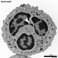 Neutrophil EM01.jpg 999 × 1,000; 402 KB
 Pancreas acinar cell em01.jpg 1,280 × 928; 496 KB
 Parvovirus 01.jpg 1,200 × 796; 275 KB
 Pig sperm capacitation 01.jpg 1,000 × 840; 204 KB
 Pig sperm capacitation 02.jpg 600 × 504; 82 KB
 Pituitary histology 010.jpg 1,005 × 961; 249 KB
 Placental imaging 03A.jpg 800 × 738; 181 KB
 Placental trophospongium.jpg 567 × 344; 94 KB
 Plasma cell EM06.jpg 595 × 600; 84 KB
 Respiratory Epithelium EM01a.jpg 800 × 584; 157 KB
 Respiratory Epithelium EM01b.jpg 220 × 161; 16 KB
 Respiratory Epithelium EM02.jpg 1,200 × 1,005; 483 KB
 Respiratory Epithelium EM02a.jpg 800 × 670; 237 KB
 Respiratory Epithelium EM02b.jpg 220 × 184; 19 KB
 Rotavirus.jpg 700 × 540; 64 KB
 Rubella virus 01.jpg 1,200 × 896; 352 KB
 Rubella virus 02.jpg 700 × 923; 135 KB
 Rubella virus 03.jpg 1,200 × 802; 224 KB
 Rubella virus.jpg 210 × 158; 5 KB
 Spermatozoa tail EM01.jpg 932 × 613; 75 KB
 Staphylococcus-aureus.jpg 320 × 240; 28 KB
 T and B lymphocytes EM09.jpg 1,196 × 627; 137 KB
 T and B lymphocytes EM10.jpg 1,196 × 677; 155 KB
 T2 lymphocyte EM13.jpg 781 × 795; 177 KB
 T2 lymphocyte EM14.jpg 728 × 771; 156 KB
 Treponema-pallidum.jpg 320 × 240; 21 KB
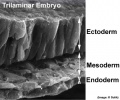 Trilaminar embryo.jpg 432 × 359; 32 KB
 Varicella zoster virus.jpg 800 × 600; 102 KB
 Vascular Sinus EM01.jpg 1,200 × 878; 279 KB
 Vascular Sinus EM01a.jpg 800 × 585; 141 KB
 Vascular Sinus EM01b.jpg 220 × 161; 15 KB
 Venule microvessel EM.jpg 600 × 626; 91 KB
 West Nile virus EM01.jpg 700 × 538; 114 KB
 Zamboni1966 fig02.jpg 1,556 × 1,000; 397 KB
 Zamboni1966 fig06.jpg 1,280 × 1,158; 419 KB
 Zamboni1966 fig20.jpg 1,526 × 1,000; 429 KB
 Zamboni1966 fig29.jpg 1,280 × 1,003; 302 KB
 Zika virus TEM01.jpg 800 × 800; 212 KB


