File:Hindbrain neural crest migration.jpg: Difference between revisions
From Embryology
mNo edit summary |
mNo edit summary |
||
| Line 1: | Line 1: | ||
==Hindbrain neural crest migration== | ==Hindbrain neural crest migration== | ||
{| | {| | ||
| A schematic diagram of a chick head at embryonic day two ([[Hamburger Hamilton Stages]]), showing pathways of neural crest migration in the chick and mouse embryo and patterns of Hox gene expression in the pharyngeal arches. Hox genes are expressed in neural crest cells, which emigrate predominantly from even-numbered rhombomeres into the pharyngeal (branchial) arches generating skeletal tissues and cranial ganglia. | | width=400px|A schematic diagram of a chick head at embryonic day two ([[Hamburger Hamilton Stages]]), showing pathways of neural crest migration in the chick and mouse embryo and patterns of Hox gene expression in the pharyngeal arches. Hox genes are expressed in neural crest cells, which emigrate predominantly from even-numbered rhombomeres into the pharyngeal (branchial) arches generating skeletal tissues and cranial ganglia. | ||
Note that the first pharyngeal arch is free of Hox expression. | Note that the first pharyngeal arch is free of Hox expression. | ||
| Line 13: | Line 13: | ||
|} | |} | ||
:'''Links:''' [[Developmental Signals - Homeobox|Homeobox]] | [[Neural Crest Development]] | |||
===Reference=== | ===Reference=== | ||
<pubmed>17948031</pubmed> | <pubmed>17948031</pubmed> | ||
Revision as of 13:39, 14 October 2014
Hindbrain neural crest migration
| A schematic diagram of a chick head at embryonic day two (Hamburger Hamilton Stages), showing pathways of neural crest migration in the chick and mouse embryo and patterns of Hox gene expression in the pharyngeal arches. Hox genes are expressed in neural crest cells, which emigrate predominantly from even-numbered rhombomeres into the pharyngeal (branchial) arches generating skeletal tissues and cranial ganglia.
Note that the first pharyngeal arch is free of Hox expression. |
Legend
|
- Links: Homeobox | Neural Crest Development
Reference
<pubmed>17948031</pubmed>
Copyright
Adapted by permission from Macmillan Publishers Ltd: Nature Reviews Neuroscience (<pubmed>17948031</pubmed>), copyright (2007)
Original Figure: 4 http://www.nature.com/nrn/journal/v8/n11/fig_tab/nrn2254_F4.html
Note original figure resized and relabeled replacing branchial arches with pharyngeal arches.
File history
Click on a date/time to view the file as it appeared at that time.
| Date/Time | Thumbnail | Dimensions | User | Comment | |
|---|---|---|---|---|---|
| current | 16:23, 31 August 2010 | 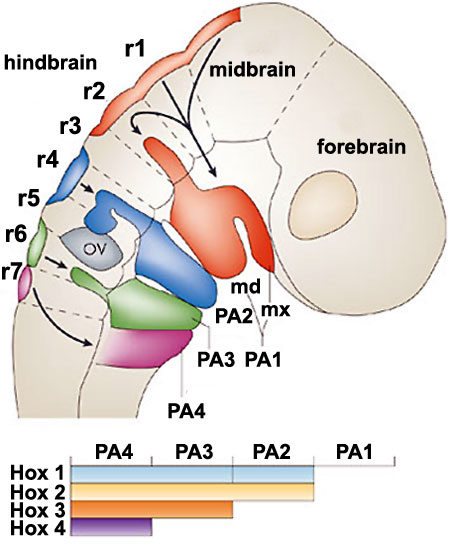 | 450 × 545 (48 KB) | S8600021 (talk | contribs) | ==Hindbrain neural crest migration== A schematic diagram of a chick head at embryonic day two, showing pathways of neural crest migration in the chick and mouse embryo and patterns of Hox gene expression in the branchial arches (BAs)42, 102, 169, 170. FB |
You cannot overwrite this file.