File:Mitochondria EM01.jpg: Difference between revisions
From Embryology
mNo edit summary |
mNo edit summary |
||
| Line 2: | Line 2: | ||
Transmission electron microscope image of a thin section cut through an area of mammalian lung tissue. The high magnification image shows a mitochondria. | Transmission electron microscope image of a thin section cut through an area of mammalian lung tissue. The high magnification image shows a mitochondria. | ||
---- | |||
{{DartmouthEM}} | |||
JEOL 100CX TEM | JEOL 100CX TEM | ||
| Line 9: | Line 14: | ||
Keywords: mitochondria, mammalian, TEM, lung | Keywords: mitochondria, mammalian, TEM, lung | ||
[[Category:Mitochondria]] [[Category:Electron Micrograph]] | [[Category:Mitochondria]] [[Category:Electron Micrograph]] | ||
Revision as of 12:55, 16 April 2014
Mitochondria Electron Micrograph
Transmission electron microscope image of a thin section cut through an area of mammalian lung tissue. The high magnification image shows a mitochondria.
Image Source: Contributed by Dartmouth College Electron Microscope Facility special thanks to Chuck Daghlian and Louisa Howard. Gallery. Original images may have been altered in size contrast and labelling. (These images are in the public domain)
JEOL 100CX TEM
Writer: Louisa Howard
Keywords: mitochondria, mammalian, TEM, lung
File history
Yi efo/eka'e gwa ebo wo le nyangagi wuncin ye kamina wunga tinya nan
| Gwalagizhi | Nyangagi | Dimensions | User | Comment | |
|---|---|---|---|---|---|
| current | 11:47, 23 Haziran 2011 | 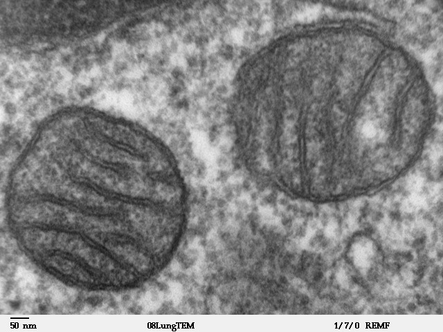 | 640 × 480 (96 KB) | S8600021 (talk | contribs) | ==Mitochondria Electron Micrograph== Transmission electron microscope image of a thin section cut through an area of mammalian lung tissue. The high magnification image shows a mitochondria. JEOL 100CX TEM Writer: Louisa Howard Keywords: mitochondria, |
You cannot overwrite this file.
File usage
The following 3 pages use this file:


