File:Hydatidiform mole 02.jpg: Difference between revisions
(==Hydatidiform mole== In this patient with a 6-weeks' gestation presenting with vaginal bleeding, transvaginal USG (A) shows a gestational sac (white arrow) on the first examination. Follow-up examination after 2 days (B) shows focal cystic changes () |
mNo edit summary |
||
| Line 6: | Line 6: | ||
(B) shows focal cystic changes (arrow) with loss of normal definition of the gestational sac, suggesting the possibility of molar changes. Investigations confirmed triploidy | (B) shows focal cystic changes (arrow) with loss of normal definition of the gestational sac, suggesting the possibility of molar changes. Investigations confirmed triploidy | ||
:'''Links:''' [[Abnormal_Development_-_Hydatidiform_Mole|Hydatidiform Mole]] | |||
===Reference=== | |||
Indian J Radiol Imaging. 2008 November; 18(4): 326–344. | Indian J Radiol Imaging. 2008 November; 18(4): 326–344. | ||
| Line 14: | Line 20: | ||
<pubmed>19774194</pubmed> | <pubmed>19774194</pubmed> | ||
Copyright © Indian Journal of Radiology and Imaging | ====Copyright==== | ||
© Indian Journal of Radiology and Imaging | |||
This is an open-access article distributed under the terms of the Creative Commons Attribution License, which permits unrestricted use, distribution, and reproduction in any medium, provided the original work is properly cited. | This is an open-access article distributed under the terms of the Creative Commons Attribution License, which permits unrestricted use, distribution, and reproduction in any medium, provided the original work is properly cited. | ||
[[Category:Placenta]] [[Category:Ultrasound]] | [[Category:Placenta]] [[Category:Ultrasound]] [[Category:Human]] [[Category:Abnormal Development]] [[Category:Uterus]] | ||
Revision as of 22:28, 10 June 2013
Hydatidiform mole
In this patient with a 6-weeks' gestation presenting with vaginal bleeding, transvaginal USG
(A) shows a gestational sac (white arrow) on the first examination. Follow-up examination after 2 days
(B) shows focal cystic changes (arrow) with loss of normal definition of the gestational sac, suggesting the possibility of molar changes. Investigations confirmed triploidy
- Links: Hydatidiform Mole
Reference
Indian J Radiol Imaging. 2008 November; 18(4): 326–344. doi: 10.4103/0971-3026.43848.
Original file name: IJRI-18-326-g013.jpg
<pubmed>19774194</pubmed>
Copyright
© Indian Journal of Radiology and Imaging
This is an open-access article distributed under the terms of the Creative Commons Attribution License, which permits unrestricted use, distribution, and reproduction in any medium, provided the original work is properly cited.
File history
Click on a date/time to view the file as it appeared at that time.
| Date/Time | Thumbnail | Dimensions | User | Comment | |
|---|---|---|---|---|---|
| current | 02:34, 27 May 2010 | 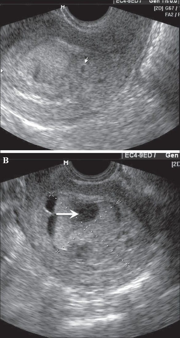 | 585 × 1,100 (237 KB) | S8600021 (talk | contribs) | ==Hydatidiform mole== In this patient with a 6-weeks' gestation presenting with vaginal bleeding, transvaginal USG (A) shows a gestational sac (white arrow) on the first examination. Follow-up examination after 2 days (B) shows focal cystic changes ( |
You cannot overwrite this file.
File usage
The following page uses this file: