File:Human right ovary and tube 1.jpg: Difference between revisions
From Embryology
mNo edit summary |
|||
| Line 16: | Line 16: | ||
Image source: UNSW Embryology, no reproduction without permission. | Image source: UNSW Embryology, no reproduction without permission. | ||
[[Category:Ovary]] [[Category:Uterus]] | [[Category:Human]] [[Category:Ovary]] [[Category:Uterus]] | ||
Revision as of 11:51, 9 May 2013
Human Ovary and Associated Uterine Tube
Adult human ovary (right) viewed by laparoscopy.
- Note the relative size and position of the ovary with respect to the uterine tube (fallopian tube, oviduct).
- The ovary surface appears white and relatively avascular
- representing the dense connective tissue layer (tunica albuginea).
- In the background the associated mesenteries and peritoneal cavity can be seen.
Image source: UNSW Embryology, no reproduction without permission.
File history
Yi efo/eka'e gwa ebo wo le nyangagi wuncin ye kamina wunga tinya nan
| Gwalagizhi | Nyangagi | Dimensions | User | Comment | |
|---|---|---|---|---|---|
| current | 09:31, 9 April 2010 | 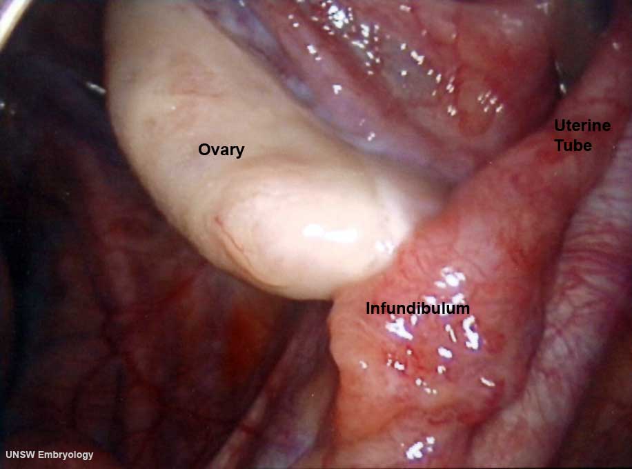 | 916 × 680 (32 KB) | S8600021 (talk | contribs) | Human right ovary and tube as viewed by laparoscopy == Image version links == Large 1000px | 800px | Medium 600px | [[:F |
You cannot overwrite this file.