External Genital Female Development Movie: Difference between revisions
No edit summary |
No edit summary |
||
| Line 1: | Line 1: | ||
{{Movie header}} | {{Movie header}} | ||
{| | {| | ||
| width=300px|<mediaplayer width='270' height='380' image="http://embryology.med.unsw.edu.au/embryology/images/3/37/Male_external_001_icon.jpg">File: | | width=300px|<mediaplayer width='270' height='380' image="http://embryology.med.unsw.edu.au/embryology/images/3/37/Male_external_001_icon.jpg">File:Female_external_001.mp4</mediaplayer> | ||
| valign="top" |[[File:Female_external_001_icon.jpg|100px|left]] | | valign="top" |[[File:Female_external_001_icon.jpg|100px|left]] | ||
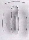
===Female External Genital Development=== | ===Female External Genital Development=== | ||
This | This animation shows the development of external female genitalia from the indifferent external structure, covering the approximate period of week 9 to 12. (Anterior to top, Posterior to bottom) | ||
Note the original cloacal membrane becomes separated into the urogenital membrane and anal membrane. | |||
The urogenital folds beneath the genital tubercle remain separate (unfused), forming the inner labia minora and second outer skin folds form the larger labia majora either side of the developing vestibule of the vagina. | |||
Note at the top of the animation, the changing relative size of the genital tubercle as it forms the glans of the clitoris. | |||
Revision as of 17:41, 6 March 2013
| Embryology - 26 Jun 2024 |
|---|
| Google Translate - select your language from the list shown below (this will open a new external page) |
|
العربية | català | 中文 | 中國傳統的 | français | Deutsche | עִברִית | हिंदी | bahasa Indonesia | italiano | 日本語 | 한국어 | မြန်မာ | Pilipino | Polskie | português | ਪੰਜਾਬੀ ਦੇ | Română | русский | Español | Swahili | Svensk | ไทย | Türkçe | اردو | ייִדיש | Tiếng Việt These external translations are automated and may not be accurate. (More? About Translations) |
| <mediaplayer width='270' height='380' image="http://embryology.med.unsw.edu.au/embryology/images/3/37/Male_external_001_icon.jpg">File:Female_external_001.mp4</mediaplayer> | Female External Genital DevelopmentThis animation shows the development of external female genitalia from the indifferent external structure, covering the approximate period of week 9 to 12. (Anterior to top, Posterior to bottom) Note the original cloacal membrane becomes separated into the urogenital membrane and anal membrane. The urogenital folds beneath the genital tubercle remain separate (unfused), forming the inner labia minora and second outer skin folds form the larger labia majora either side of the developing vestibule of the vagina. Note at the top of the animation, the changing relative size of the genital tubercle as it forms the glans of the clitoris.
|
Glossary Links: A | B | C | D | E | F | G | H | I | J | K | L | M | N | O | P | Q | R | S | T | U | V | W | X | Y | Z | Numbers | Symbols | Movies
Cite this page: Hill, M.A. (2024, June 26) Embryology External Genital Female Development Movie. Retrieved from https://embryology.med.unsw.edu.au/embryology/index.php/External_Genital_Female_Development_Movie
- © Dr Mark Hill 2024, UNSW Embryology ISBN: 978 0 7334 2609 4 - UNSW CRICOS Provider Code No. 00098G