File:Renal histology 03.jpg: Difference between revisions
From Embryology
No edit summary |
|||
| Line 1: | Line 1: | ||
==Renal Histology== | ==Renal Histology== | ||
* distal-tubule (x40) | * distal-tubule (x40) {{HE}} | ||
* collecting duct | * collecting duct | ||
===Tubule Histology=== | ===Tubule Histology=== | ||
Revision as of 14:01, 23 February 2013
Renal Histology
- distal-tubule (x40) (Stain - Haematoxylin Eosin)
- collecting duct
Tubule Histology
Proximal Convoluted Tubules (PCT)
- brush border
- star-shaped
- larger outside diameter
Distal Convoluted Tubules (DCT)
- clean lumen surface
- apical nuclei
Collecting Tubules (CT)
- larger lumen than DCT (about size of PCT)
- cuboidal cells and smaller than DCT
- Renal System Histology: Nephron tubule overview | glomerulus structure | vascular and renal poles | Medullary rays | Nephron tubules
Links: Histology | Histology Stains | Blue Histology images copyright Lutz Slomianka 1998-2009. The literary and artistic works on the original Blue Histology website may be reproduced, adapted, published and distributed for non-commercial purposes. See also the page Histology Stains.
Cite this page: Hill, M.A. (2024, June 27) Embryology Renal histology 03.jpg. Retrieved from https://embryology.med.unsw.edu.au/embryology/index.php/File:Renal_histology_03.jpg
- © Dr Mark Hill 2024, UNSW Embryology ISBN: 978 0 7334 2609 4 - UNSW CRICOS Provider Code No. 00098G
Original File name: kid40he.jpg
File history
Yi efo/eka'e gwa ebo wo le nyangagi wuncin ye kamina wunga tinya nan
| Gwalagizhi | Nyangagi | Dimensions | User | Comment | |
|---|---|---|---|---|---|
| current | 11:24, 4 September 2011 | 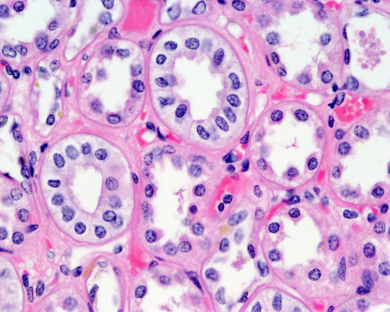 | 1,280 × 1,024 (280 KB) | S8600021 (talk | contribs) | Human-Kidney-kid40he.jpg-collecting-duct,-distal-tubule-x40.jpg |
You cannot overwrite this file.
File usage
The following 2 pages use this file: