File:Mouse E18.5 cochlea sem01.jpg: Difference between revisions
No edit summary |
No edit summary |
||
| Line 1: | Line 1: | ||
==Mouse Embryo | ==Mouse Embryo (E18.5) Cochlea SEM== | ||
Mouse | {{Mouse cochlea sem links}} | ||
| Line 12: | Line 12: | ||
Scale bars = 500 μm. | Scale bars = 500 μm. | ||
doi:10.1371/journal.pgen.0020004.g005 | doi:10.1371/journal.pgen.0020004.g005 | ||
Revision as of 14:45, 17 February 2013
Mouse Embryo (E18.5) Cochlea SEM
- Links: Cochlea overview SEM | Base region SEM | Mid-base and Apex region SEM | Mid-base region SEM | Mid-base hair cells SEM | Inner Ear Development | Hearing | Mouse Development
Reference
Kiernan AE, Xu J & Gridley T. (2006). The Notch ligand JAG1 is required for sensory progenitor development in the mammalian inner ear. PLoS Genet. , 2, e4. PMID: 16410827 DOI.
Copyright
© 2006 Kiernan et al. This is an open-access article distributed under the terms of the Creative Commons Attribution License, which permits unrestricted use, distribution, and reproduction in any medium, provided the original author and source are credited.
Figure 5. doi:10.1371/journal.pgen.0020004.g005 (images cropped and relabelled from full figure)
Cite this page: Hill, M.A. (2024, June 14) Embryology Mouse E18.5 cochlea sem01.jpg. Retrieved from https://embryology.med.unsw.edu.au/embryology/index.php/File:Mouse_E18.5_cochlea_sem01.jpg
- © Dr Mark Hill 2024, UNSW Embryology ISBN: 978 0 7334 2609 4 - UNSW CRICOS Provider Code No. 00098G
Figure 5. Hair Cell Patterning Defects in the Cochlea
Scanning electron micrographs demonstrating the different patterns of hair cell production along the length of the cochlea in E18.5 mouse embryos.
Scale bars = 500 μm.
doi:10.1371/journal.pgen.0020004.g005
File history
Click on a date/time to view the file as it appeared at that time.
| Date/Time | Thumbnail | Dimensions | User | Comment | |
|---|---|---|---|---|---|
| current | 14:29, 17 February 2013 | 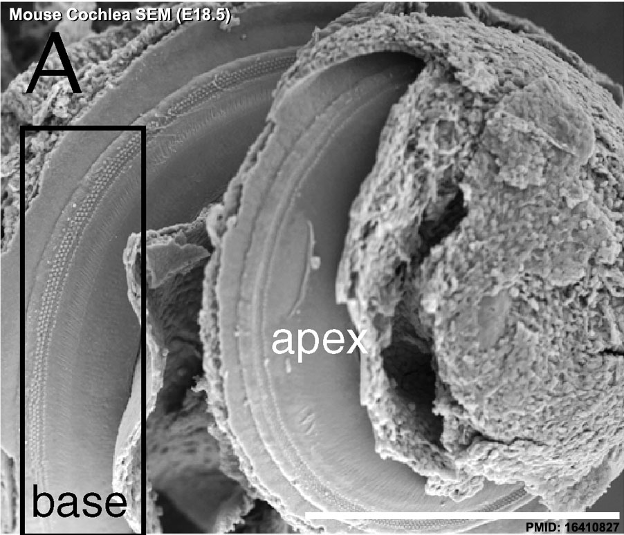 | 902 × 774 (150 KB) | Z8600021 (talk | contribs) | Mouse E18.5 cochlea sem01.jpg {{Mouse cochlea sem links}} |
You cannot overwrite this file.
File usage
The following page uses this file: