File:Adrenal histology 001.jpg: Difference between revisions
| Line 1: | Line 1: | ||
==Adrenal Histology - Cortex and Medulla== | ==Adrenal Histology - Cortex and Medulla== | ||
'''Capsule''' - a fibrous capsule. | |||
* surface of the gland is surrounded by tissue containing much fat | |||
* vessels enter the organ through the furrows on its anterior surface and base. | |||
'''Adrenal Cortex''' - divided into three zones (Adrenocorticotropic hormone (ACTH) required for zones 2 and 3) | '''Adrenal Cortex''' - divided into three zones (Adrenocorticotropic hormone (ACTH) required for zones 2 and 3) | ||
Revision as of 17:35, 16 May 2012
Adrenal Histology - Cortex and Medulla
Capsule - a fibrous capsule.
- surface of the gland is surrounded by tissue containing much fat
- vessels enter the organ through the furrows on its anterior surface and base.
Adrenal Cortex - divided into three zones (Adrenocorticotropic hormone (ACTH) required for zones 2 and 3)
- zona glomerulosa (about 15%)
- zona fasciculata (about 75%)
- zone reticularis is (about 10%)
Adrenal Medulla - 2 indistinguishable cell types, neural crest in origin.
- Adrenaline producing cells - epinephrine (80%)
- Noradrenaline producing cells - norepinephrine (20%)
- cells arranged in strands or small clusters with capillaries and venules in the intervening spaces.
- cell cytoplasm weakly basophilic.
- chromaffin cells because granules can be stained with potassium bichromate.
- innervated by preganglionic sympathetic fibres.
Adrenal gland, monkey - H&E
- Adrenal Histology: Cortex and Medulla | Unlabelled Overview | Cortical Zones | Zona Glomerulosa and Fasciculata | Zona Glomerulosa | Zona Fasciculata | Zona Reticularis and Medulla | Zona Reticularis | Medulla | Fetal Cortex | Developing Adult Cortex | BGD - Endocrine Histology | Histology Stains | Adrenal Development
Links: Histology | Histology Stains | Blue Histology images copyright Lutz Slomianka 1998-2009. The literary and artistic works on the original Blue Histology website may be reproduced, adapted, published and distributed for non-commercial purposes. See also the page Histology Stains.
Cite this page: Hill, M.A. (2024, June 2) Embryology Adrenal histology 001.jpg. Retrieved from https://embryology.med.unsw.edu.au/embryology/index.php/File:Adrenal_histology_001.jpg
- © Dr Mark Hill 2024, UNSW Embryology ISBN: 978 0 7334 2609 4 - UNSW CRICOS Provider Code No. 00098G
Original File Name: Adr041he.jpg
File history
Click on a date/time to view the file as it appeared at that time.
| Date/Time | Thumbnail | Dimensions | User | Comment | |
|---|---|---|---|---|---|
| current | 14:41, 12 May 2012 | 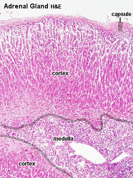 | 450 × 600 (151 KB) | Z8600021 (talk | contribs) | |
| 13:31, 5 October 2009 | 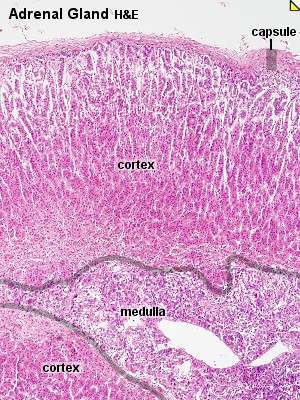 | 300 × 400 (78 KB) | S8600021 (talk | contribs) | Adr041he.jpg |
You cannot overwrite this file.