File:Hertwig266.jpg: Difference between revisions
No edit summary |
No edit summary |
||
| Line 5: | Line 5: | ||
pi, Pigmented epithelium of the eye (outer lamella of the optic cup, or secondary optic vesicle) ; r, retina (inner lamella of the optic cup) ; /M, marginal zone of the optic cup, which forms the pars ciliaris et iridis retinas ; y, vitreous body with blood-vessels ; tv, tunica vasculosa lentis ; bk, blood-corpuscles ; ch, choroidea ; If, lens-fibres ; le, lens-epithelium ; I' ', zone of the lens-fibre nuclei ; It, fundament of the cornea ; he, external corneal epithelium. | pi, Pigmented epithelium of the eye (outer lamella of the optic cup, or secondary optic vesicle) ; r, retina (inner lamella of the optic cup) ; /M, marginal zone of the optic cup, which forms the pars ciliaris et iridis retinas ; y, vitreous body with blood-vessels ; tv, tunica vasculosa lentis ; bk, blood-corpuscles ; ch, choroidea ; If, lens-fibres ; le, lens-epithelium ; I' ', zone of the lens-fibre nuclei ; It, fundament of the cornea ; he, external corneal epithelium. | ||
---- | |||
{{Hertwig images}} | {{Hertwig images}} | ||
[[Category:Vision]] [[Category:Mouse]] | |||
Latest revision as of 21:48, 28 March 2012
Fig. 266. Section through the optic fundament of an embryo Mouse
after KESSLER.
pi, Pigmented epithelium of the eye (outer lamella of the optic cup, or secondary optic vesicle) ; r, retina (inner lamella of the optic cup) ; /M, marginal zone of the optic cup, which forms the pars ciliaris et iridis retinas ; y, vitreous body with blood-vessels ; tv, tunica vasculosa lentis ; bk, blood-corpuscles ; ch, choroidea ; If, lens-fibres ; le, lens-epithelium ; I' ', zone of the lens-fibre nuclei ; It, fundament of the cornea ; he, external corneal epithelium.
| Historic Disclaimer - information about historic embryology pages |
|---|
 |
Reference
Hertwig O. Text-book of the embryology of man and mammals. (1892) Translated 1901 by Mark EL. from 3rd German Edition. S. Sonnenschein, London.
Cite this page: Hill, M.A. (2024, June 15) Embryology Hertwig266.jpg. Retrieved from https://embryology.med.unsw.edu.au/embryology/index.php/File:Hertwig266.jpg
- © Dr Mark Hill 2024, UNSW Embryology ISBN: 978 0 7334 2609 4 - UNSW CRICOS Provider Code No. 00098G
File history
Click on a date/time to view the file as it appeared at that time.
| Date/Time | Thumbnail | Dimensions | User | Comment | |
|---|---|---|---|---|---|
| current | 18:00, 12 May 2011 | 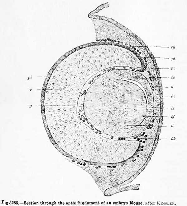 | 600 × 663 (83 KB) | S8600021 (talk | contribs) | {{Hertwig images}} |
You cannot overwrite this file.
File usage
The following page uses this file: