File:Human fetal pancreas anatomy cartoon.jpg: Difference between revisions
(===Fetal topographical anatomy of the pancreatic head and duodenum with special reference to courses of the pancreaticoduodenal arteries=== A diagram showing joining processes between the dorsal and ventral primordia of the pancreas as well as the hypoth) |
mNo edit summary |
||
| Line 1: | Line 1: | ||
===Fetal topographical anatomy of the pancreatic head and duodenum with special reference to courses of the pancreaticoduodenal arteries | ==Human fetal pancreas anatomy== | ||
Fetal topographical anatomy of the pancreatic head and duodenum with special reference to courses of the pancreaticoduodenal arteries. | |||
A diagram showing joining processes between the dorsal and ventral primordia of the pancreas as well as the hypothetical rotation of the duodenum along a left-right axis. Viewed from the posterosuperior side of the body. A horizontal plane including most parts of the duodenum is shown to emphasize, in contrast to adults, the course of the second portion (D2) directing posteriorly rather than inferiorly. The topographical anatomy of the duodenum and pancreatic head is based on observations of fetuses of 20-30 weeks (e.g., Figs. 3,4,5 and 6). The anterior mass of the pancreatic head (labeled by dots) seems to correspond to the dorsal primordium of the pancreas, while an oblique or perpendicular posterior mass [labeled by sharps (#)] seems to primarily correspond to the ventral primordium. The portal vein and right hepatic artery run in the superior sides of and along their border between the anterior and posterior masses. Notably, tongue-like protrusions of the anterior mass or dorsal pancreas (stars) surround the primary posterior mass or the ventral pancreas. In particular, the right anterior protrusion (open star) often reaches the inferior end of the ventral pancreas. The common bile duct crosses the superior side of the first portion of the duodenum. We hypothesize a rotation along a left-right axis through the third portion of the duodenum in the later stage of development. In addition, the fourth portion is connected by a fascia (asterisk) to a thick fascia along the left renal vein. | A diagram showing joining processes between the dorsal and ventral primordia of the pancreas as well as the hypothetical rotation of the duodenum along a left-right axis. Viewed from the posterosuperior side of the body. A horizontal plane including most parts of the duodenum is shown to emphasize, in contrast to adults, the course of the second portion (D2) directing posteriorly rather than inferiorly. The topographical anatomy of the duodenum and pancreatic head is based on observations of fetuses of 20-30 weeks (e.g., Figs. 3,4,5 and 6). The anterior mass of the pancreatic head (labeled by dots) seems to correspond to the dorsal primordium of the pancreas, while an oblique or perpendicular posterior mass [labeled by sharps (#)] seems to primarily correspond to the ventral primordium. The portal vein and right hepatic artery run in the superior sides of and along their border between the anterior and posterior masses. Notably, tongue-like protrusions of the anterior mass or dorsal pancreas (stars) surround the primary posterior mass or the ventral pancreas. In particular, the right anterior protrusion (open star) often reaches the inferior end of the ventral pancreas. The common bile duct crosses the superior side of the first portion of the duodenum. We hypothesize a rotation along a left-right axis through the third portion of the duodenum in the later stage of development. In addition, the fourth portion is connected by a fascia (asterisk) to a thick fascia along the left renal vein. | ||
===Reference=== | |||
<pubmed>20376893</pubmed>| [http://www.eymj.org/search.php?where=aview&id=128080&code=0069YMJ&vmode=AFTR Yonsei Med J.] | |||
== | ====Copyright==== | ||
This is an Open Access article distributed under the terms of the Creative Commons Attribution Non-Commercial License (http://creativecommons.org/licenses/by-nc/3.0) which permits unrestricted noncommercial use, distribution, and reproduction in any medium, provided the original work is properly cited. | |||
Original File Name: Fig. 7 Ymj-51-398-g007-l.jpg | |||
{{Footer}} | |||
[[Category:Pancreas]] | [[Category:Pancreas]] | ||
Revision as of 18:33, 23 Mayıs 2016
Human fetal pancreas anatomy
Fetal topographical anatomy of the pancreatic head and duodenum with special reference to courses of the pancreaticoduodenal arteries.
A diagram showing joining processes between the dorsal and ventral primordia of the pancreas as well as the hypothetical rotation of the duodenum along a left-right axis. Viewed from the posterosuperior side of the body. A horizontal plane including most parts of the duodenum is shown to emphasize, in contrast to adults, the course of the second portion (D2) directing posteriorly rather than inferiorly. The topographical anatomy of the duodenum and pancreatic head is based on observations of fetuses of 20-30 weeks (e.g., Figs. 3,4,5 and 6). The anterior mass of the pancreatic head (labeled by dots) seems to correspond to the dorsal primordium of the pancreas, while an oblique or perpendicular posterior mass [labeled by sharps (#)] seems to primarily correspond to the ventral primordium. The portal vein and right hepatic artery run in the superior sides of and along their border between the anterior and posterior masses. Notably, tongue-like protrusions of the anterior mass or dorsal pancreas (stars) surround the primary posterior mass or the ventral pancreas. In particular, the right anterior protrusion (open star) often reaches the inferior end of the ventral pancreas. The common bile duct crosses the superior side of the first portion of the duodenum. We hypothesize a rotation along a left-right axis through the third portion of the duodenum in the later stage of development. In addition, the fourth portion is connected by a fascia (asterisk) to a thick fascia along the left renal vein.
Reference
<pubmed>20376893</pubmed>| Yonsei Med J.
Copyright
This is an Open Access article distributed under the terms of the Creative Commons Attribution Non-Commercial License (http://creativecommons.org/licenses/by-nc/3.0) which permits unrestricted noncommercial use, distribution, and reproduction in any medium, provided the original work is properly cited.
Original File Name: Fig. 7 Ymj-51-398-g007-l.jpg
Cite this page: Hill, M.A. (2026, October 11) Embryology Human fetal pancreas anatomy cartoon.jpg. Retrieved from https://embryology.med.unsw.edu.au/embryology/index.php/File:Human_fetal_pancreas_anatomy_cartoon.jpg
- © Dr Mark Hill 2026, UNSW Embryology ISBN: 978 0 7334 2609 4 - UNSW CRICOS Provider Code No. 00098G
File history
Yi efo/eka'e gwa ebo wo le nyangagi wuncin ye kamina wunga tinya nan
| Gwalagizhi | Nyangagi | Dimensions | User | Comment | |
|---|---|---|---|---|---|
| current | 00:26, 19 October 2010 | 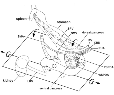 | 455 × 376 (93 KB) | S8600021 (talk | contribs) | ===Fetal topographical anatomy of the pancreatic head and duodenum with special reference to courses of the pancreaticoduodenal arteries=== A diagram showing joining processes between the dorsal and ventral primordia of the pancreas as well as the hypoth |
You cannot overwrite this file.
File usage
The following page uses this file:


