|
|
| Line 81: |
Line 81: |
| * Capsule (thin) with trabecular or septa (dense connective tissue) | | * Capsule (thin) with trabecular or septa (dense connective tissue) |
|
| |
|
| [[File:Thymus histology 01.jpg|600px]] | | [[File:Lymph node cartoon 03.jpg|600px]] |
|
| |
|
| [[File:Thymus histology 06.jpg|600px]]
| |
|
| |
|
| Infant thymus
| |
|
| |
|
| {| | | {| |
| Line 97: |
Line 95: |
| |} | | |} |
|
| |
|
|
| |
| {|
| |
| ! Adult Thymus
| |
| |-
| |
| | [[File:Thymus adult.jpg|300px]]
| |
| |
| |
| * Cortical lymphoid tissue is replaced by adipose tissue (involution)
| |
| * Increase in size of thymic corpuscles
| |
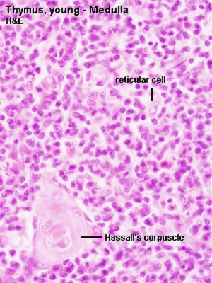
| * '''Thymic corpuscle''' - (Hassall’s corpuscle) mass of concentric epithelioreticular cells.
| |
| |}
| |
|
| |
|
|
| |
|
| {{Thymus Histology}} | | {{Lymph Node Histology}} |
| |} | | |} |
|
| |
|
| |
|
| |
|
| |
|
| |
|
| ==Lymph Nodes== | | ==Lymph Nodes== |
Revision as of 18:17, 26 February 2016
Lymph Nodes
Lymph Node Anatomy
| Lymph Node Anatomy
|
- Location throughout the entire body - concentrated in axilla, groin, lung, gastrointestinal tract mesenteries
- Small (1 mm - 2 cm) encapsulated organ (diffuse lymphoid tissue - no capsule)
- Antigen transformed lymphocytes from the blood
- In lymph vessel pathways “filter” lymph
- Afferent - towards node (A - arrives at the node)
- Efferent - away from node (E - exits the node)
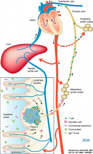
Mesenteric lymph nodes
|

|
Lymph Node Functions
| Lymph Node Functions
|
- In lymph vessel pathways “filter” lymph
- Immune - detect infections from peripheral tissues
- skin, respiratory tract, gastrointestinal tract, etc.
- secondary lymphoid organ
- return extracellular fluid to circulation
|
|
Lymph Node Structure
| Lymph Node Structure
|
Structure Overview
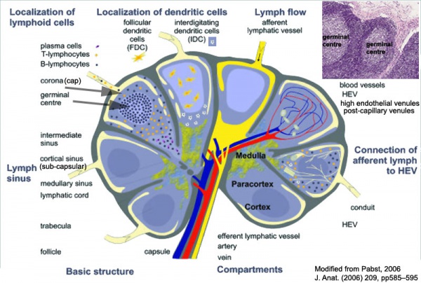
- Connective tissue capsule (thin) with numerous trabeculae (septa)
- major blood vessels run in CT
- Lobules containing cortex and medulla regions
- medullary regions are continuous (connected together)
- NOT supplied by afferent lymph vessels
Blood-Thymic Barrier
- Blood vessels are separated from thymus cortex by epithelioreticular cells.
- impermeable to most macromolecules.
- Barrier layers: capillary endothelium - endothelial basal lamina- perivascular CT sheath - basal lamina of epithelioreticular cells - epithelioreticular cell sheath
| Thymus Epithelioreticular cells (TEC)
|
Macrophages
|
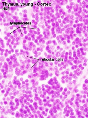
Lymphocytes
|
- Abundant, eosinophilic, large, ovoid and light nucleus 1-2 nucleoli
- sheathe cortical capillaries
- form an epitheloid layer
- maintain microenvironment for development of T-lymphocytes in cortex (thymic epitheliocytes)
|
- cortex and medulla
- difficult to distinguish from reticular cells in (Stain - Haematoxylin Eosin)
- remove auto-reactive T-lymphocytes
|
- located in cortex and medulla
- more numerous (denser) in cortex
- majority are developing T-lymphocytes (= thymic lymphocytes or thymocytes)
|
|
Lymph Node Histology
| Lymph Node Histology
|
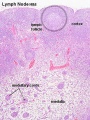
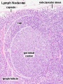
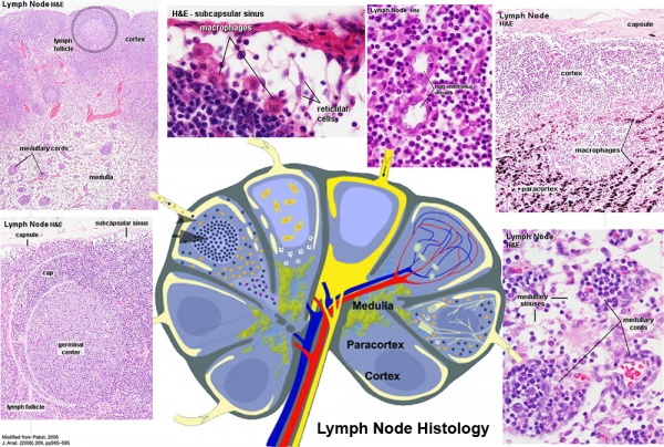
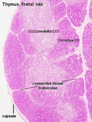
- Capsule (thin) with trabecular or septa (dense connective tissue)
| Fetal thymus
|
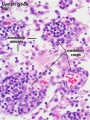
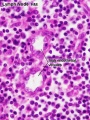
Young medulla
|
Young cortex
|
|
|
|
Template:Lymph Node Histology
|
Lymph Nodes
Lymph Node Structure
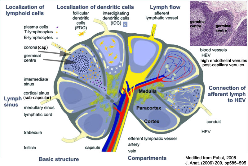
Connective Tissue
- Capsule - dense connective tissue (irregular CT, some adipocytes))
- Trabeculae - dense connective tissue
- Reticular Tissue - Reticular cells and fibers, supporting meshwork (collagen type III)
- Reticular cell produces reticular fibers (collagen type III) and surrounds the fibers with its cytoplasm
- reticular fibbers can also be produced by fibroblasts
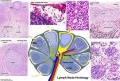
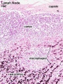
Cartoon with histology features
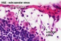
Subcapsular Sinus (marginal sinus, continuation of trabecular sinus)
Medullary Cords and Sinuses
Links: Immunobiology - Figure 1.8. Organization of a lymph node