Talk:Draft 2016: Difference between revisions
From Embryology
mNo edit summary |
|||
| Line 24: | Line 24: | ||
|- | |- | ||
| valign=top| | | valign=top| | ||
* In lymph vessel pathways “filter” lymph | |||
* '''Immune''' - detect infections from peripheral tissues | |||
** skin, respiratory tract, gastrointestinal tract, etc. | |||
* secondary lymphoid organ | |||
* return extracellular fluid to circulation | |||
| [[File:Lymph_node_cartoon_01.jpg|Lymph_node_cartoon_01.jpg|600px]] | | [[File:Lymph_node_cartoon_01.jpg|Lymph_node_cartoon_01.jpg|600px]] | ||
|- | |- | ||
Revision as of 18:15, 26 February 2016
Lymph Nodes
Lymph Node Anatomy
| Lymph Node Anatomy | |
|---|---|
Mesenteric lymph nodes |

|

Lymph Node Functions
| Lymph Node Functions | |
|---|---|
|

|
Lymph Node Structure
| Lymph Node Structure | ||||||
|---|---|---|---|---|---|---|
Structure Overview

|
Lymph Node Histology
| Lymph Node Histology | |||||||||
|---|---|---|---|---|---|---|---|---|---|
Infant thymus
|


Lymph Nodes
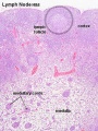
Lymph Node Structure
Connective Tissue
- Capsule - dense connective tissue (irregular CT, some adipocytes))
- Trabeculae - dense connective tissue
- Reticular Tissue - Reticular cells and fibers, supporting meshwork (collagen type III)
- Reticular cell produces reticular fibers (collagen type III) and surrounds the fibers with its cytoplasm
- reticular fibbers can also be produced by fibroblasts
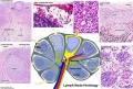
Cartoon with histology features
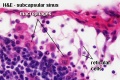
Subcapsular Sinus (marginal sinus, continuation of trabecular sinus)
Follicle
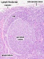
Germinal Centre
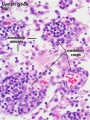
Medullary Cords and Sinuses
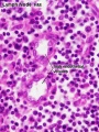
High Endothelial Venules
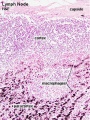
Macrophages


Lymphocyte (T and B) Traffic
Important - Please note that the lymphocytes exit from the node by efferent lymphatic vessels. See also Image - Cell Trafficking into and out of Lymph Nodes. |
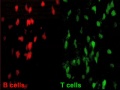
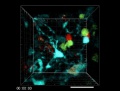
T and B motility |
T and B interaction |

High Endothelial Venules See also Lymphocyte Migration at High Endothelial Venule Model |
Links: Immunobiology - Figure 1.8. Organization of a lymph node


