File:Pituitary histology 003.jpg: Difference between revisions
From Embryology
No edit summary |
No edit summary |
||
| Line 6: | Line 6: | ||
Original File Name: Hyn40pp.jpg | Original File Name: Hyn40pp.jpg | ||
Image Source: UWA Blue Histology Copyright Lutz Slomianka 1998-2009. The literary and artistic works (excluding the UWA logo) on this web-site may be reproduced, adapted, published and distributed for noncommercial purposes. | |||
http://www.lab.anhb.uwa.edu.au/mb140/CorePages/Endocrines/endocrin.htm | |||
[[Category:Histology]] [[Category:Endocrine]] [[Category:Pituitary]] | |||
Revision as of 13:35, 5 October 2009
Pituitary histology - Neurohypophysis
Pituitary, sheep - PAAB/PAS/Orange G
Identify nuclei of pituicytes and Herring bodies.
Original File Name: Hyn40pp.jpg
Image Source: UWA Blue Histology Copyright Lutz Slomianka 1998-2009. The literary and artistic works (excluding the UWA logo) on this web-site may be reproduced, adapted, published and distributed for noncommercial purposes.
http://www.lab.anhb.uwa.edu.au/mb140/CorePages/Endocrines/endocrin.htm
File history
Click on a date/time to view the file as it appeared at that time.
| Date/Time | Thumbnail | Dimensions | User | Comment | |
|---|---|---|---|---|---|
| current | 16:53, 12 May 2012 | 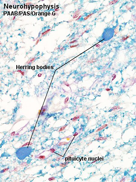 | 450 × 600 (94 KB) | Z8600021 (talk | contribs) | larger image |
| 13:26, 5 October 2009 |  | 300 × 400 (59 KB) | S8600021 (talk | contribs) | Hyn40pp.jpg |
You cannot overwrite this file.