File:Human extraocular muscles 01.jpg: Difference between revisions
(==Human Extraocular Muscles== Illustration of human eye showing 6 EOMs inserting on the globe in what is referred to as the Spiral of Tillaux. ===Reference== <pubmed>22132088</pubmed>| [http://www.plosone.org/article/info%3Adoi%2F10.1371%2Fjournal.pone) |
|||
| (18 intermediate revisions by the same user not shown) | |||
| Line 1: | Line 1: | ||
==Human Extraocular Muscles== | ==Human Extraocular Muscles== | ||
Cartoon showing attachment of the human 6 extraocular muscles to the eyeball. | |||
{| | |||
! Legend | |||
! About the Muscles | |||
|- | |||
| width=230px| | |||
* '''IR''' - inferior rectus | |||
* '''SR''' - superior rectus | |||
* '''LR''' - lateral rectus | |||
* '''MR''' - medial rectus | |||
* '''SO''' - superior oblique | |||
* '''IO''' - inferior oblique | |||
| width=500px valign=top| | |||
* Five of the six muscles (inferior rectus, superior rectus, lateral rectus, medial rectus, and superior oblique) originate at a common tendinous ring of fibrous tissue (the Annulus of Zinn). | |||
** The Annulus of Zinn surrounds the optic nerve, ophthalmic artery, and ophthalmic vein at their entrance through the apex of the orbit. | |||
* The sixth muscle (inferior oblique) has a separate origin point on the orbital side of the bony maxilla at the anterior inferomedial strut. | |||
|} | |||
<br> | |||
{| | |||
! Muscle Structure | |||
|- | |||
| colspan=2|Each muscle has 2 equally sized layers. | |||
|- | |||
! Layer | |||
! Inserts | |||
! Single innervation (%) | |||
|- | |||
| outer orbital | |||
| connective tissue ring | |||
| 80 | |||
|- | |||
| inner global | |||
| eye sclera | |||
| 90 | |||
|} | |||
===Reference== | |||
{{Vision Links}} | |||
<br> | |||
[[Musculoskeletal_System_-_Muscle_Development|'''Muscle Development''']] | |||
===Reference=== | |||
<pubmed>22132088</pubmed>| [http://www.plosone.org/article/info%3Adoi%2F10.1371%2Fjournal.pone.0027095 PLoS One.] | <pubmed>22132088</pubmed>| [http://www.plosone.org/article/info%3Adoi%2F10.1371%2Fjournal.pone.0027095 PLoS One.] | ||
Citation: Kasprick DS, Kish PE, Junttila TL, Ward LA, Bohnsack BL, et al. (2011) Microanatomy of Adult Zebrafish Extraocular Muscles. PLoS ONE 6(11): e27095. doi:10.1371/journal.pone.0027095 | Citation: Kasprick DS, Kish PE, Junttila TL, Ward LA, Bohnsack BL, et al. (2011) Microanatomy of Adult Zebrafish Extraocular Muscles. PLoS ONE 6(11): e27095. doi:10.1371/journal.pone.0027095 | ||
====Copyright==== | |||
© 2011 Kasprick et al. This is an open-access article distributed under the terms of the Creative Commons Attribution License, which permits unrestricted use, distribution, and reproduction in any medium, provided the original author and source are credited. | |||
Figure 1. doi:10.1371/journal.pone.0027095.g001 Pone.0027095.g001.jpg Text modified from figure legend and paper text. | |||
Figure 1. doi:10.1371/journal.pone.0027095.g001 | |||
Pone.0027095.g001.jpg | |||
{{Footer}} | |||
[[Category:Human]] [[Category:Vision]] [[Category:Muscle]] [[Category:Cartoon]] | [[Category:Human]] [[Category:Vision]] [[Category:Muscle]] [[Category:Cartoon]] | ||
Latest revision as of 09:00, 30 October 2017
Human Extraocular Muscles
Cartoon showing attachment of the human 6 extraocular muscles to the eyeball.
| Legend | About the Muscles |
|---|---|
|
|
| Muscle Structure | ||
|---|---|---|
| Each muscle has 2 equally sized layers. | ||
| Layer | Inserts | Single innervation (%) |
| outer orbital | connective tissue ring | 80 |
| inner global | eye sclera | 90 |
Reference
<pubmed>22132088</pubmed>| PLoS One.
Citation: Kasprick DS, Kish PE, Junttila TL, Ward LA, Bohnsack BL, et al. (2011) Microanatomy of Adult Zebrafish Extraocular Muscles. PLoS ONE 6(11): e27095. doi:10.1371/journal.pone.0027095
Copyright
© 2011 Kasprick et al. This is an open-access article distributed under the terms of the Creative Commons Attribution License, which permits unrestricted use, distribution, and reproduction in any medium, provided the original author and source are credited.
Figure 1. doi:10.1371/journal.pone.0027095.g001 Pone.0027095.g001.jpg Text modified from figure legend and paper text.
Cite this page: Hill, M.A. (2024, June 10) Embryology Human extraocular muscles 01.jpg. Retrieved from https://embryology.med.unsw.edu.au/embryology/index.php/File:Human_extraocular_muscles_01.jpg
- © Dr Mark Hill 2024, UNSW Embryology ISBN: 978 0 7334 2609 4 - UNSW CRICOS Provider Code No. 00098G
File history
Click on a date/time to view the file as it appeared at that time.
| Date/Time | Thumbnail | Dimensions | User | Comment | |
|---|---|---|---|---|---|
| current | 12:04, 8 June 2012 | 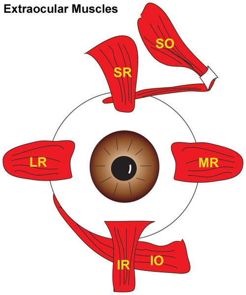 | 500 × 600 (47 KB) | Z8600021 (talk | contribs) | ==Human Extraocular Muscles== Illustration of human eye showing 6 EOMs inserting on the globe in what is referred to as the Spiral of Tillaux. ===Reference== <pubmed>22132088</pubmed>| [http://www.plosone.org/article/info%3Adoi%2F10.1371%2Fjournal.pone |
You cannot overwrite this file.
File usage
The following 4 pages use this file: