File:Lymph node histology 06.jpg: Difference between revisions
| (3 intermediate revisions by the same user not shown) | |||
| Line 1: | Line 1: | ||
==Lymph Node Histology== | ==Lymph Node Histology - Macrophages== | ||
{{HE}} carbon injected. | |||
Macrophages take up the carbon and appear black with this technique. | |||
Slide shows the distribution of macrophages in lymph nodes. | |||
{{ | |||
Most of the macrophages are located in the paracortex and medulla. | |||
{{Lymph Node Images}} | |||
{{Blue Histology}} | |||
[[Category:Immune]] | [[Category:Immune]] | ||
Latest revision as of 09:03, 26 February 2018
Lymph Node Histology - Macrophages
(Stain - Haematoxylin Eosin) carbon injected.
Macrophages take up the carbon and appear black with this technique.
Slide shows the distribution of macrophages in lymph nodes.
Most of the macrophages are located in the paracortex and medulla.
- Lymph Node Histology: Subcapsular Sinus | Follicle | Germinal Centre | Medullary Cords and Sinuses | High Endothelial Venules | Macrophages | Node cartoons
Links: Histology | Histology Stains | Blue Histology images copyright Lutz Slomianka 1998-2009. The literary and artistic works on the original Blue Histology website may be reproduced, adapted, published and distributed for non-commercial purposes. See also the page Histology Stains.
Cite this page: Hill, M.A. (2026, November 16) Embryology Lymph node histology 06.jpg. Retrieved from https://embryology.med.unsw.edu.au/embryology/index.php/File:Lymph_node_histology_06.jpg
- © Dr Mark Hill 2026, UNSW Embryology ISBN: 978 0 7334 2609 4 - UNSW CRICOS Provider Code No. 00098G
File history
Yi efo/eka'e gwa ebo wo le nyangagi wuncin ye kamina wunga tinya nan
| Gwalagizhi | Nyangagi | Dimensions | User | Comment | |
|---|---|---|---|---|---|
| current | 13:11, 25 February 2012 | 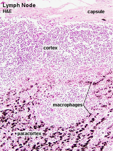 | 450 × 600 (141 KB) | Z8600021 (talk | contribs) | increase size |
| 10:03, 14 February 2011 |  | 300 × 400 (142 KB) | S8600021 (talk | contribs) | ==Lymph Node Histology== Original file name: Lyn42he.jpg http://www.lab.anhb.uwa.edu.au/mb140/CorePages/Lymphoid1/lymph1.htm#Lymph Lymph node histology 06.jpg {{Template:Blue Histology}} Category:Immune |
You cannot overwrite this file.
File usage
The following 5 pages use this file:


