File:Mouse eye TGF-beta model.jpg: Difference between revisions
From Embryology
mNo edit summary |
|||
| (6 intermediate revisions by 2 users not shown) | |||
| Line 1: | Line 1: | ||
==Mouse Eye TGF-beta Model== | ==Mouse Eye TGF-beta Model== | ||
Summary of the TGFβ-dependent development of anterior and posterior ocular structures. | {| | ||
! colspan=2|Summary of the TGFβ-dependent development of anterior and posterior ocular structures. | |||
|- | |||
| valign=top|'''a''' Neural crest-derived cells (NC, blue) contribute to structures of the anterior eye segment and the primary vitreous (PV). | |||
* TGFβ signaling is involved in the formation of the ciliary body (CB) and the trabecular meshwork (TM), and in control of PV growth. | * TGFβ signaling is involved in the formation of the ciliary body (CB) and the trabecular meshwork (TM), and in control of PV growth. | ||
* Moreover, normal PV development and/or TGFβ signaling are important for correct retinal patterning. | * Moreover, normal PV development and/or TGFβ signaling are important for correct retinal patterning. | ||
| valign=top|'''b''' In the cornea, prospective stromal keratocytes and endothelial cells are of neural crest origin. | |||
* Here, TGFβ signaling is needed for the expression of the transcription factors Foxc1 and Pitx2 and for normal differentiation of NC-derived cells into collagen-synthesizing stromal keratocytes. | |||
* Moreover, in forming corneal endothelial cells (and in the TM), expression of Foxc1 and cell survival requires TGFβ signalling. | |||
|} | |||
:'''Links:''' [[:File:Mouse eye neural crest.jpg|Image - Mouse eye neural crest]] | [[:File:Mouse_eye_TGF-beta_model.jpg|Image - Mouse eye TGF-beta model]] [[Developmental_Signals_-_TGF-beta|TGF-beta]] | [[Vision_-_Cornea_Development|Cornea Development]] | [[Sensory - Vision Development|Vision Development]] | [[Neural Crest Development]] | [[Head Development]] | |||
===Reference=== | ===Reference=== | ||
<pubmed>16403239</pubmed>| [http://jbiol.com/content/4/3/11 J Biol.] | <pubmed>16403239</pubmed>| [http://jbiol.com/content/4/3/11 J Biol.] | ||
====Copyright==== | |||
© 2005 Ittner et al.; licensee BioMed Central Ltd. | © 2005 Ittner et al.; licensee BioMed Central Ltd. | ||
This is an open access article distributed under the terms of the Creative Commons Attribution License (http://creativecommons.org/licenses/by/2.0), which permits unrestricted use, distribution, and reproduction in any medium, provided the original work is properly cited. | This is an open access article distributed under the terms of the Creative Commons Attribution License (http://creativecommons.org/licenses/by/2.0), which permits unrestricted use, distribution, and reproduction in any medium, provided the original work is properly cited. | ||
[[Category:Mouse]] [[Category:Neural Crest]] [[Category:Vision]] [[Category:Cartoon]] | Ittner et al. Journal of Biology 2005 4:11 doi:10.1186/jbiol29 | ||
Original file name: Figure 9. http://jbiol.com/content/4/3/11/figure/F9 | |||
[[Category:Mouse]] [[Category:Neural Crest]] [[Category:Vision]] [[Category:Cartoon]][[Category:TGF-beta]][[Category:Cornea]] | |||
Latest revision as of 14:16, 30 August 2014
Mouse Eye TGF-beta Model
| Summary of the TGFβ-dependent development of anterior and posterior ocular structures. | |
|---|---|
a Neural crest-derived cells (NC, blue) contribute to structures of the anterior eye segment and the primary vitreous (PV).
|
b In the cornea, prospective stromal keratocytes and endothelial cells are of neural crest origin.
|
- Links: Image - Mouse eye neural crest | Image - Mouse eye TGF-beta model TGF-beta | Cornea Development | Vision Development | Neural Crest Development | Head Development
Reference
<pubmed>16403239</pubmed>| J Biol.
Copyright
© 2005 Ittner et al.; licensee BioMed Central Ltd. This is an open access article distributed under the terms of the Creative Commons Attribution License (http://creativecommons.org/licenses/by/2.0), which permits unrestricted use, distribution, and reproduction in any medium, provided the original work is properly cited.
Ittner et al. Journal of Biology 2005 4:11 doi:10.1186/jbiol29
Original file name: Figure 9. http://jbiol.com/content/4/3/11/figure/F9
File history
Click on a date/time to view the file as it appeared at that time.
| Date/Time | Thumbnail | Dimensions | User | Comment | |
|---|---|---|---|---|---|
| current | 18:32, 30 August 2011 | 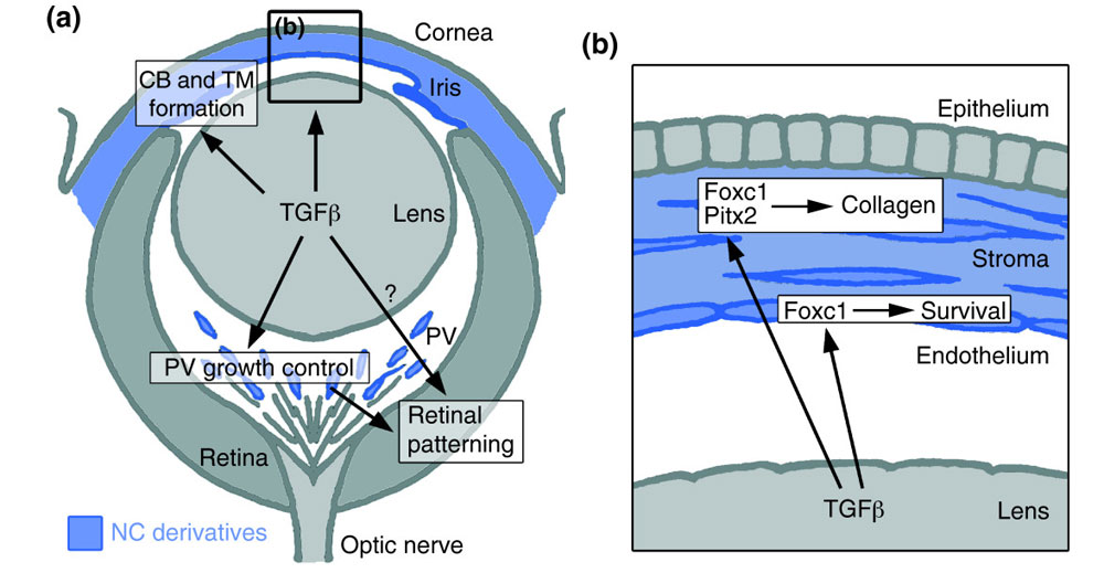 | 1,000 × 519 (88 KB) | S8600021 (talk | contribs) | ==Mouse eye TGF-beta model== Summary of the TGFβ-dependent development of anterior and posterior ocular structures. (a) NC-derived cells (blue) contribute to structures of the anterior eye segment and the primary vitreous (PV). TGFβ signaling is invo |
You cannot overwrite this file.
File usage
The following 5 pages use this file: