File:Establishment of HD hybrid cell line.jpg: Difference between revisions
((A) First polar body of mature rhesus macaque oocyte was removed by gentle squeezing through a slit of zona pellucida (A-a). Staining of 1st polar body DNA (arrowhead) and oocyte DNA (arrow) (A-b). HD monkey skin cell was placed under the zona pellucida () |
mNo edit summary |
||
| (One intermediate revision by one other user not shown) | |||
| Line 1: | Line 1: | ||
==Establishment of HD hybrid cell line== | |||
(A) First polar body of mature rhesus macaque oocyte was removed by gentle squeezing through a slit of zona pellucida (A-a). Staining of 1st polar body DNA (arrowhead) and oocyte DNA (arrow) (A-b). HD monkey skin cell was placed under the zona pellucida (black arrow) (A-c). Reconstructed oocyte with HD monkey skin cell (A-d; yellow arrow) was placed between two electrodes for electrofusion (A-d). | |||
(B) Day 12 hatching blastocyst derived from HD monkey hybrid embryo (B-a; arrow indicated ICM). HD monkey hybrid blastocyst outgrowth at six days after attached onto feeder cells (B-b). High magnification of selected region (inset) of the ICM outgrowth (arrowhead). HD monkey hybrid cell line (TrES1) at passage 10 (B-c). | |||
(C) G-banding analysis of TrES1. Cytogenetic analysis of TrES1 demonstrated tetraploid chromosome (84; XXXY). | |||
(D) Expression of ES-cell specific markers: Alkaline phosphatase, Oct4, SSEA4 and TRA-1-60. | |||
===Reference=== | |||
http://www.ncbi.nlm.nih.gov/pmc/articles/PMC2833146/?tool=pmcentrez | |||
====Copyright==== | |||
This is an Open Access article distributed under the terms of the Creative Commons Attribution License (http://creativecommons.org/licenses/by/2.0), which permits unrestricted use, distribution, and reproduction in any medium, provided the original work is properly cited. | This is an Open Access article distributed under the terms of the Creative Commons Attribution License (http://creativecommons.org/licenses/by/2.0), which permits unrestricted use, distribution, and reproduction in any medium, provided the original work is properly cited. | ||
[[Category:Monkey]] | |||
[[Category:Polar Body]] | |||
Latest revision as of 10:22, 17 June 2014
Establishment of HD hybrid cell line
(A) First polar body of mature rhesus macaque oocyte was removed by gentle squeezing through a slit of zona pellucida (A-a). Staining of 1st polar body DNA (arrowhead) and oocyte DNA (arrow) (A-b). HD monkey skin cell was placed under the zona pellucida (black arrow) (A-c). Reconstructed oocyte with HD monkey skin cell (A-d; yellow arrow) was placed between two electrodes for electrofusion (A-d).
(B) Day 12 hatching blastocyst derived from HD monkey hybrid embryo (B-a; arrow indicated ICM). HD monkey hybrid blastocyst outgrowth at six days after attached onto feeder cells (B-b). High magnification of selected region (inset) of the ICM outgrowth (arrowhead). HD monkey hybrid cell line (TrES1) at passage 10 (B-c).
(C) G-banding analysis of TrES1. Cytogenetic analysis of TrES1 demonstrated tetraploid chromosome (84; XXXY).
(D) Expression of ES-cell specific markers: Alkaline phosphatase, Oct4, SSEA4 and TRA-1-60.
Reference
http://www.ncbi.nlm.nih.gov/pmc/articles/PMC2833146/?tool=pmcentrez
Copyright
This is an Open Access article distributed under the terms of the Creative Commons Attribution License (http://creativecommons.org/licenses/by/2.0), which permits unrestricted use, distribution, and reproduction in any medium, provided the original work is properly cited.
File history
Yi efo/eka'e gwa ebo wo le nyangagi wuncin ye kamina wunga tinya nan
| Gwalagizhi | Nyangagi | Dimensions | User | Comment | |
|---|---|---|---|---|---|
| current | 21:09, 17 August 2011 | 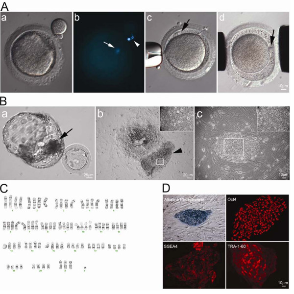 | 600 × 600 (93 KB) | Z3290270 (talk | contribs) | (A) First polar body of mature rhesus macaque oocyte was removed by gentle squeezing through a slit of zona pellucida (A-a). Staining of 1st polar body DNA (arrowhead) and oocyte DNA (arrow) (A-b). HD monkey skin cell was placed under the zona pellucida ( |
You cannot overwrite this file.
File usage
The following 2 pages use this file: