File:Heart innervation 02.jpeg: Difference between revisions
(Fig. 2. Schematic representation of the origin and distribution of nerves to the heart, viewed from behind, and as if the nerves and ganglia occupied the same coronal plane. Superior and middle cervical ganglia and a cervicothoracic ganglion are presen...) |
mNo edit summary |
||
| (2 intermediate revisions by the same user not shown) | |||
| Line 2: | Line 2: | ||
Fig. 2. Schematic representation of the origin and distribution of nerves to the heart, viewed | Fig. 2. Schematic representation of the origin and distribution of nerves to the heart, viewed | ||
from behind, and as if the nerves and ganglia occupied the same coronal plane. Superior and | from behind, and as if the nerves and ganglia occupied the same coronal plane. | ||
middle cervical ganglia and a cervicothoracic ganglion are present on the left side, whereas on | |||
Superior and middle cervical ganglia and a cervicothoracic ganglion are present on the left side, whereas on | |||
the right there is a superior cervical ganglion and a long ganglionated mass that extends into | the right there is a superior cervical ganglion and a long ganglionated mass that extends into | ||
the thorax. Cardiac nerves arising from the vagus nerves, and sympathetic ganglia and trunks, | the thorax. | ||
are shown as solid lines | |||
Cardiac nerves arising from the vagus nerves ({{CN X}}), and sympathetic ganglia and trunks, | |||
are shown as solid lines. | |||
The solid circles on or related to the cardiac nerves represent ganglia. | |||
===Reference=== | |||
{{#pmid:1018009}} | {{#pmid:1018009}} | ||
Latest revision as of 05:33, 10 December 2019
Summary
Fig. 2. Schematic representation of the origin and distribution of nerves to the heart, viewed from behind, and as if the nerves and ganglia occupied the same coronal plane.
Superior and middle cervical ganglia and a cervicothoracic ganglion are present on the left side, whereas on the right there is a superior cervical ganglion and a long ganglionated mass that extends into the thorax.
Cardiac nerves arising from the vagus nerves (CN X), and sympathetic ganglia and trunks, are shown as solid lines.
The solid circles on or related to the cardiac nerves represent ganglia.
Reference
Gardner E & O'Rahilly R. (1976). The nerve supply and conducting system of the human heart at the end of the embryonic period proper. J. Anat. , 121, 571-87. PMID: 1018009
File history
Yi efo/eka'e gwa ebo wo le nyangagi wuncin ye kamina wunga tinya nan
| Gwalagizhi | Nyangagi | Dimensions | User | Comment | |
|---|---|---|---|---|---|
| current | 05:23, 10 December 2019 |  | 732 × 816 (123 KB) | Z8600021 (talk | contribs) | |
| 05:23, 10 December 2019 | 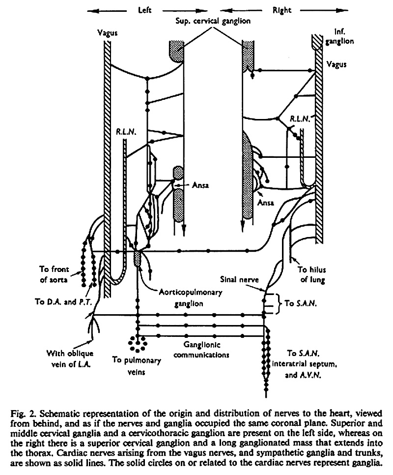 | 800 × 966 (194 KB) | Z8600021 (talk | contribs) | Fig. 2. Schematic representation of the origin and distribution of nerves to the heart, viewed from behind, and as if the nerves and ganglia occupied the same coronal plane. Superior and middle cervical ganglia and a cervicothoracic ganglion are presen... |
You cannot overwrite this file.
File usage
The following page uses this file:


