ANAT2341 Lab 7: Difference between revisions
m (→Group Project) |
No edit summary |
||
| (58 intermediate revisions by 2 users not shown) | |||
| Line 1: | Line 1: | ||
= | ==Organogenesis Lab== | ||
Please note the different location for this week’s practical class: Wallace Wurth Teaching Lab 116. | |||
'''PRACTICAL CLASS PROGRAM''' | |||
* Weekly Quiz + revision (15 minutes) | |||
* Practical class activities (90 minutes) | |||
* Practical Class Revision (15 minutes) | |||
'''PRACTICAL CLASS ACTIVITIES (90 minutes)''' | |||
* Fertile egg dissections, stage definition, and annotations of structures (first 60 minutes) | |||
* Observation of skeletal preparations of chicken and mouse foetuses (first 60 minutes) | |||
* Group presentation of annotated embryo images (final 30 minutes) | |||
'''LEARNING OBJECTIVES''' | |||
* Understanding early neurulation, mesoderm and heart development, and being able to identify the defining structures in the chicken embryo. | |||
* Understanding craniofacial and limb development and being able to identify the defining structures in chicken embryos. | |||
* Understanding the development of the musculoskeletal system and being able to identify the defining structures in chicken embryos. | |||
* Be able to apply basic practical laboratory skills and work with embryo and regeneration models. | |||
* Be able to work effectively within a small team to complete academic tasks. | |||
* Be able to present embryonic observations effectively and appropriately to an audience | |||
* Be able to self-manage and work independently with an ability to take responsibility for their own learning, and an appreciation of the value of learning. | |||
[[File:Chicken_Embryo_Hamburger_stages.jpg|600px|link=Hamburger Hamilton Stages]] | |||
''These are the Hamburger stages of chicken development'' | |||
See also the [https://www.jove.com/video/306/windowing-chicken-eggs-for-developmental-studies JoVE article on chicken egg preparation]: <pubmed>18989413</pubmed> | |||
{{ | ===Additional Chicken Links=== | ||
[[File:Viktor Hamburger.jpg|thumb|alt=Viktor Hamburger|link=Embryology History - Viktor Hamburger|Viktor Hamburger (1900 – 2001)]] | |||
More about chicken embryogenesis: [[Chicken Development]] | [[Hamburger Hamilton Stages]] | |||
<br> | |||
{{Chicken links}} | |||
<br> | |||
<gallery> | |||
File:HHstage1-4.jpg|stage 1-4 | |||
File:HHstage5-10.jpg|stages 5-10 | |||
File:HHstage11-14.jpg|stages 11-14 | |||
File:HHstage15-18.jpg|stages 15-18 | |||
File:HHstage19-21.jpg|stages 19-21 | |||
File:HHstage22-25.jpg|stages 22-25 | |||
File:HHstage26-28.jpg|stages 26-28 | |||
File:HHstage29-32.jpg|stage 29-32 | |||
</gallery> | |||
[[File:Mouse_vs_Human_embryogenesis.jpg]] | |||
''This figure compares the human and mouse developmental stages'' | |||
More about Mouse embryogenesis: [[Mouse Timeline Detailed]] | |||
{{Chicken}} | |||
===External Links=== | |||
{{External Links}} | |||
* JOVE - [http://www.jove.com/science-education/5153/an-introduction-to-the-chick-gallus-gallus-domesticus An Introduction to the Chicken] | |||
Latest revision as of 11:38, 22 October 2019
Organogenesis Lab
Please note the different location for this week’s practical class: Wallace Wurth Teaching Lab 116.
PRACTICAL CLASS PROGRAM
- Weekly Quiz + revision (15 minutes)
- Practical class activities (90 minutes)
- Practical Class Revision (15 minutes)
PRACTICAL CLASS ACTIVITIES (90 minutes)
- Fertile egg dissections, stage definition, and annotations of structures (first 60 minutes)
- Observation of skeletal preparations of chicken and mouse foetuses (first 60 minutes)
- Group presentation of annotated embryo images (final 30 minutes)
LEARNING OBJECTIVES
- Understanding early neurulation, mesoderm and heart development, and being able to identify the defining structures in the chicken embryo.
- Understanding craniofacial and limb development and being able to identify the defining structures in chicken embryos.
- Understanding the development of the musculoskeletal system and being able to identify the defining structures in chicken embryos.
- Be able to apply basic practical laboratory skills and work with embryo and regeneration models.
- Be able to work effectively within a small team to complete academic tasks.
- Be able to present embryonic observations effectively and appropriately to an audience
- Be able to self-manage and work independently with an ability to take responsibility for their own learning, and an appreciation of the value of learning.
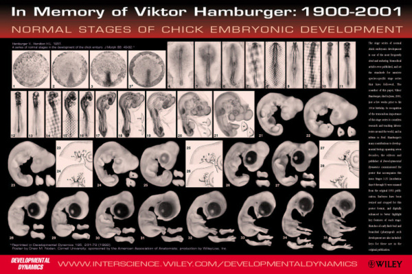
These are the Hamburger stages of chicken development
See also the JoVE article on chicken egg preparation: <pubmed>18989413</pubmed>
Additional Chicken Links
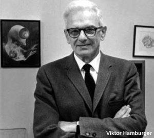
More about chicken embryogenesis: Chicken Development | Hamburger Hamilton Stages
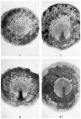
stage 1-4
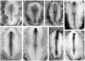
stages 5-10
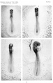
stages 11-14
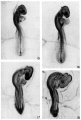
stages 15-18
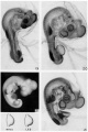
stages 19-21
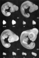
stages 22-25
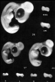
stages 26-28
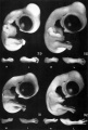
stage 29-32


This figure compares the human and mouse developmental stages
More about Mouse embryogenesis: Mouse Timeline Detailed
External Links
External Links Notice - The dynamic nature of the internet may mean that some of these listed links may no longer function. If the link no longer works search the web with the link text or name. Links to any external commercial sites are provided for information purposes only and should never be considered an endorsement. UNSW Embryology is provided as an educational resource with no clinical information or commercial affiliation.

