File:Myelination animation.gif: Difference between revisions
From Embryology
(==Myelination animation== {{Blue Histology}} Category:Animation) |
|||
| (9 intermediate revisions by the same user not shown) | |||
| Line 1: | Line 1: | ||
==Myelination animation== | ==Myelination animation== | ||
This simple animation shows how during development a glial cell myelinates a neural axon. | |||
* '''Grey''' - axon | |||
* '''Brown''' - glia cytoplasm | |||
* '''Blue''' - glia nucleus | |||
* '''Black''' - glia cell membrane, forming myelin sheath. | |||
* mesaxon - site of glial membrane initial contact. | |||
===Myelination process=== | |||
* Peripheral glial cell - Schwann cell. | |||
* Central glial cell - Oligodendrocyte. | |||
* leads to an increased conduction velocity within the axon that is myelinated compared to an unmyelinated axon. | |||
* occurs late in neural development in both the central and peripheral nervous system. | |||
** begins in third trimester and continues postnatally. (More? [[:File:Neural-development.jpg|Neural development overview table]]) | |||
* glia develop (later) from the same stem cell population as neurons. | |||
:[[Neural System Development|'''Links''']]: [[:File:Neural-development.jpg|Neural development overview table]] | [[:File:Peripheral_nerve_histology_05.jpg|Image - myelinated axons]] | [[AE Practical - Neural Histology|Medicine Histology]] | [[ANAT2241 Nervous Tissue|Science Histology]] | [[Neural System Development]] | | |||
{{Blue Histology}} | {{Blue Histology}} | ||
[[Category:Animation]] | [[Category:Neural]] [[Category:Animation]] | ||
Latest revision as of 12:23, 24 March 2014
Myelination animation
This simple animation shows how during development a glial cell myelinates a neural axon.
- Grey - axon
- Brown - glia cytoplasm
- Blue - glia nucleus
- Black - glia cell membrane, forming myelin sheath.
- mesaxon - site of glial membrane initial contact.
Myelination process
- Peripheral glial cell - Schwann cell.
- Central glial cell - Oligodendrocyte.
- leads to an increased conduction velocity within the axon that is myelinated compared to an unmyelinated axon.
- occurs late in neural development in both the central and peripheral nervous system.
- begins in third trimester and continues postnatally. (More? Neural development overview table)
- glia develop (later) from the same stem cell population as neurons.
- Links: Neural development overview table | Image - myelinated axons | Medicine Histology | Science Histology | Neural System Development |
Links: Histology | Histology Stains | Blue Histology images copyright Lutz Slomianka 1998-2009. The literary and artistic works on the original Blue Histology website may be reproduced, adapted, published and distributed for non-commercial purposes. See also the page Histology Stains.
Cite this page: Hill, M.A. (2026, November 19) Embryology Myelination animation.gif. Retrieved from https://embryology.med.unsw.edu.au/embryology/index.php/File:Myelination_animation.gif
- © Dr Mark Hill 2026, UNSW Embryology ISBN: 978 0 7334 2609 4 - UNSW CRICOS Provider Code No. 00098G
File history
Yi efo/eka'e gwa ebo wo le nyangagi wuncin ye kamina wunga tinya nan
| Gwalagizhi | Nyangagi | Dimensions | User | Comment | |
|---|---|---|---|---|---|
| current | 13:47, 24 September 2012 | 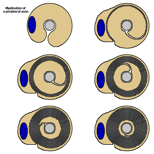 | 300 × 200 (77 KB) | Z8600021 (talk | contribs) | ==Myelination animation== {{Blue Histology}} Category:Animation |
You cannot overwrite this file.
File usage
The following 5 pages use this file:


