File:Stage7-sem4.jpg: Difference between revisions
No edit summary |
No edit summary |
||
| (One intermediate revision by one other user not shown) | |||
| Line 1: | Line 1: | ||
Human Embryo | ==Human Embryo Carnegie stage 7== | ||
17 days, pre-somite, scanning electron micrograph image | |||
Embryonic disc (epiblast/ectoderm layer) dorsolateral view, with amniotic membrane partially removed. | Embryonic disc (epiblast/ectoderm layer) dorsolateral view, with amniotic membrane partially removed. | ||
| Line 13: | Line 13: | ||
{{Template:SEM}} | {{Template:SEM}} | ||
[[Category:Human Embryo]] [[Category:Carnegie Stage]] [[Category:Week 3]] [[Category:Gastrulation]] | [[Category:Human Embryo]] [[Category:Carnegie Stage]] [[Category:Carnegie Stage 7]] [[Category:Week 3]] [[Category:Gastrulation]] | ||
Latest revision as of 15:35, 28 July 2012
Human Embryo Carnegie stage 7
17 days, pre-somite, scanning electron micrograph image
Embryonic disc (epiblast/ectoderm layer) dorsolateral view, with amniotic membrane partially removed.
Primitive node (Henson's node) in centre of disc and primitive streak, shown as indentation in disc is extending to the left. Connecting stalk to the left.
Scale bar 100 microns
See also: Stage7-sem5.jpg selected region of primitive streak | Stage7-sem3.jpg original image not cropped.
Image Source: Scanning electron micrographs of the Carnegie stages of the early human embryos are reproduced with the permission of Prof Kathy Sulik, from embryos collected by Dr. Vekemans and Tania Attié-Bitach. Images are for educational purposes only and cannot be reproduced electronically or in writing without permission.
File history
Yi efo/eka'e gwa ebo wo le nyangagi wuncin ye kamina wunga tinya nan
| Gwalagizhi | Nyangagi | Dimensions | User | Comment | |
|---|---|---|---|---|---|
| current | 13:31, 21 August 2009 | 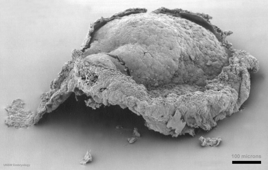 | 937 × 595 (79 KB) | MarkHill (talk | contribs) | Human Embryo Carnegie stage 7, 17 days, pre-somite, scanning electron micrograph image Embryonic disc (epiblast/ectoderm layer) dorsolateral view, with amniotic membrane partially removed. Primitive node (Henson's node) in centre of disc and primitive |
You cannot overwrite this file.
File usage
The following 10 pages use this file:
- 2010 Foundations Lecture - Introduction to Human Development
- 2011 Lab 2 - Week 3
- Carnegie stage 7
- Embryonic Development
- Foundations Lecture - Introduction to Human Development
- Foundations Practical - Week 3 and 4
- Gastrulation
- Human Embryo - Scanning electron microscopy
- Human Embryo SEM
- Pre-Medicine Program - Embryology