File:Historic-testis.jpg: Difference between revisions
No edit summary |
|||
| Line 1: | Line 1: | ||
==Testis Historic Drawing in Cross-section== | ==Testis Historic Drawing in Cross-section== | ||
Vertical section of the testis, to show the arrangement of the ducts. | |||
The glandular structure of the testis consists of numerous lobules. Their number, in a single testis, is estimated by Berres at 250, and by Krause at 400. They differ in size according to their position, those in the middle of the gland being larger and longer. | The glandular structure of the testis consists of numerous lobules. Their number, in a single testis, is estimated by Berres at 250, and by Krause at 400. They differ in size according to their position, those in the middle of the gland being larger and longer. | ||
The lobules are conical in shape, the base being directed toward the circumference of the organ, the apex toward the mediastinum. Each lobule is contained in one of the intervals between the fibrous septa which extend between the mediastinum testis and the tunica albuginea, and consists of from one to three, or more, minute convoluted tubes, the tubuli seminiferi. The tubules may be separately unravelled, by careful dissection under water, and may be seen to commence either by free cecal ends or by anastomotic loops. They are supported by loose connective tissue which contains here and there groups of “interstitial cells” containing yellow pigment granules. The total number of tubules is estimated by Lauth at 840, and the average length of each is 70 to 80 cm. Their diameter varies from 0.12 to 0.3 mm. The tubules are pale in color in early life, but in old age they acquire a deep yellow tinge from containing much fatty matter. Each tubule consists of a basement layer formed of laminated connective tissue containing numerous elastic fibers with flattened cells between the layers and covered externally by a layer of flattened epithelioid cells. Within the basement membrane are epithelial cells arranged in several irregular layers, which are not always clearly separated, but which may be arranged in three different groups. Among these cells may be seen the spermatozoa in different stages of development. | The lobules are conical in shape, the base being directed toward the circumference of the organ, the apex toward the mediastinum. Each lobule is contained in one of the intervals between the fibrous septa which extend between the mediastinum testis and the tunica albuginea, and consists of from one to three, or more, minute convoluted tubes, the ''tubuli seminiferi''. | ||
The tubules may be separately unravelled, by careful dissection under water, and may be seen to commence either by free cecal ends or by anastomotic loops. They are supported by loose connective tissue which contains here and there groups of “interstitial cells” containing yellow pigment granules. The total number of tubules is estimated by Lauth at 840, and the average length of each is 70 to 80 cm. Their diameter varies from 0.12 to 0.3 mm. The tubules are pale in color in early life, but in old age they acquire a deep yellow tinge from containing much fatty matter. Each tubule consists of a basement layer formed of laminated connective tissue containing numerous elastic fibers with flattened cells between the layers and covered externally by a layer of flattened epithelioid cells. Within the basement membrane are epithelial cells arranged in several irregular layers, which are not always clearly separated, but which may be arranged in three different groups. Among these cells may be seen the spermatozoa in different stages of development. | |||
# Lining the basement membrane and forming the outer zone is a layer of cubical cells, with small nuclei; some of these enlarge to become spermatogonia. The nuclei of some of the spermatogonia may be seen to be in process of indirect division, and in consequence of this daughter cells are formed, which constitute the second zone. | # Lining the basement membrane and forming the outer zone is a layer of cubical cells, with small nuclei; some of these enlarge to become spermatogonia. The nuclei of some of the spermatogonia may be seen to be in process of indirect division, and in consequence of this daughter cells are formed, which constitute the second zone. | ||
Revision as of 16:46, 3 May 2012
Testis Historic Drawing in Cross-section
Vertical section of the testis, to show the arrangement of the ducts.
The glandular structure of the testis consists of numerous lobules. Their number, in a single testis, is estimated by Berres at 250, and by Krause at 400. They differ in size according to their position, those in the middle of the gland being larger and longer.
The lobules are conical in shape, the base being directed toward the circumference of the organ, the apex toward the mediastinum. Each lobule is contained in one of the intervals between the fibrous septa which extend between the mediastinum testis and the tunica albuginea, and consists of from one to three, or more, minute convoluted tubes, the tubuli seminiferi.
The tubules may be separately unravelled, by careful dissection under water, and may be seen to commence either by free cecal ends or by anastomotic loops. They are supported by loose connective tissue which contains here and there groups of “interstitial cells” containing yellow pigment granules. The total number of tubules is estimated by Lauth at 840, and the average length of each is 70 to 80 cm. Their diameter varies from 0.12 to 0.3 mm. The tubules are pale in color in early life, but in old age they acquire a deep yellow tinge from containing much fatty matter. Each tubule consists of a basement layer formed of laminated connective tissue containing numerous elastic fibers with flattened cells between the layers and covered externally by a layer of flattened epithelioid cells. Within the basement membrane are epithelial cells arranged in several irregular layers, which are not always clearly separated, but which may be arranged in three different groups. Among these cells may be seen the spermatozoa in different stages of development.
- Lining the basement membrane and forming the outer zone is a layer of cubical cells, with small nuclei; some of these enlarge to become spermatogonia. The nuclei of some of the spermatogonia may be seen to be in process of indirect division, and in consequence of this daughter cells are formed, which constitute the second zone.
- Within this first layer is to be seen a number of larger polyhedral cells, with clear nuclei, arranged in two or three layers; these are the intermediate cells or spermatocytes. Most of these cells are in a condition of karyokinetic division, and the cells which result from this division form those of the next layer, the spermatoblasts or spermatids.
- The third layer of cells consists of the spermatoblasts or spermatids, and each of these, without further subdivision, becomes a spermatozoön. The spermatids are small polyhedral cells, the nucleus of each of which contains half the usual number of chromosomes. In addition to these three layers of cells others are seen, which are termed the supporting cells (cells of Sertoli). They are elongated and columnar, and project inward from the basement membrane toward the lumen of the tube. As development of the spermatozoa proceeds the latter group themselves around the inner extremities of the supporting cells. The nuclear portion of the spermatid, which is partly imbedded in the supporting cell, is differentiated to form the head of the spermatozoön, while part of the cell protoplasm forms the middle piece and the tail is produced by an outgrowth from the double centriole of the cell. Ultimately the heads are liberated and the spermatozoa are set free.
(text from Gray's Anatomy, 1918)
Gray's figure 1149
| Historic Disclaimer - information about historic embryology pages |
|---|
 |
File history
Click on a date/time to view the file as it appeared at that time.
| Date/Time | Thumbnail | Dimensions | User | Comment | |
|---|---|---|---|---|---|
| current | 16:43, 3 May 2012 | 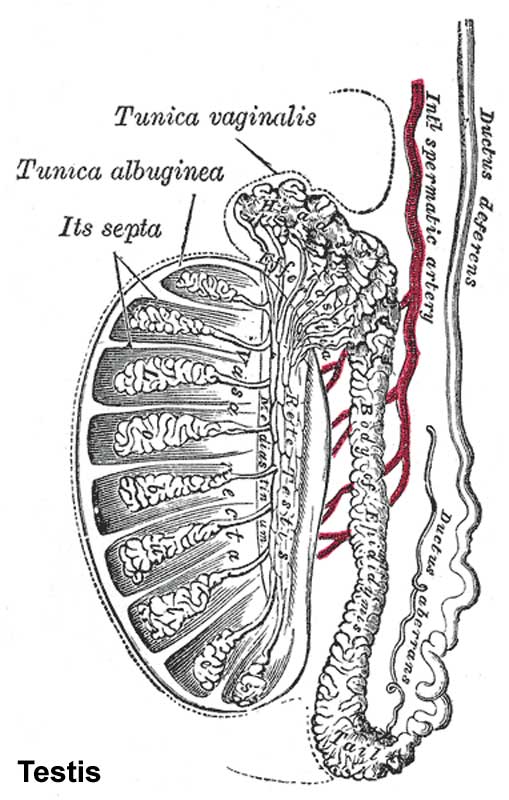 | 509 × 800 (67 KB) | Z8600021 (talk | contribs) | |
| 16:52, 21 September 2009 |  | 279 × 400 (47 KB) | S8600021 (talk | contribs) |
You cannot overwrite this file.
File usage
The following 25 pages use this file:
- 2009 Lecture 16
- 2010 BGD Lecture - Development of the Embryo/Fetus 1
- 2010 BGD Practical 3 - Gametogenesis
- 2010 Lecture 16
- 2011 Lab 1 - Gametogenesis
- 2011 Lab 8
- 2011 Lecture 16
- ANAT2341 Lab 1 - Gametogenesis
- BGDA Lecture - Development of the Embryo/Fetus 1
- BGDA Practical - Male Reproductive Tract Histology
- BGDA Practical 3 - Gametogenesis
- BGDB Practical - Sexual Differentiation
- BGD Lecture - Sexual Differentiation
- Cell Division - Meiosis
- Ductus Deferens Development
- Genital - Male Development
- Genital System Development
- Lecture - Fertilization
- Lecture - Genital Development
- Primordial Germ Cell Development
- Sertoli cell
- Spermatozoa Development
- Testis Development
- Testis Development Movie
- Talk:BGDA Practical 3 - Gametogenesis