File:Spleen histology 04.jpg: Difference between revisions
From Embryology
No edit summary |
|||
| Line 4: | Line 4: | ||
* peripheral localisation of the central arteries in nodules is quite distinct. | * peripheral localisation of the central arteries in nodules is quite distinct. | ||
{{Spleen Histology}} | |||
Revision as of 12:49, 26 February 2012
Spleen Histology
- reticular fibres of the white pulp appear somewhat finer and, at times, they are arranged as concentric rings.
- peripheral localisation of the central arteries in nodules is quite distinct.
Links: Histology | Histology Stains | Blue Histology images copyright Lutz Slomianka 1998-2009. The literary and artistic works on the original Blue Histology website may be reproduced, adapted, published and distributed for non-commercial purposes. See also the page Histology Stains.
Cite this page: Hill, M.A. (2024, June 20) Embryology Spleen histology 04.jpg. Retrieved from https://embryology.med.unsw.edu.au/embryology/index.php/File:Spleen_histology_04.jpg
- © Dr Mark Hill 2024, UNSW Embryology ISBN: 978 0 7334 2609 4 - UNSW CRICOS Provider Code No. 00098G
Original File name: Spl43re.jpg
File history
Yi efo/eka'e gwa ebo wo le nyangagi wuncin ye kamina wunga tinya nan
| Gwalagizhi | Nyangagi | Dimensions | User | Comment | |
|---|---|---|---|---|---|
| current | 18:41, 23 February 2012 | 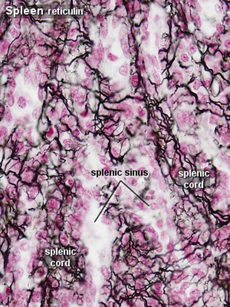 | 450 × 600 (108 KB) | Z8600021 (talk | contribs) | |
| 15:00, 21 February 2011 | 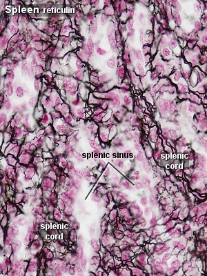 | 300 × 400 (70 KB) | S8600021 (talk | contribs) | ==Spleen Histology== Spleen histology 04.jpg Original File name: Spl43re.jpg {{Blue Histology}} Category:Spleen Category:Endocrine Category:Histology Category:Immune |
You cannot overwrite this file.
File usage
The following 8 pages use this file: