File:Blood capillary EM 01.jpg: Difference between revisions
From Embryology
(==Blood Capillary Electron Micrograph== A blood capillary lined by an endothelial cell (en) is surrounded by a continuous basal lamina (arrowhead) in which is incorporated a pericyte (p) Panel D cropped from Figure 2. (1471-2121-12-29-2.jpg) Contrast an) |
(No difference)
|
Revision as of 10:25, 5 February 2012
Blood Capillary Electron Micrograph
A blood capillary lined by an endothelial cell (en) is surrounded by a continuous basal lamina (arrowhead) in which is incorporated a pericyte (p)
Panel D cropped from Figure 2. (1471-2121-12-29-2.jpg) Contrast and size adjusted.
Reference
<ref><pubmed>21702933</pubmed>| PMC3141733 | BMC Cell Biol.
Original file name: 1471-2121-12-29-2.jpg
File history
Yi efo/eka'e gwa ebo wo le nyangagi wuncin ye kamina wunga tinya nan
| Gwalagizhi | Nyangagi | Dimensions | User | Comment | |
|---|---|---|---|---|---|
| current | 10:25, 5 February 2012 | 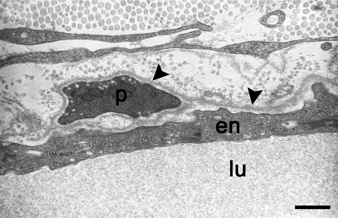 | 1,107 × 714 (260 KB) | S8600021 (talk | contribs) | ==Blood Capillary Electron Micrograph== A blood capillary lined by an endothelial cell (en) is surrounded by a continuous basal lamina (arrowhead) in which is incorporated a pericyte (p) Panel D cropped from Figure 2. (1471-2121-12-29-2.jpg) Contrast an |
You cannot overwrite this file.
File usage
The following 2 pages use this file: