Atlas of the Development of Man 2 - Cardiovascular: Difference between revisions
(Created page with "Handatlas der entwicklungsgeschichte des menschen: Volume 2 (Atlas of the Development of Man Volume 2) {{Template:KollmannAtlas2}} {{Glossary}} {{Footer}}") |
No edit summary |
||
| Line 1: | Line 1: | ||
Handatlas der entwicklungsgeschichte des menschen: Volume 2 (Atlas of the Development of Man Volume 2) | Handatlas der entwicklungsgeschichte des menschen: Volume 2 (Atlas of the Development of Man Volume 2) | ||
{{Template:KollmannAtlas2}} | |||
==Cardiovascular Images== | |||
Embryology the heart and vessels (Embryologia cordis et vasorum) | |||
For the embryology of the heart, wherever possible, human hearts were used. Where this was not possible, the models of the mammalian heart by Born been used since the development of the heart of man | |||
coincides with that of the rabbit. | |||
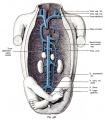
The coloring of the vessels is the same as in representations of the circulation is otherwise common, that is, those vessels that lead to the birth of venous blood are blue, those who after the birth of arterial | |||
blood are painted red. | |||
The embryonic vessels had received clear walls to represent graphically. In reality they have only limited endothelial lining, as indicated in many sections. | |||
<gallery> | |||
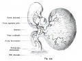
File:Kollmann511.jpg|Fig. 511. Vascular system of a human embryo of 1.3 mm. | |||
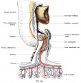
File:Kollmann512.jpg|Fig. 512. Vascular system of a human embryo of 1.3 mm. | |||
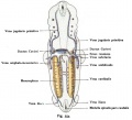
File:Kollmann513.jpg|Fig. 513. Human embryo heart of 2.11 mm in length. | |||
File:Kollmann514.jpg | |||
File:Kollmann515.jpg | |||
File:Kollmann516.jpg | |||
File:Kollmann517.jpg | |||
File:Kollmann518.jpg | |||
File:Kollmann519.jpg | |||
File:Kollmann520.jpg | |||
File:Kollmann521.jpg | |||
File:Kollmann522.jpg | |||
File:Kollmann523.jpg | |||
File:Kollmann524.jpg | |||
File:Kollmann525.jpg | |||
File:Kollmann526.jpg | |||
File:Kollmann527.jpg | |||
File:Kollmann528.jpg|Fig. 528. Heart of a Rabbit Embryo seen from behind at 3.4 mm head length | |||
File:Kollmann529.jpg|Fig. 529. The heart of a 24 mm Embryo | |||
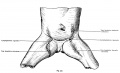
File:Kollmann530.jpg|Fig. 530. Fetal heart (6 months) in normal situation | |||
File:Kollmann531.jpg|Fig. 531. Heart included in the pericardium of a human embryo of 7.5 mm body length | |||
File:Kollmann532.jpg|Fig. 532. Development of the heart chambers and septa | |||
File:Kollmann533.jpg|Fig. 533. Heart of a newborn viewed from the front and placed in the vertical direction | |||
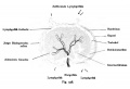
File:Kollmann534.jpg|Fig. 534. Fetal heart, dorsal half with the afferent paths, open and colored according to the physiological condition of the blood | |||
File:Kollmann535.jpg|Fig. 535. The aortic arch in the shark embryo (Pristiurus) | |||
File:Kollmann536.jpg|Fig. 536. The arteries of the gill arch region of a shark embryo (Pristiurus) | |||
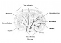
File:Kollmann537.jpg|Fig. 537. Aortic arch of mammals and man | |||
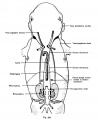
File:Kollmann538.jpg|Fig. 538. Arteries in mammals and humans from the Aortic Arch | |||
File:Kollmann539.jpg | |||
File:Kollmann540.jpg | |||
File:Kollmann541.jpg | |||
File:Kollmann542.jpg | |||
File:Kollmann543.jpg | |||
File:Kollmann544.jpg | |||
File:Kollmann545.jpg | |||
File:Kollmann546.jpg | |||
File:Kollmann547.jpg | |||
File:Kollmann548.jpg | |||
File:Kollmann549.jpg | |||
File:Kollmann550.jpg | |||
File:Kollmann551.jpg | |||
File:Kollmann552.jpg | |||
File:Kollmann553.jpg | |||
File:Kollmann554.jpg | |||
File:Kollmann555.jpg | |||
File:Kollmann556.jpg | |||
File:Kollmann557.jpg | |||
File:Kollmann558.jpg | |||
File:Kollmann559.jpg | |||
File:Kollmann560.jpg | |||
File:Kollmann561.jpg | |||
File:Kollmann562.jpg | |||
File:Kollmann563.jpg | |||
File:Kollmann564.jpg | |||
Kollmann565-569.jpg|Fig. 565-569. | |||
File:Kollmann570.jpg | |||
File:Kollmann571.jpg | |||
File:Kollmann572.jpg | |||
File:Kollmann573.jpg | |||
File:Kollmann574.jpg | |||
File:Kollmann575.jpg | |||
File:Kollmann576.jpg | |||
File:Kollmann577.jpg | |||
File:Kollmann578.jpg | |||
File:Kollmann579.jpg | |||
File:Kollmann580.jpg | |||
File:Kollmann581.jpg | |||
File:Kollmann582.jpg | |||
</gallery> | |||
{{Template:KollmannAtlas2}} | {{Template:KollmannAtlas2}} | ||
Revision as of 14:10, 21 October 2011
Handatlas der entwicklungsgeschichte des menschen: Volume 2 (Atlas of the Development of Man Volume 2)
- Kollmann Atlas 2: Gastrointestinal | Respiratory | Urogenital | Cardiovascular | Neural | Integumentary | Smell | Vision | Hearing | Kollmann Atlas 1 | Kollmann Atlas 2 | Julius Kollmann
Cardiovascular Images
Embryology the heart and vessels (Embryologia cordis et vasorum)
For the embryology of the heart, wherever possible, human hearts were used. Where this was not possible, the models of the mammalian heart by Born been used since the development of the heart of man coincides with that of the rabbit.
The coloring of the vessels is the same as in representations of the circulation is otherwise common, that is, those vessels that lead to the birth of venous blood are blue, those who after the birth of arterial blood are painted red.
The embryonic vessels had received clear walls to represent graphically. In reality they have only limited endothelial lining, as indicated in many sections.
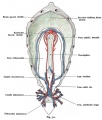
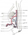
Fig. 511. Vascular system of a human embryo of 1.3 mm.
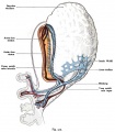
Fig. 512. Vascular system of a human embryo of 1.3 mm.

Fig. 513. Human embryo heart of 2.11 mm in length.


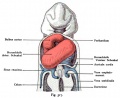
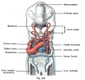
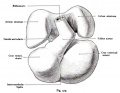

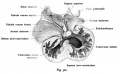

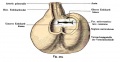

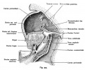
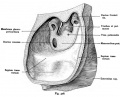

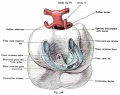
Fig. 528. Heart of a Rabbit Embryo seen from behind at 3.4 mm head length
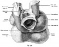
Fig. 529. The heart of a 24 mm Embryo
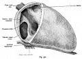
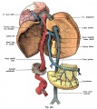
Fig. 530. Fetal heart (6 months) in normal situation
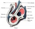
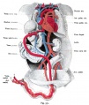
Fig. 531. Heart included in the pericardium of a human embryo of 7.5 mm body length
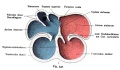
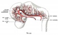
Fig. 532. Development of the heart chambers and septa
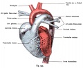
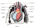
Fig. 533. Heart of a newborn viewed from the front and placed in the vertical direction
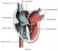
Fig. 534. Fetal heart, dorsal half with the afferent paths, open and colored according to the physiological condition of the blood
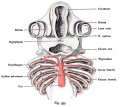
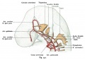
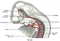
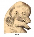
Fig. 535. The aortic arch in the shark embryo (Pristiurus)
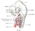
Fig. 536. The arteries of the gill arch region of a shark embryo (Pristiurus)
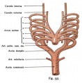
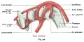
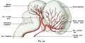
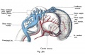
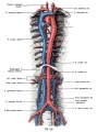
Fig. 537. Aortic arch of mammals and man
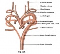
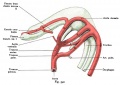
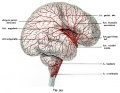
Fig. 538. Arteries in mammals and humans from the Aortic Arch

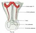


- Kollmann565-569.jpg
Fig. 565-569.


- Kollmann Atlas 2: Gastrointestinal | Respiratory | Urogenital | Cardiovascular | Neural | Integumentary | Smell | Vision | Hearing | Kollmann Atlas 1 | Kollmann Atlas 2 | Julius Kollmann
Glossary Links
- Glossary: A | B | C | D | E | F | G | H | I | J | K | L | M | N | O | P | Q | R | S | T | U | V | W | X | Y | Z | Numbers | Symbols | Term Link
Cite this page: Hill, M.A. (2024, June 27) Embryology Atlas of the Development of Man 2 - Cardiovascular. Retrieved from https://embryology.med.unsw.edu.au/embryology/index.php/Atlas_of_the_Development_of_Man_2_-_Cardiovascular
- © Dr Mark Hill 2024, UNSW Embryology ISBN: 978 0 7334 2609 4 - UNSW CRICOS Provider Code No. 00098G