File:Fetal 10wk urogenital 2.jpg: Difference between revisions
From Embryology
(These are images from an early fetus (female, 10 week, 40 mm). This stage of development is after the embryonic period (up to week 8) but still only 2 weeks into early fetal development. Section A is the most sagittal (lateral towards right) of all sectio) |
No edit summary |
||
| (3 intermediate revisions by the same user not shown) | |||
| Line 1: | Line 1: | ||
==Human Fetus (week 10) Female== | |||
Section | |||
female, 10 week, 40 mm CRL, early fetal, sagittal section, pelvic region | |||
This stage of development is after the embryonic period (up to week 8) but still only 2 weeks into early fetal development. | |||
Section B is the less lateral than plane A and more medial (towards the midline). Sections, planes B, C and D move towards the midline. | |||
Original file name: H10wkUrogenBL.jpg | Original file name: H10wkUrogenBL.jpg | ||
{{Template:10wkFetus}} | |||
[[Category:Renal]] [[Category:Genital]] [[Category:Musculoskeletal]] | |||
Latest revision as of 21:17, 29 May 2011
Human Fetus (week 10) Female
female, 10 week, 40 mm CRL, early fetal, sagittal section, pelvic region
This stage of development is after the embryonic period (up to week 8) but still only 2 weeks into early fetal development.
Section B is the less lateral than plane A and more medial (towards the midline). Sections, planes B, C and D move towards the midline.
Original file name: H10wkUrogenBL.jpg
Related Images
Fetus (week 10) Planes A (most lateral), B (lateral), C (medial) and D (midline) from lateral towards the midline.
- Human Fetus - most lateral | lateral | medial | midline
- Head - most lateral | lateral | medial | midline
- Cerebellum - most lateral | lateral | medial | midline
- Urogenital Unlabelled - most lateral | lateral | medial | midline
- Urogenital Labelled - most lateral | lateral | medial | midline
- Large Images - midline
- Image Source: UNSW Embryology, no reproduction without permission.
File history
Yi efo/eka'e gwa ebo wo le nyangagi wuncin ye kamina wunga tinya nan
| Gwalagizhi | Nyangagi | Dimensions | User | Comment | |
|---|---|---|---|---|---|
| current | 21:11, 29 May 2011 | 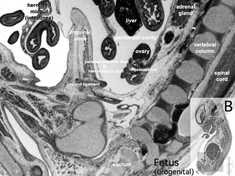 | 800 × 600 (110 KB) | S8600021 (talk | contribs) | |
| 22:57, 20 September 2009 | 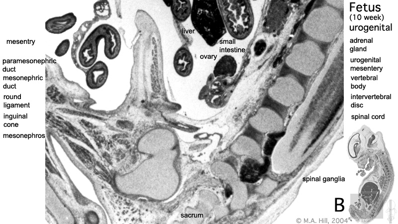 | 800 × 450 (136 KB) | S8600021 (talk | contribs) | These are images from an early fetus (female, 10 week, 40 mm). This stage of development is after the embryonic period (up to week 8) but still only 2 weeks into early fetal development. Section A is the most sagittal (lateral towards right) of all sectio |
You cannot overwrite this file.
File usage
The following 14 pages use this file:
- 2009 Lecture 15
- 2010 Lab 8
- 2010 Lecture 15
- 2011 Lab 8 - Fetal
- ANAT2241 Urinary System
- ANAT2341 Lab 8 - Fetal
- BGDB Sexual Differentiation - Fetal
- Fetal Development - 10 Weeks
- Genital - Female Development
- Lecture - Renal Development
- Renal System - Fetal
- Renal System Development
- Renal System Histology
- Urinary Bladder Development