File:Liver plasmodium infection cartoon.jpg: Difference between revisions
m (→Reference) |
|||
| (2 intermediate revisions by 2 users not shown) | |||
| Line 2: | Line 2: | ||
The dual blood supply of the liver, consisting of branches of the portal vein and the hepatic artery, merges upon entry into the liver lobule at the portal field. The blood flows along the sinusoid and exits at the central vein. | The dual blood supply of the liver, consisting of branches of the portal vein and the hepatic artery, merges upon entry into the liver lobule at the portal field. The blood flows along the sinusoid and exits at the central vein. | ||
# First sporozoites enter the liver lobule either via the portal vein or the hepatic artery, and then are abruptly arrested by binding to the sinusoidal cell layer. The initial binding is presumably mediated by stellate-cell-derived | # First sporozoites enter the liver lobule either via the portal vein or the hepatic artery, and then are abruptly arrested by binding to the sinusoidal cell layer. The initial binding is presumably mediated by stellate-cell-derived extracellular matrix proteoglycans that protrude from the space of Disse across the endothelial sieve plates into the sinusoidal lumen. | ||
# After a pause, the parasites begin to glide along the sinusoid, frequently moving against the bloodstream, until they then encounter a Kupffer cell, on the surface of which they recognize selected chondroitin and heparan sulfate proteoglycans. Sporozoites position themselves with their apical cell pole facing the phagocyte. | # After a pause, the parasites begin to glide along the sinusoid, frequently moving against the bloodstream, until they then encounter a Kupffer cell, on the surface of which they recognize selected chondroitin and heparan sulfate proteoglycans. Sporozoites position themselves with their apical cell pole facing the phagocyte. | ||
# After a considerable pause, they slowly pass through the Kupffer cell and cross the space of Disse beyond it, exhibiting a clearly visible constriction. | # After a considerable pause, they slowly pass through the Kupffer cell and cross the space of Disse beyond it, exhibiting a clearly visible constriction. | ||
# Once inside the liver parenchyma, the parasites increase their velocity and migrate for many minutes through several hepatocytes, before they eventually settle down in a final one for | # Once inside the liver parenchyma, the parasites increase their velocity and migrate for many minutes through several hepatocytes, before they eventually settle down in a final one for exoerythrocytic form development. | ||
Sporozoite transmigration results in a trail of necrotic hepatocytes, whose remains are subsequently removed by infiltrating inflammatory cells. | Sporozoite transmigration results in a trail of necrotic hepatocytes, whose remains are subsequently removed by infiltrating inflammatory cells. | ||
| Line 15: | Line 15: | ||
===Reference=== | |||
{{#pmid:15901208}} | |||
====Copyright==== | |||
© 2005 Frevert et al. This is an open-access article distributed under the terms of the Creative Commons Attribution License, which permits unrestricted use, distribution, and reproduction in any medium, provided the original work is properly cited. | |||
Original Image name: Figure 11. Journal.pbio.0030192.g011.jpg doi:10.1371/journal.pbio.0030192.g011 | |||
http://www.plosbiology.org/article/slideshow.action?uri=info:doi/10.1371/journal.pbio.0030192&imageURI=info:doi/10.1371/journal.pbio.0030192.g011 | |||
{{Footer}} | |||
[[Category:Liver]] [[Category:Cartoon]] | |||
Latest revision as of 10:33, 27 January 2019
Model of Plasmodium Sporozoite Infection of the Mammalian Liver
The dual blood supply of the liver, consisting of branches of the portal vein and the hepatic artery, merges upon entry into the liver lobule at the portal field. The blood flows along the sinusoid and exits at the central vein.
- First sporozoites enter the liver lobule either via the portal vein or the hepatic artery, and then are abruptly arrested by binding to the sinusoidal cell layer. The initial binding is presumably mediated by stellate-cell-derived extracellular matrix proteoglycans that protrude from the space of Disse across the endothelial sieve plates into the sinusoidal lumen.
- After a pause, the parasites begin to glide along the sinusoid, frequently moving against the bloodstream, until they then encounter a Kupffer cell, on the surface of which they recognize selected chondroitin and heparan sulfate proteoglycans. Sporozoites position themselves with their apical cell pole facing the phagocyte.
- After a considerable pause, they slowly pass through the Kupffer cell and cross the space of Disse beyond it, exhibiting a clearly visible constriction.
- Once inside the liver parenchyma, the parasites increase their velocity and migrate for many minutes through several hepatocytes, before they eventually settle down in a final one for exoerythrocytic form development.
Sporozoite transmigration results in a trail of necrotic hepatocytes, whose remains are subsequently removed by infiltrating inflammatory cells.
(text from article legend)
- Links: Liver Development | Image- Model of Plasmodium Sporozoite Infection of the Mammalian Liver | Image- Liver structure cartoon
Reference
Frevert U, Engelmann S, Zougbédé S, Stange J, Ng B, Matuschewski K, Liebes L & Yee H. (2005). Intravital observation of Plasmodium berghei sporozoite infection of the liver. PLoS Biol. , 3, e192. PMID: 15901208 DOI.
Copyright
© 2005 Frevert et al. This is an open-access article distributed under the terms of the Creative Commons Attribution License, which permits unrestricted use, distribution, and reproduction in any medium, provided the original work is properly cited.
Original Image name: Figure 11. Journal.pbio.0030192.g011.jpg doi:10.1371/journal.pbio.0030192.g011
Cite this page: Hill, M.A. (2024, June 21) Embryology Liver plasmodium infection cartoon.jpg. Retrieved from https://embryology.med.unsw.edu.au/embryology/index.php/File:Liver_plasmodium_infection_cartoon.jpg
- © Dr Mark Hill 2024, UNSW Embryology ISBN: 978 0 7334 2609 4 - UNSW CRICOS Provider Code No. 00098G
File history
Yi efo/eka'e gwa ebo wo le nyangagi wuncin ye kamina wunga tinya nan
| Gwalagizhi | Nyangagi | Dimensions | User | Comment | |
|---|---|---|---|---|---|
| current | 09:21, 4 May 2011 | 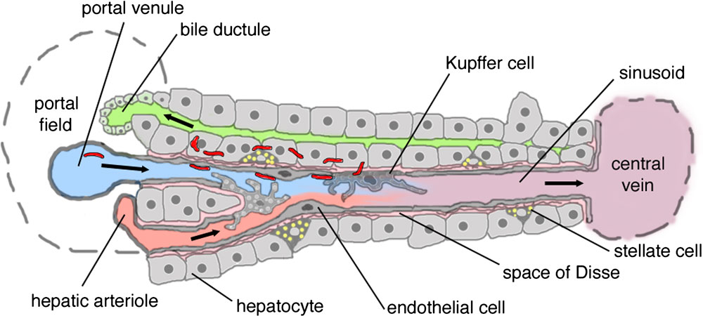 | 1,000 × 450 (78 KB) | S8600021 (talk | contribs) | ==Model of Plasmodium Sporozoite Infection of the Mammalian Liver== Dual blood supply of the liver merges upon entry into the liver lobule at the portal field. # branches of the portal vein # branches of the hepatic artery The blood flows along the si |
You cannot overwrite this file.
File usage
There are no pages that use this file.