File:Melanocyte1.jpg: Difference between revisions
No edit summary |
No edit summary |
||
| Line 1: | Line 1: | ||
==Description== | ==Description== | ||
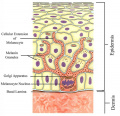
Melanocyte depicted in orange at the bottom of the epidermis, just above the basal lamina. Structures such as melanocytic extensions into the epidermal cells, also known as dendrites, can be seen, as well as the melanin granules. | |||
==Copyright== | ==Copyright== | ||
Allowed to use non-commercially according to http://slideplayer.com/support/terms/ "Except as otherwise provided, the Content published on this Website may be reproduced or distributed in unmodified form for personal non-commercial use only." | Allowed to use non-commercially according to http://slideplayer.com/support/terms/ "Except as otherwise provided, the Content published on this Website may be reproduced or distributed in unmodified form for personal non-commercial use only." | ||
==Reference== | |||
Warren, D. (2017). Melanin is created by melanocytes and packaged in melanosomes © 2014 Pearson Education, Inc. Retrieved from https://slideplayer.com/slide/11716447/ [Accessed 11 Sep. 2018] | |||
Revision as of 12:00, 11 September 2018
Description
Melanocyte depicted in orange at the bottom of the epidermis, just above the basal lamina. Structures such as melanocytic extensions into the epidermal cells, also known as dendrites, can be seen, as well as the melanin granules.
Copyright
Allowed to use non-commercially according to http://slideplayer.com/support/terms/ "Except as otherwise provided, the Content published on this Website may be reproduced or distributed in unmodified form for personal non-commercial use only."
Reference
Warren, D. (2017). Melanin is created by melanocytes and packaged in melanosomes © 2014 Pearson Education, Inc. Retrieved from https://slideplayer.com/slide/11716447/ [Accessed 11 Sep. 2018]
File history
Yi efo/eka'e gwa ebo wo le nyangagi wuncin ye kamina wunga tinya nan
| Gwalagizhi | Nyangagi | Dimensions | User | Comment | |
|---|---|---|---|---|---|
| current | 15:46, 30 October 2018 |  | 505 × 243 (18 KB) | Z8600021 (talk | contribs) | |
| 18:53, 8 September 2018 |  | 510 × 495 (75 KB) | Z5229549 (talk | contribs) | Diagram of an epidermal melanocyte and its structures. =Copyright= Allowed to use non-commercially according to http://slideplayer.com/support/terms/ "Except as otherwise provided, the Content published on this Website may be reproduced or distribute... |
You cannot overwrite this file.
File usage
The following 5 files are duplicates of this file (more details):
The following 2 pages use this file: