File:Joint development 02.jpg: Difference between revisions
No edit summary |
mNo edit summary |
||
| Line 1: | Line 1: | ||
== Joint Development== | == Joint Development== | ||
A Genetic System to Drive Gene Recombination in Developing Joints | A Genetic System to Drive Gene Recombination in Developing Joints | ||
(A) A 140-kb BAC from the Gdf5 locus was modified by inserting Cre-IRES-hPLAP into the translation start site of Gdf5 and used to make transgenic mice. Not to scale. See Materials and Methods for details. | (A) A 140-kb BAC from the Gdf5 locus was modified by inserting Cre-IRES-hPLAP into the translation start site of Gdf5 and used to make transgenic mice. Not to scale. See Materials and Methods for details. | ||
| Line 13: | Line 13: | ||
(D) Newborn (P0) forelimb with skin partially removed showing LACZ activity expressed in all phalangeal joints (red Salmon gal staining, black arrowheads) and regions of some tendons (asterisk). (E) Section through the most distal phalangeal joint of a P0 hindlimb stained with Alcian blue to mark cartilage showing LACZ expression (stained red) in all tissues of developing joints: articular cartilage (black arrowhead), precursors of ligaments and synovial membranes (black arrow), and cells where cavitation is occurring (asterisk). | (D) Newborn (P0) forelimb with skin partially removed showing LACZ activity expressed in all phalangeal joints (red Salmon gal staining, black arrowheads) and regions of some tendons (asterisk). (E) Section through the most distal phalangeal joint of a P0 hindlimb stained with Alcian blue to mark cartilage showing LACZ expression (stained red) in all tissues of developing joints: articular cartilage (black arrowhead), precursors of ligaments and synovial membranes (black arrow), and cells where cavitation is occurring (asterisk). | ||
===References=== | |||
{{#pmid:15492776}} | |||
====Copyright==== | |||
© 2004 Rountree et al. This is an open-access article distributed under the terms of the Creative Commons Attribution License, which permits unrestricted use, distribution, and reproduction in any medium, provided the original work is properly cited. | |||
{{Footer}} | |||
[[Category:Mouse]] [[Category:Musculoskeletal]] [[Category:Joint]] | [[Category:Mouse]] [[Category:Musculoskeletal]] [[Category:Joint]] | ||
Latest revision as of 23:05, 21 March 2018
Joint Development
A Genetic System to Drive Gene Recombination in Developing Joints
(A) A 140-kb BAC from the Gdf5 locus was modified by inserting Cre-IRES-hPLAP into the translation start site of Gdf5 and used to make transgenic mice. Not to scale. See Materials and Methods for details.
(B–E) Visualization of Gdf5-Cre driven recombination patterns based on activation of lacZ expression from the R26R Cre reporter allele.
(B) LACZ activity is visible as blue staining in the ear (ea) and the joints of the shoulder (s), elbow (eb), wrist (w), knee (k), ankle (a), vertebra (vj), and phalanges (black arrowheads) of an E14.5 mouse embryo.
(C) E14.5 hindlimb double-stained to show both HPLAP expression from the transgene (grey/purple staining) and LACZ expression from the rearranged R26R allele (blue staining). Note that both markers are visible in the oldest, proximal interphalangeal joint (black arrowhead), only HPLAP activity is visible in the more recently formed medial interphalangeal joint (black arrow), and neither HPLAP nor LACZ expression is visible in the youngest, most distal joint of the digit (white arrowhead).
(D) Newborn (P0) forelimb with skin partially removed showing LACZ activity expressed in all phalangeal joints (red Salmon gal staining, black arrowheads) and regions of some tendons (asterisk). (E) Section through the most distal phalangeal joint of a P0 hindlimb stained with Alcian blue to mark cartilage showing LACZ expression (stained red) in all tissues of developing joints: articular cartilage (black arrowhead), precursors of ligaments and synovial membranes (black arrow), and cells where cavitation is occurring (asterisk).
References
Rountree RB, Schoor M, Chen H, Marks ME, Harley V, Mishina Y & Kingsley DM. (2004). BMP receptor signaling is required for postnatal maintenance of articular cartilage. PLoS Biol. , 2, e355. PMID: 15492776 DOI.
Copyright
© 2004 Rountree et al. This is an open-access article distributed under the terms of the Creative Commons Attribution License, which permits unrestricted use, distribution, and reproduction in any medium, provided the original work is properly cited.
Cite this page: Hill, M.A. (2024, June 27) Embryology Joint development 02.jpg. Retrieved from https://embryology.med.unsw.edu.au/embryology/index.php/File:Joint_development_02.jpg
- © Dr Mark Hill 2024, UNSW Embryology ISBN: 978 0 7334 2609 4 - UNSW CRICOS Provider Code No. 00098G
File history
Yi efo/eka'e gwa ebo wo le nyangagi wuncin ye kamina wunga tinya nan
| Gwalagizhi | Nyangagi | Dimensions | User | Comment | |
|---|---|---|---|---|---|
| current | 08:43, 12 June 2010 | 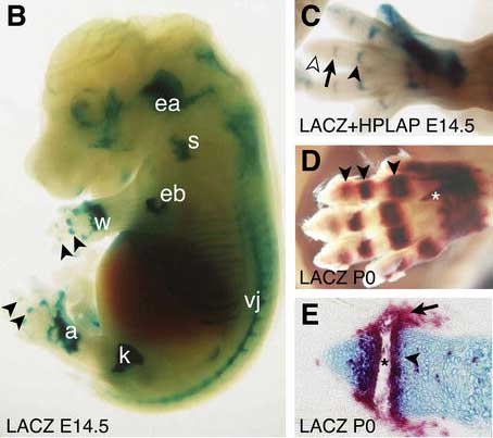 | 454 × 403 (22 KB) | S8600021 (talk | contribs) | == Joint Development== A Genetic System to Drive Gene Recombination in Developing Joints<ref><pubmed>15492776</pubmed> | [http://www.plosbiology.org/article/info%3Adoi%2F10.1371%2Fjournal.pbio.0020355 PLoS]</ref> (A) A 140-kb BAC from the Gdf5 locus was |
You cannot overwrite this file.
File usage
The following page uses this file: