File:1899 Cajal 01.jpg: Difference between revisions
No edit summary |
mNo edit summary |
||
| Line 1: | Line 1: | ||
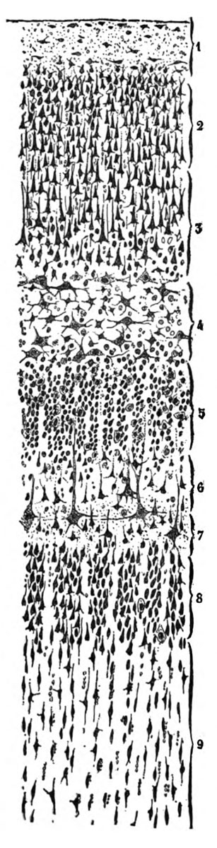
==Fig. 1. Vertical section of the visual cortex of man== | ==Fig. 1. Vertical section of the visual cortex of man== | ||
Calcarine sulcus, stained by Nissl's method — | Calcarine sulcus, stained by Nissl's method — semi-schematlc. | ||
1. Pleziform layer. 2. Layer of small pyramids. 3. Layer of medium-sized pyramids. 4. Layer of large stellate cells. 6. Layer of small stellate cells. 6. Second plexiform layer, or layer of small pyramids with arched axon. 7. Layer of giant pyramids. 8. Layer of medium-sized pyramidal cells with ascending axon. 9. Layer of fusiform and triangular cells. | 1. Pleziform layer. 2. Layer of small pyramids. 3. Layer of medium-sized pyramids. 4. Layer of large stellate cells. 6. Layer of small stellate cells. 6. Second plexiform layer, or layer of small pyramids with arched axon. 7. Layer of giant pyramids. 8. Layer of medium-sized pyramidal cells with ascending axon. 9. Layer of fusiform and triangular cells. | ||
| Line 8: | Line 8: | ||
{{Cajal lectures 1899}} | {{Cajal lectures 1899}} | ||
{{Historic Disclaimer}} | {{Historic Disclaimer}} | ||
===Reference=== | |||
{{ | {{Ref-Cajal1899a}} | ||
{{Footer}} | {{Footer}} | ||
Latest revision as of 16:19, 30 October 2017
Fig. 1. Vertical section of the visual cortex of man
Calcarine sulcus, stained by Nissl's method — semi-schematlc.
1. Pleziform layer. 2. Layer of small pyramids. 3. Layer of medium-sized pyramids. 4. Layer of large stellate cells. 6. Layer of small stellate cells. 6. Second plexiform layer, or layer of small pyramids with arched axon. 7. Layer of giant pyramids. 8. Layer of medium-sized pyramidal cells with ascending axon. 9. Layer of fusiform and triangular cells.
- 1899 Human Sensory Cortex: 1. The Visual Cortex | 2. Layer of the Large Stellate Cells | 3. The Sensori-Motor Cortex | Figures | Cajal
| Historic Disclaimer - information about historic embryology pages |
|---|
 |
Reference
Cajal SR. Comparative Study of the Sensory Areas of the Human Cortex (1899)
Cite this page: Hill, M.A. (2024, June 16) Embryology 1899 Cajal 01.jpg. Retrieved from https://embryology.med.unsw.edu.au/embryology/index.php/File:1899_Cajal_01.jpg
- © Dr Mark Hill 2024, UNSW Embryology ISBN: 978 0 7334 2609 4 - UNSW CRICOS Provider Code No. 00098G
File history
Click on a date/time to view the file as it appeared at that time.
| Date/Time | Thumbnail | Dimensions | User | Comment | |
|---|---|---|---|---|---|
| current | 17:00, 15 October 2012 | 307 × 1,200 (121 KB) | Z8600021 (talk | contribs) |
You cannot overwrite this file.
File usage
The following 2 pages use this file: