|
|
| (12 intermediate revisions by the same user not shown) |
| Line 1: |
Line 1: |
| {{Header}} | | {{Header}} |
| {{Ref-Patten1951}} | | {{Ref-Patten1951}}<br> |
| | |
| {{Patten1951 TOC}} | | {{Patten1951 TOC}} |
| {| class="wikitable mw-collapsible mw-collapsed" | | {| class="wikitable mw-collapsible mw-collapsed" |
| Line 20: |
Line 19: |
| {{Historic Disclaimer}} | | {{Historic Disclaimer}} |
| =Embryology of the Pig= | | =Embryology of the Pig= |
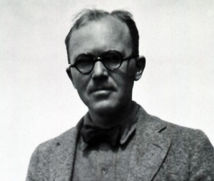
| Frontispiece | | [[File:Bradley M. Patten.jpg|thumb|alt=Bradley M. Patten|link=Embryology History - Bradley Patten|Bradley Patten ( -1971)]] |
| | Frontispiece |
|
| |
|
| Reconstruction (X 17.5) showing the organ systems of a 9.4 mm. pig embryo. For explanation see figures 60 and 66. | | Reconstruction (X 17.5) showing the organ systems of a 9.4 mm. pig embryo. For explanation see figures 60 and 66. |
|
| |
|
| By BRADLEY M. PATTEN | | By Bradley M. Patten |
|
| |
|
| Professor of Anatomy in the University of Michigan Medical School | | Professor of Anatomy in the University of Michigan Medical School |
|
| |
|
|
| |
|
| THIRD EDITION
| | Third Edition |
|
| |
|
|
| |
|
| WITH COLORED FRONTISPIECE
| | With Colored Frontispiece |
|
| |
|
| AND 186 ILLUSTRATIONS IN THE TEXT (CONTAINING 412 FIGURES)
| | And 186 Illustrations In The Text (Containing 412 Figures) Of Which 6 Are In Color |
| OF WHICH 6 ARE IN COLOR
| |
|
| |
|
|
| |
|
| | Philadelphia : THE BLAKISTON COMPANY : Toronto |
|
| |
|
| Philadelphia : THE BLAKISTON COMPANY : Toronto
| |
|
| |
|
| | Third Edition |
|
| |
|
|
| |
|
| Third Edition
| | Copyright, October 1948, by The Blakiston Company |
|
| |
|
|
| |
|
| Copyright, October 1948, by The Blakiston Company
| | By P. Blakiston's Son & Co. |
|
| |
|
|
| |
|
| BY P. Blakiston ’s Son & Co.
| | Copyright, 1951, by P Blakiston's Son & Co , Inc. |
|
| |
|
|
| |
|
| Copyright, 1951, by P Blakiston’s Son & Co , Inc.
| | {{Patten1951 TOC}} |
|
| |
|
| | ==Preface to Third Edition== |
|
| |
|
| Preface to Third Edition
| | In making the revision for a new edition of this book it did not seem desirable essentially to change its form or scope. Effort has been concentrated on improving the presentation of the original subject matter and bringing it up to date, rather than on its expansion. The entire book has been reset to a greater page width which has permitted enlarging certain of the illustrations that, in earlier editions, had proved to be too greatly reduced. With the generous cooperation of the publishers several of the important plates on the cardiovascular system have been remade to a larger scale and with color. A number of new illustrations have been drawn for sections where experience has shown that students needed additional graphic assistance in interpreting their laboratory material. It is hoped that these changes will all contribute toward making the book as a whole more serviceable. |
|
| |
|
|
| |
|
| In making the revision for a new edition of this book it did not
| | Bradley M. Patten |
| seem desirable essentially to change its form or scope. Effort has been
| |
| concentrated on improving the presentation of the original subject
| |
| matter and bringing it up to date, rather than on its expansion. The
| |
| entire book has been reset to a greater page width which has permitted enlarging certain of the illustrations that, in earlier editions,
| |
| had proved to be too greatly reduced. With the generous cooperation
| |
| of the publishers several of the important plates on the cardiovascular
| |
| system have been remade to a larger scale and with color. A number
| |
| of new illustrations have been drawn for sections where experience
| |
| has shown that students needed additional graphic assistance in
| |
| interpreting their laboratory material. It is hoped that these changes
| |
| will all contribute toward making the book as a whole more serviceable.
| |
|
| |
|
| Bradley M. Patten
| | August 1048 |
|
| |
|
| August 1048
| |
|
| |
|
| | ==Preface to First Edition== |
|
| |
|
| Preface to First Edition
| | This book represents an endeavor to set forth in brief and simple form the fundamental facts of mammalian development. The thread of the story and the illustrations have been based on pig embryos because of their value and availability as laboratory material. But special stress has been laid on the embryological phenomena involved instead of on the details of specific conditions existing in the pig. Throughout the book, every efTort has been made to present developmental processes as dynamic events with emphasis on their sequence and significance, rather than as a series of still pictures of selected stages. |
|
| |
|
|
| |
|
| This book represents an endeavor to set forth in brief and simple
| | Obviously no book can deal fully with all phases of development, even in a single form, and still remain serviceable as a text. As this book is for the student, it has seemed expedient, for the sake of clearness and simplicity, to omit many things which I should like to have included. My primary aim has been to write an account in which the essentials stand out adequately interpreted and unobscured by a multiplicity of details — to lay a foundation which can be further built upon in accordance with special needs or individual desires. |
| form the fundamental facts of mammalian development. The thread
| |
| of the story and the illustrations have been based on pig embryos
| |
| because of their value and availability as laboratory material. But
| |
| special stress has been laid on the embryological phenomena involved
| |
| instead of on the details of specific conditions existing in the pig.
| |
| Throughout the book, every efTort has been made to present developmental processes as dynamic events with emphasis on their sequence
| |
| and significance, rather than as a series of still pictures of selected
| |
| stages.
| |
|
| |
|
| Obviously no book can deal fully with all phases of development,
| |
| even in a single form, and still remain serviceable as a text. As this
| |
| book is for the student, it has seemed expedient, for the sake of clearness and simplicity, to omit many things which I should like to have
| |
| included. My primary aim has been to write an account in which the
| |
| essentials stand out adequately interpreted and unobscured by a
| |
| multiplicity of details — to lay a foundation which can be further
| |
| built upon in accordance with special needs or individual desires.
| |
|
| |
|
| Bradley M. Patten | | Bradley M. Patten |
| | |
| January 1927
| |
|
| |
|
| | January 1927 |
|
| |
|
| ==Acknowledgment== | | ==Acknowledgment== |
| | The pleasantest thing about working on this book has been the generous aid I have received from many sources. Throughout the preparation of the initial edition the encouragement, criticism, and suggestions of my colleagues, Dr. F. C. Waite and Dr. S. W. Chase, were of the greatest help. In the preparation of material and in making the illustrations for the first edition the beautifully accurate work of Miss Kathryn Toulmin was of inestimable value. In making the new drawings added in the third edition I was fortunate in securing the unusually able assistance of Mrs. Dorothy Van Eck. |
|
| |
|
|
| |
|
| The pleasantest thing about working on this book has been the
| | Dr. G. L. Streeter and Dr. C. H. Heuser of the Carnegie Institute allowed me free use of their extensive collection of young embryos and generously gave me many photographs made from their material. To Mrs. Charles S, Minot I am indebted for permission to use several figures from the late Professor Minot's works. The accrediting in the figure legends of these and other borrowed illustrations by no means covers my obligation to other writers. Practically the entire bibliography is a statement of indebtedness for information and ideas. |
| generous aid I have received from many sources. Throughout the
| |
| preparation of the initial edition the encouragement, criticism, and
| |
| suggestions of my colleagues, Dr. F. C. Waite and Dr. S. W. Chase,
| |
| were of the greatest help. In the preparation of material and in making
| |
| the illustrations for the first edition the beautifully accurate work of
| |
| Miss Kathryn Toulmin was of inestimable value. In making the new
| |
| drawings added in the third edition I was fortunate in securing the
| |
| unusually able assistance of Mrs. Dorothy Van Eck.
| |
|
| |
|
| Dr. G. L. Streeter and Dr. C. H. Heuser of the Carnegie Institute
| |
| allowed me free use of their extensive collection of young embryos
| |
| and generously gave me many photographs made from their material.
| |
| To Mrs. Charles S, Minot I am indebted for permission to use several
| |
| figures from the late Professor Minot’s works. The accrediting in the
| |
| figure legends of these and other borrowed illustrations by no means
| |
| covers my obligation to other writers. Practically the entire bibliography is a statement of indebtedness for information and ideas.
| |
|
| |
|
| I wish I might acknowledge individually the helpful services | | I wish I might acknowledge individually the helpful services rendered by many of my students, but they are too numerous. Several reconstructions which I have used directly or indirectly have been largely their work. Of even greater assistance have been their suggestions during the shaping of the work — suggestions of especial value because they were made from a point of view difficult for an instructor to appreciate without such aid. |
| rendered by many of my students, but they are too numerous. Several | |
| reconstructions which I have used directly or indirectly have been | |
| largely their work. Of even greater assistance have been their suggestions during the shaping of the work — suggestions of especial value | |
| because they were made from a point of view difficult for an instructor | |
| to appreciate without such aid. | |
|
| |
|
| To The Blakiston Company, I am indebted for much helpful
| |
| cooperation and especially for their liberality with regard to the
| |
| number and quality of the illustrations.
| |
|
| |
|
| No person other than my wife could have deciphered and put
| | To The Blakiston Company, I am indebted for much helpful cooperation and especially for their liberality with regard to the number and quality of the illustrations. |
| into usable form manuscript of the character I frequently turned over
| |
| to her for revision and typing. Without her generous help the preparation of the text would have been much more arduous and long
| |
| delayed.
| |
|
| |
|
| Bradley M. Patten
| |
|
| |
|
| | No person other than my wife could have deciphered and put into usable form manuscript of the character I frequently turned over to her for revision and typing. Without her generous help the preparation of the text would have been much more arduous and long delayed. |
|
| |
|
|
| |
|
| ==Chapter 7==
| | Bradley M. Patten |
|
| |
|
|
| |
|
| Tke Structure of Emkryos from Nine to
| |
| Twelve Millimeters in Lengtk
| |
|
| |
|
| | {{Footer}} |
|
| |
|
| Pig embryos of about 10 mm. crown-rump length are especially
| | {{Patten1951 TOC}} |
| valuable as material for laboratory study. They are neither so small
| |
| and delicate that procuring and preparing them involves unusually
| |
| skillful manipulation, nor so large that complete series of sections are
| |
| tedious to make and difficult to study. Developmentally they are not
| |
| so far advanced that their structure is hard to comprehend if the
| |
| student is familiar with the early stages of development in any of the
| |
| vertebrates. Yet if their morphology is thoroughly mastered it forms a
| |
| starting point from which the later developmental processes in
| |
| mammals may readily be traced. It seems advantageous, therefore, to
| |
| deal with this, the most commonly utilized stage of development,
| |
| somewhat less in narrative fashion than has been done heretofore. We
| |
| shall, as it were, pause for a while and look over carefully the conditions which thus far have been established so that we may follow the
| |
| story of the later phases of development more understandingly.
| |
| | |
| I. External Features
| |
| | |
| Flexion. ^ Like all forms which develop in the limited confines
| |
| of an egg shell or within the uterine cavity, the body of a young pig
| |
| embryo is curled up on itself. This flexion of the spinal axis is more
| |
| marked in some regions than others. The points of conspicuous bending have received special designations from the region in which
| |
| they appear. In a pig of 10 mm. (Fig. 58) the cranial flexure, cervical
| |
| flexure, dorsal flexure, and lumbosacral flexure are all well developed.
| |
| | |
| The Head. In the cephalic region the thin skin leaves the contours
| |
| of the brain clearly suggested (cf. Figs. 58 and 59). The nasal pits
| |
| have appeared as definitely circumscribed depressions at the rostral
| |
| end of the head and the bulging caused by the growing optic cups
| |
| clearly marks the position of the eyes. Especially in fresh specimens
| |
| the retinal pigment can be seen through the overlying skin.
| |
| | |
| 107
| |
| | |
| | |
| | |
| | |
| 108 STRUCTURE OF NINE- TO TWELVE-MILLIMETER EMBRYOS
| |
| | |
| | |
| Mesencephalon
| |
| | |
| | |
| Maxillary process'
| |
| Nasal pit
| |
| Cardiac prominence
| |
| Hepatic prominence
| |
| Belly-stalk
| |
| | |
| | |
| Posterior,
| |
| appendage bud
| |
| | |
| | |
| Metencephalon
| |
| | |
| Mandibular arch
| |
| | |
| ^Hyomandibular cleft
| |
| | |
| Hyoid arch
| |
| | |
| | |
| | |
| Cervical sinus
| |
| | |
| | |
| Anterior
| |
| appendage bud
| |
| | |
| | |
| Mesonephric
| |
| | |
| prominence
| |
| | |
| | |
| Somite
| |
| | |
| | |
| Fig. 58. Drawing (X 7) showing external appearance of 10 min. pig embryo.
| |
| | |
| (Modified from Minot.)
| |
| | |
| | |
| The Gill Arches. Flanking the oral cavity and caudal to it, in the
| |
| region which will be under the chin, the gill (branchial) arches appear
| |
| as strongly marked local elevations. This entire region is at this stage
| |
| so compressed against the thorax that one gets a very incomplete view
| |
| of its structure unless an embryo is decapitated and the head viewed
| |
| in ventral aspect (Figs. 168 and 169). It is then seen that the maxillary
| |
| processes form the lateral parts of the upper jaw, and that the two
| |
| mandibular elevations meet each other in the mid-ventral line to form
| |
| the arch (mandibular arch) of the lower jaw. Posterior to the mandibular are three similar arches, the hyoid and and the unnamed
| |
| third and fourth post-oral arches. Later in development the arches
| |
| posterior to the mandible become less conspicuous and are incorporated into the neck.
| |
| | |
| The Gill Clefts. Between the branchial arches are deep furrows
| |
| which mark the position of ancestral gill clefts. Although in mammalian embryos these furrows do not ordinarily break through into
| |
| the pharynx they are commonly called clefts because of their phylogenetic significance. Only the most cephalic of these clefts is named
| |
| (hyomandibular cleft, Fig. 58); the others are designated by their
| |
| | |
| | |
| | |
| EXTERNAL FEATURES
| |
| | |
| | |
| 109
| |
| | |
| | |
| post-oral numbers. The entire region about the third and fourth postoral clefts becomes especially deeply depressed and is known as the
| |
| cervical sinus.
| |
| | |
| The Appendage Buds. Both anterior and posterior appendage
| |
| buds in 10 mm, pigs are still paddle-shaped (Fig. 58). Not until
| |
| embryos have grown to a length of between 15 and 20 mm. do the five
| |
| digits show as terminal enlargements (Figs. 33 and 34). It is significant
| |
| that the ancestral five-digit type of appendage appears transitorily
| |
| in the pig embryo, before its characteristic highly specialized hoof is
| |
| even suggested.
| |
| | |
| The Trunk. At this stage the superficial bulges due to the somites
| |
| are evident all the way from the cervical to the caudal region of the
| |
| | |
| | |
| | |
| | |
| | |
| | |
| 110 STRUCTURE OF NINE- TO TWELVE-MILLIMETER EMBRYOS
| |
| | |
| body. Well-marked prominences in the lateral and ventral portions
| |
| of the body-wall indicate the position of the heart, t^e liver, and the
| |
| mesonephros (cf. Figs. 58 and 59).
| |
| | |
| The band of tissue (“milk ridgeâ€) which gives rise to the mammary
| |
| glands is not ordinarily developed in embryos of the 9 to 12 mm.
| |
| range. It usually becomes clearly marked by 15 mm. (Fig. 33), and
| |
| by 20 mm. the nipples can generallv be recognized (Fig. 34).
| |
| | |
| II. The Nervous System
| |
| | |
| The Brain. In younger embryos the brain consisted of three
| |
| regions, fore-brain (prosencephalon), mid-brain (mesencephalon),
| |
| and hind-brain (rhombencephalon) (Fig. 36, E). Now we find five
| |
| regional divisions or vesicles, as they are called. The prosencephalon
| |
| has divided to form the telencephalon and diencephalon; the mesencephalon has remained undivided; and the rhombencephalon has
| |
| become differentiated into metencephalon and myelencephalon (Figs. 59,
| |
| 60, 65, and 88).
| |
| | |
| The telencephalon consists of the most anterior median part of the
| |
| brain and two lateral outgrowths from it called the lateral telencephalic
| |
| vesicles (Figs. 60, 67, and 69). Its posterior boundary is conventionally
| |
| established by drawing a line from a fold in the roof of the brain called
| |
| the velum transversum (see Fig. 65 where the leader to Seessel’s
| |
| pocket crosses the dorsal wall of the neural tube) to the recessus
| |
| opticus, a depression in the floor of the brain at the level of the optic
| |
| stalks (Figs, 65 and 88).
| |
| | |
| The diencephalon is the hinder portion of the old prosencephalon.
| |
| Its caudal boundary is conventionally established by drawing a line
| |
| from an elevation in the floor of the neural tube called the tuberculum
| |
| posterius (Fig. 88) to a depression in the roof of the neural tube which
| |
| is just appearing at this stage of development, and may (Fig. 60) or
| |
| may not (Fig. 65) be evident in the particular embryo under observation. The most conspicuous special features of the diencephalon are
| |
| the paired lateral outgrowths from it which form the optic vesicles
| |
| (Figs. 60 and 63) and the median ventral diverticulum which constitutes the infundibulum (Fig. 65). The median dorsal outgrowth of
| |
| the diencephalon known as the epiphysis, which is so conspicuous a
| |
| feature in chick embryos of the third and fourth day, appears relatively late in the pig. No suggestion of an epiphyseal evagination has
| |
| appeared in 9 to 12 mm. embryos.
| |
| | |
| The mesencephalon shows little change from its condition in
| |
| | |
| | |
| | |
| THE NERVOUS SYSTEM
| |
| | |
| | |
| 111
| |
| | |
| | |
| younger embryos. Its demarcation from the metencephalon posteriorly
| |
| is indicated by a conspicuous constriction in the neural tube (Figs.
| |
| 59, 60, and 65).
| |
| | |
| The division of the primitive hind-brain (rhombencephalon) into
| |
| metencephalon and myelencephalon is indicated at this stage,
| |
| though not conspicuous or definite. The dorsal wall of the neural tube
| |
| just caudal to the meso-rhombencephalic constriction is markedly
| |
| thick, contrasting strikingly with the very thin roof of the more
| |
| posterior part of the hind-brain (Fig. 65). The zone of the neural tube
| |
| where the dorsal thickening exists is the metencephalon ; the posterior,
| |
| thin-roofed portion of the hind-brain is the myelencephalon. Although
| |
| all external indications of individual neuromeres have by this time
| |
| disappeared, the internal face of the myelencephalic wall still shows
| |
| definite neuromeric markings (Figs. 61 and 67).
| |
| | |
| The Cranial Nerves. The peripheral relations of the cranial
| |
| nerves to cephalic structures and their central relations to the brain
| |
| are strikingly constant throughout the vertebrate series. In fishes we
| |
| recognize 10 pairs of cranial nerves. In the mammals we encounter
| |
| these same 10 cranial nerves with essentially similar relations both
| |
| centrally and peripherally. But the mammalian brain in its progressive
| |
| specialization has incorporated a part of the neural tube which in
| |
| primitive fishes was unmodified spinal cord. One of the clearest evidences of this process is the fact that we find in the mammals 12 pairs
| |
| of cranial nerves, the first, 10 of which are homologous with the 10
| |
| cranial nerves of fishes and the last two of which represent a modification of nerves which in fishes were the most anterior of the spinal
| |
| nerves.
| |
| | |
| The 12 cranial nerves of mammals are designated by numbers
| |
| almost as commonly as they are referred to by name. Beginning with
| |
| the most rostral they are: (I) olfactory, (II) optic, (III) oculomotor,
| |
| (IV) trochlear, (V) trigeminal, (VI) abducens, (VII) facial, (VIII)
| |
| acoustic, (IX) glossopharyngeal, (X) vagus, (XI) spinal accessory,
| |
| and (XII) hypoglossal. In 9 to 12 mm. pig embryos all of the cranial
| |
| nerves except the olfactory and optic are readily recognizable (Figs.
| |
| 59-63, and 92). Those carrying sensory fibers show conspicuous
| |
| ganglia near their point of connection with the brain (see nerves
| |
| V, VII, VIII, IX, and X in Fig. 92). Except for the acoustic (VIII)
| |
| all these ganglionated nerves carry also some motor fibers — that is,
| |
| they are mixed nerves. The cranial nerves composed practically
| |
| entirely of motor fibers have no external ganglia (nerves III, IV, VI,
| |
| and XII, Fig. 92).
| |
| | |
| | |
| | |
| 112 STRUCTURE OF NINE- TO TWELVE-MILLIMETER EMBRYOS
| |
| | |
| | |
| | |
| Fig. 60. Reconstruction (X 14) of the nervous, digestive, respiratory, and
| |
| urinary systems of a 9.4 mm. pig embryo. Compare with Frontispiece and
| |
| with figure 66. By laying a straight-edge across the numbered lines in the
| |
| margin the locations of the accompanying cross-sections may be determined.
| |
| | |
| Abbreviations
| |
| | |
| All. St., allantoic stalk, C.I., dorsal root ganglion of first
| |
| | |
| Br. ep., eparterial bronchus. cervical nerve.
| |
| | |
| | |
| | |
| EXTERNAL FEATURES
| |
| | |
| | |
| 113
| |
| | |
| | |
| Fig. 60 — (Continued)
| |
| Abbreviations
| |
| | |
| | |
| Ch. t., chorda tympani branch of
| |
| facial nerve.
| |
| | |
| Dien., diencephalon.
| |
| | |
| G.acc., accessory ganglion.
| |
| | |
| G.acou., acoustic ganglion of eighth
| |
| nerve.
| |
| | |
| G.Fro., Froriep’s ganglion.
| |
| | |
| G.gas., Gasserian (semilunar) ganglion of fifth nerve.
| |
| | |
| (bgen., geniculate ganglion of
| |
| seventh nerve.
| |
| | |
| G.jug., jugular ganglion of tenth
| |
| nerve.
| |
| | |
| G.nod., nodose ganglion of tenth
| |
| nerve.
| |
| | |
| G.pet., petrosal ganglion of ninth
| |
| nerve.
| |
| | |
| G.pv., prevertebral sympathetic ganglion.
| |
| | |
| G.sup., superior ganglion of ninth
| |
| nerve.
| |
| | |
| Gen. Em. genital eminence.
| |
| | |
| Gl. B., gall bladder.
| |
| | |
| Glom.j glomerulus.
| |
| | |
| L. , lung.
| |
| | |
| Lg. Int., large intestine.
| |
| | |
| M. Mes.j splanchnic mesoderm cut
| |
| where reflected over mesonephros
| |
| from mesentery.
| |
| | |
| Mand.V., mancfibular branch of fifth
| |
| (trigeminal) nerve.
| |
| | |
| Max.V., maxillary branch of fifth
| |
| (trigeminal) nerve.
| |
| | |
| Mes., mesencephalon.
| |
| | |
| Mes. D., mesonephric duct.
| |
| | |
| Met., metencephalon.
| |
| | |
| Met.D., metanephric diverticulum.
| |
| | |
| Myel.j myelencephalon.
| |
| | |
| N. IIL, third cranial (oculomotor)
| |
| nerve.
| |
| | |
| N.IV., fourth cranial (trochlear)
| |
| nerve.
| |
| | |
| N.VI., sixth cranial (abducens)
| |
| nerve.
| |
| | |
| | |
| N.VI I., seventh cranial (facial)
| |
| nerve.
| |
| | |
| N.X., tenth cranial (vagus) nerve.
| |
| | |
| N.XI., eleventh cranial (accessory)
| |
| nerve.
| |
| | |
| N.XI I., twelfth cranial (hypoglossal)
| |
| nerve.
| |
| | |
| N.cerv.d., descending cervical nerve.
| |
| | |
| N.Ph., phrenic nerve.
| |
| | |
| Na., location of nasal (olfactory) pit.
| |
| | |
| Nch., notochord.
| |
| | |
| Neur. Cr., neural crest.
| |
| | |
| Neph., nephrogenous tissue of metanephros.
| |
| | |
| Oes., esophagus.
| |
| | |
| Op.c., optic cup.
| |
| | |
| Oph.V., ophthalmic branch of fifth
| |
| (trigeminal) nerve.
| |
| | |
| Ot., auditory vesicle (otocyst).
| |
| | |
| P.C.G., post-cloacal gut.
| |
| | |
| P.C.V., posterior cardinal vein.
| |
| | |
| Panc.d., dorsal pancreas.
| |
| | |
| Panc.v., ventral pancreas.
| |
| | |
| Pelvic Dil., pelvic dilation of metanephric diverticulum.
| |
| | |
| Ph.4., fourth pharyngeal pouch.
| |
| | |
| Rath, p., Rathke’s pocket.
| |
| | |
| Sm. Int., small intestine.
| |
| | |
| St., stomach.
| |
| | |
| Sub. Card. Sin., large venous sinus
| |
| formed by the transverse anastomosis of the subcardinal veins
| |
| (subcardinal sinus).
| |
| | |
| T.I., first thoracic spinal ganglion.
| |
| | |
| Tel., telencephalon (specifically the
| |
| label is on the right lateral telencephalic vesicle).
| |
| | |
| Thym., thymus.
| |
| | |
| Thyr., thyroid.
| |
| | |
| Tr., trachea.
| |
| | |
| V.T.M., superficial mesonephric
| |
| veins connecting post- and subcardinals (named venae transversales mediales by Sabin).
| |
| | |
| | |
| | |
| 114 STRUCTURE OF NINE- TO TWELVE-MILLIMETER EMBRYOS
| |
| | |
| | |
| ganglion VIII ganglion VII
| |
| | |
| | |
| | |
| myelencephalon
| |
| | |
| | |
| Fig. 61. Transverse section of 9.4 mm. pig embryo passing through the
| |
| myelencephalic region (X 15). This and the following drawings of crosssections were taken from the same series used in making the reconstructions
| |
| which appear as figures 60 and 66. The serial number of this section is 96. Its
| |
| location on the reconstructions may be determined by laying a straight-edge
| |
| on the marginal lines numbered 96.
| |
| | |
| | |
| phanmgeal pouch I mandibular branch nerve V
| |
| | |
| pharyngeal pouch II \ / int ca
| |
| | |
| | |
| 1st cervical
| |
| ganglion
| |
| | |
| | |
| | |
| | |
| int carotid a.
| |
| | |
| / optic cup
| |
| | |
| diencephalon
| |
| | |
| | |
| br, am.
| |
| w ^^ard V
| |
| | |
| | |
| | |
| | |
| | |
| | |
| 4 ^
| |
| | |
| | |
| | |
| | |
| | |
| ant cardinal v. / /
| |
| | |
| nerve XL /
| |
| ganglion nod.X
| |
| | |
| | |
| N. Ratbfcra
| |
| | |
| \ pocket
| |
| | |
| . ophth branch of
| |
| \ nerve V
| |
| | |
| nerve 111
| |
| | |
| | |
| I ^ branch of ant cardinal
| |
| | |
| «erve VII Imaxillary branch nerve V
| |
| | |
| | |
| Fig. 62. Transverse section of 9.4 mm. pig embryo passing through the
| |
| pharynx (X 15). (Its serial number on reconstructions appearing as figures
| |
| 60 and 66 is 142.)
| |
| | |
| | |
| | |
| THE NERVOUS SYSTEM
| |
| | |
| | |
| 115
| |
| | |
| | |
| The Spinal Ckird and Spinal Nerves. The caudal part of the
| |
| myelenccphalon merges without any definite line of demarcation
| |
| into the spinal cord. The walls of the neural tube in the cord region
| |
| have already begun to become differentiated. Dorsally and ventrally
| |
| they remain thin, but laterally rapid proliferation of primordial nerve
| |
| cells has caused them to increase greatly in thickness so that the
| |
| originally oval lumen of the tube becomes slit-like (cf. Figs. 42, B-D,
| |
| and 83).
| |
| | |
| The cells arising from the neural crests (Fig. 35) have become
| |
| aggregated on either side of the cord into segmentally arranged
| |
| clusters, the spinal ganglia (Fig. 59). From nerve cells in each of these
| |
| ganglia, fibers grow both toward the cord and peripherally, establishing the dorsal root (afferent root) (sensory root) of a spinal nerve
| |
| (Figs. 73, 84, and 85). The ventral root (efferent root) (motor root) of a
| |
| spinal nerve is composed of fibers which grow out from cells lying
| |
| in the wall of the neural tube (Fig. 84). Outside the cord the dorsal
| |
| and the ventral root unite to form a spinal nerve trunk.
| |
| | |
| Immediately distal to the union of the dorsal and the ventral root
| |
| of a spinal nerve, the nerve trunk breaks up into three main branches :
| |
| a dorsal ramus carrying the fibers associated with the dorsal part of the
| |
| body, a ventral ramus composed of fibers terminating in ventral parts
| |
| | |
| | |
| hyoid arch hyomandibular cleft
| |
| | |
| | |
| | |
| Fig. 63. Transverse section of 9.4 mm. pig at the level of the eyes (X 15).
| |
| (Serial number on reconstructions, 159.)
| |
| | |
| | |
| | |
| 116 STRUCTURE OF NINE- TO TWELVE -MII.LIMETER EMBRYOS
| |
| | |
| of the body, and a ramus commtmicans containing the fibers which extend
| |
| by way of the prevertebral sympathetic chain to the viscera (Fig. 85).
| |
| | |
| Primarily the spinal nerves are strictly metameric in arrangement,
| |
| each nerve carrying the sensory fibers from, and the motor fibers to,
| |
| that segment of the body in which it arises. But the underlying
| |
| metamerism of the body in the adult is greatly modified, almost
| |
| obliterated in many regions, by such processes as the fusion of primordial tissues from several metameres to form new, highly specialized
| |
| structures, or by the migration of entire organs from their place of
| |
| origin to new positions in the body. Since the spinal nerves arise very
| |
| early in development and are at first associated with structures at
| |
| their own metameric level, their final arrangement constitutes a
| |
| valuable record of evolutionary and developmental history. The
| |
| appendages, for example, arise by the coalescence and organization
| |
| of primordial tissue from several adjacent metameres. The corresponding spinal nerves innervate these tissues. Originally entirely separate,
| |
| these nerves merge peripherally to form the nerves to the appendages.
| |
| But the story of polymetameric development is permanently recorded
| |
| by the series of nerve roots which retain their independent origin from
| |
| the spinal cord in spite of their peripheral fusion in the brachial and
| |
| sacral plexuses.
| |
| | |
| Furthermore, the caudal migration of the appendages during
| |
| development is clearly evidenced by the fact that their nerves arise
| |
| from the cord at a more cephalic level than that occupied by the
| |
| appendages themselves. (Note the involvement of cervical nerves
| |
| in the formation of the brachial plexus, Fig. 60.) Similarly the caudal
| |
| migration of the diaphragm from its place of first appearance at what
| |
| is destined to be the level of the neck, is indicated by the cervical
| |
| origin from the cord of the phrenic nerve to the diaphragm. In a pig
| |
| embryo of about 10 mm. the phrenic nerve (Fig. 60) can be seen
| |
| extending directly from its level of origin toward the septum transversum (Fig. 59) which is the beginning of the diaphragm. Later in
| |
| development, as the diaphragm moves caudad, the terminal portion
| |
| of the phrenic nerve will be pulled caudad with it, constituting a
| |
| permanent record of the migration of the diaphragm.
| |
| | |
| The Sense Organs. Although the cranial nerves associated with
| |
| the nose and the eye are not as yet well developed in 10 mm. pig
| |
| embryos, the primordia of the sense organs themselves are established.
| |
| The olfactory organs are represented by a pair of depressions situated
| |
| at the rostral end of the head (Figs. 58, 168, and 169). From special
| |
| | |
| | |
| THE NERVOUS SYSTEM
| |
| | |
| | |
| 117
| |
| | |
| | |
| ized cells in the ectodermal lining of these nasal pits, nerve fibers will
| |
| later arise and grow into the telencephalon establishing the olfactory
| |
| nerves.
| |
| | |
| The single-walled spheroidal optic vesicle seen in younger embryos (Fig. 36, E) has been converted by invagination of its distal
| |
| portion into a double-walled cup (Figs. 41, D, and 63), The inner
| |
| layer of the optic cup is already much thickened, foreshadowing its
| |
| development into the highly specialized sensory layer of the retina.
| |
| The outer layer remains thinner and becomes the pigmented layer
| |
| of the retina. The invagination of the primary optic vesicle to form
| |
| the optic cup takers place eccentrically. As a result the lip of the cup
| |
| is not, at first, complete. It shows a ventral gap, called the choroid
| |
| fissure^ which does not ( lose until much later in development (Figs. 47
| |
| and 60, choroid fissure shown but not labeled; Fig. 41, D, section cut
| |
| somewhat on a slant so that it passes through the choroid fissure in one
| |
| eye but not in the other). The choroid fissure is continued as a groove
| |
| on the ventral surface of the optic stalk. Wh(m the fibers which constitute the optic nerve grow from cells in the sensory layer of the retina
| |
| to the brain, they pass along this groove in the optic stalk.
| |
| | |
| While these changes have been occurring in the optic vesicle, the
| |
| lens has been established by invagination of the superficial ectoderm
| |
| overlying the optic cup (Fig. 41, D). By the time the embryo has
| |
| attained a length of 10 mm., the lens has been completely separated
| |
| from the parent cctodcTm and appears as a spheroidal vesicle lying
| |
| in the opening of the optic cup (Figs. 60 and 63).
| |
| | |
| The primordium of the internal ear mechanism is as yet very
| |
| simple in form. It makes its first appearance as a loc:al thickening of
| |
| the superficial ectoderm overlying the hind-brain. This thickened
| |
| area, the olfactory placode, then sinks in to form a pit (Figs. 30 and 36,
| |
| E) which soon becomes closed over to form the auditory (otic) vesicle.
| |
| By 10 mm. the auditory vesicle has entirely lost all connection with
| |
| the ectoderm from which it was derived. The only indication of its
| |
| origin is a slender stalk, the endolymphatic duct, which extends
| |
| dorsally toward the site of the original invagination.
| |
| | |
| Although the nerve connections of the auditory apparatus with
| |
| the brain have not yet been definitely established, they are clearly
| |
| indicated by the nerve fibers growing from cells in the acoustic
| |
| ganglion to the brain on the one hand, and toward the otic vesicle on
| |
| the other. Of significance, also, is the close proximity of the first
| |
| pharyngeal (hyomandibular) pouch to the otic vesicle (Fig. 60).
| |
| | |
| | |
| | |
| 118 STRUCTURE OF NINE- TO TWELVE -MILLIMETER EMBRYOS
| |
| | |
| | |
| This pouch is destined to give rise to the middle ear chamber and the
| |
| Eustachian tube.
| |
| | |
| III. The Digestive and Respiratory Organs
| |
| | |
| The Oral Region. The digestive system of 10 mm. pigs still shows
| |
| most of the primary landmarks seen in younger embryos. The stomodaeal depression has broken through into the fore-gut establishing
| |
| the oral opening, but a small diverticulum called Seessel’s pocket
| |
| persists as a vestige of the pre-oral gut (cf. Figs. 37, 40, and 65). SeessePs pocket is of no especial interest in itself for it gives rise to no adult
| |
| structure. In embryos of this age, however, it is a valuable landmark
| |
| indicating as it does precisely the point at which stomodaeal ectoderm
| |
| and fore-gut entoderm became continuous when the oral plate
| |
| ruptured.
| |
| | |
| The part played by the growth of nasal, maxillary, and mandibular processes in deepening the original stomodaeal depression and in
| |
| the formation of the face and jaws is discussed elsewhere (Chap. 13).
| |
| But mention should be made here of another stomodaeal structure
| |
| called Rathke’s pocket. Rathke’s pocket arises in the mid-line as a
| |
| slender diverticulum of stomodaeal ectoderm growing toward the
| |
| infundibulum (Fig. 65). Later in development it separates from the
| |
| ectoderm and its deep portion becomes fused with the infundibulum
| |
| to form an endocrine gland known as the hypophysis.
| |
| | |
| The Pharynx. Caudal to the oral opening, the fore-gut becomes
| |
| very broad and considerably flattened dorso-ventrally to form the
| |
| pharynx. A series of four pairs of pocket-like diverticula, the pharyngeal pouches, arise from it laterally (Fig. 60). Each pharyngeal pouch
| |
| is situated opposite one of the external gill furrows, representing, as
| |
| it were, an abortive attempt at establishing an open gill cleft. In
| |
| mammalian embryos this process ordinarily stops just short of completion, the gill clefts remaining closed by a thin membrane (Figs.
| |
| 41, B, and 62).
| |
| | |
| The Trachea and Lung Buds. In the floor of the pharynx at the
| |
| level of the most posterior pair of pharyngeal pouches, a median
| |
| ventral groove appears which is rapidly converted into a tubular outgrowth parallel to the digestive tract. This groove is the tracheal
| |
| (Iciryngo-tracheal) groove, and the tubular outgrowth which is formed
| |
| by its prolongation caudad is the trachea. In 10 mm. embryos the
| |
| caudal end of the trachea has become enlarged and bifurcated to form
| |
| the lung buds. Thus the original evagination from the pharynx is the
| |
| | |
| | |
| | |
| 9
| |
| | |
| | |
| | |
| Fig. 64. Model' of 10 mm. pig embryo dissected to the sagittal plane. (After
| |
| | |
| Prentiss.)
| |
| | |
| | |
| primordium of larynx, trachea, bronchi, and lungs, but for the sake
| |
| of brevity it is often referred to as “the lung bud†(Figs. 40, 60, 65,
| |
| and 69-74),
| |
| | |
| The Esophagus and Stomach. Posterior to the pharynx the
| |
| digestive tube is distinctly narrowed to form the esophagus (Figs. 65
| |
| and 69—74). A marked local dilation, already of suggestive shape,
| |
| indicates the beginning of the stomach (Figs, 59, 60, 64, and 67).
| |
| | |
| The Liver and Pancreas. Immediately caudal to the stomach
| |
| are the outgrowths of the gut which constitute the primordia of the
| |
| pancreas and of the liver and gall-bladder. The pancreas at this stage
| |
| | |
| | |
| | |
| | |
| | |
| | |
| | |
| 120 STRUCTURE OF NINE- TO TWELVE -MIIXIMETER EMBRYOS
| |
| | |
| | |
| consists of two independent parts, a large dorsal bud and a small
| |
| ventral bud (Figs. 60, 76, and 104). The original hepatic diverticulum
| |
| (Figs. 40 and 103, A) has given rise to a very extensive mass of glandular tissue which is crowded ventrally and cephalically from its point
| |
| of origin to constitute the liver (Figs. 59, 64, and 65). The narrowed
| |
| proximal portion of the original evagination from the gut persists
| |
| as the duct draining the liver, and a diverticulum of it becomes
| |
| enlarged to form the gall-bladder (Fig. 104).
| |
| | |
| The Intestines. The elongation of the intestines which later
| |
| results in their characteristic coiling has just commenced in 10 mm.
| |
| embryos. The gut has become relatively thinner than in earlier stages
| |
| and protrudes into the belly-stalk in the form of a slender U-shaped
| |
| loop (cf. Figs. 37, D, 40, 64, and 65). Communicating with the gut at
| |
| the apex of the loop is the yolk-stalk. The yolk-stalk by this time has
| |
| become greatly attenuated and the yolk-sac with which it communicates is reduced to a shriveled vesicle embedded in the belly-stalk
| |
| (Figs. 64 and 65).
| |
| | |
| In some of the older embryos in the 9 to 12 mm. range the Ushaped bend of the gut has been twisted to form a loop and a slight
| |
| enlargement of the gut just caudal to the yolk-stalk will be found
| |
| indicating the beginning of the cecum (Fig. 64). Cephalic to this
| |
| enlargement the gut will become small intestine, and caudal to it,
| |
| large intestine.
| |
| | |
| The Cloaca. The dilated caudal end of the gut where the allantoic
| |
| stalk and the mesonephric ducts enter is called the cloaca (Fig. 60).
| |
| It is in this region that the proctodaeal depression ruptures into the gut
| |
| establishing its posterior opening to the outside. In 10 mm. embryor
| |
| the tissue intervening between the gut and the proctodaeum is nevet
| |
| thick and usually shows definite signs of impending disintegration if
| |
| not an actual rupture (Fig. 65). Caudal to the level of the proctodaeum a variable portion of the hind-gut persists for a time as the
| |
| so-called post-cloacal (post-anal) gut (Fig. 60).
| |
| | |
| IV. The Coelomic Cavity
| |
| | |
| When first established the coelom consisted of paired cavities
| |
| bounded by the splanchnic and somatic layers of the lateral mesoderm (Fig. 108, A). With the folding off of the body from the extraembryonic membranes and the closure of the embryonic body
| |
| ventrally, the primary right and left coelomic chambers are carried
| |
| toward each other in the mid-line (Fig. 108, B~D). In this process the
| |
| | |
| | |
| | |
| posterior choroid plexus^
| |
| | |
| | |
| I’HE GOKI.OMIC CAVITY
| |
| | |
| | |
| metencephalon
| |
| | |
| | |
| basilar artery .
| |
| | |
| | |
| myelencephalon
| |
| | |
| | |
| | |
| | |
| mesencephalon
| |
| | |
| | |
| tuberculum
| |
| posterious
| |
| | |
| | |
| infundibulum
| |
| | |
| | |
| aortic arch IV â–
| |
| | |
| | |
| aortic arch VI
| |
| pericardial
| |
| | |
| coelom —
| |
| | |
| | |
| esophagus right atrium
| |
| | |
| | |
| lung bud
| |
| | |
| | |
| | |
| | |
| | |
| | |
| | |
| | |
| | |
| | |
| | |
| | |
| | |
| | |
| | |
| | |
| Rathke’s pocket
| |
| | |
| | |
| Seessel’s pocket
| |
| | |
| | |
| ' optic chiaama
| |
| | |
| | |
| recessus opticus
| |
| | |
| | |
| lamina terminalis
| |
| | |
| | |
| extra-embryonic
| |
| Wif V. coelom
| |
| | |
| | |
| ^genital eminence
| |
| | |
| | |
| . proctodaeum
| |
| | |
| | |
| dorsal pancreas
| |
| | |
| omphalomesenteric
| |
| | |
| (sup. mesenteric) artery \
| |
| | |
| omphalomes. vein
| |
| ( portal )
| |
| | |
| subcardinal anastomosis^
| |
| | |
| | |
| >oiii i
| |
| | |
| / A #aripB%r **V • I
| |
| | |
| | |
| gut loop
| |
| | |
| | |
| allantoic stalk
| |
| | |
| | |
| Fig. 65. Sagittal section of 10 mm. pig embryo (X 16)
| |
| | |
| | |
| | |
| 122 STRUCTURE OF NINE- TO TWELVE -MILLIMETER EMBRYOS
| |
| | |
| gut tract is caught between the layers of splanchnic mesoderm which
| |
| form the mesial boundaries of the coelomic chambers. The double
| |
| layer of splanchnic mesoderm thus formed serves as a supporting
| |
| membrane for the gut and is known as the mesentery. Shortly after its
| |
| formation the part of the mesentery ventral to the gut breaks through,
| |
| bringing the right and left coelomic chambers into confluence and
| |
| thus establishing a single body cavity within the embryo (Fig. 108, F).
| |
| | |
| Only in the mid-body region do these changes take place exactly
| |
| as described above. While they are essentially similar elsewhere there
| |
| are certain local modifications of the process which are of special
| |
| interest. At the level of the pancreas and liver the ventral mesentery
| |
| persists, supporting the liver (Figs. 108, E, and 111). Where the extraembryonic membranes are continuous with the embryo at the bellystalk, the body cavity remains for a long time continuous with the
| |
| extra-embryonic coelom (Figs. 108, C, and 111). In the cardiac region
| |
| the digestive tract develops in the dorsal body-wall so that no dorsal
| |
| mesentery is formed. Here the heart is formed ventral to the gut and
| |
| suspended in the coelom by a double layer of splanchnic mesoderm
| |
| in a manner quite suggestive of that in which the liver is suspended in
| |
| the ventral mesentery farther caudally in the body (cf. Figs. 43, D,
| |
| and 108, E).
| |
| | |
| In 10 mm. pig embryos the body cavity is not yet divided into
| |
| separate pericardial, pleural, and peritoneal chambers. Between the
| |
| liver and the heart, however, there has appeared a shelf-like structure
| |
| which partially separates the thoracic region of the coelom from the
| |
| abdominal. This incomplete partition is called the septum transversum. At this stage it is an ingrowth of ventral body-wall tissue fused
| |
| to the cephalic face of the liver (Fig. 64). The septum transversum
| |
| itself never extends all the way across the coelom. Later in development we shall see it supplemented by the pleuroperitoneal folds
| |
| which arise from the dorsal body-wall, and complete the diaphragmatic partition across the coelom.
| |
| | |
| V. The Urinary System
| |
| | |
| The Pronephros. In mammalian embryos the pronephros is a
| |
| vestigial organ appearing only transitorily in the form of a few rudimentary tubules. In 10 mm. pig embryos even these tubules have
| |
| almost completely disappeared. The paired ducts which originally
| |
| appeared in connection with the pronephric tubules persist, however,
| |
| and are appropriated by the developing mesonephric tubules. After
| |
| | |
| | |
| | |
| THE CIRCULATORY SYSTEM
| |
| | |
| | |
| 123
| |
| | |
| | |
| forming this new association they are called the mesonephric (Wolffian) ducts.
| |
| | |
| The Mesonephros. In younger embryos we saw the development
| |
| of mesonephric tubules from the intermediate mesoderm and the
| |
| manner in which they attained connection with the mesonephric duct
| |
| (Figs. 38, E, F, 40, and 41, G, H). In pigs of 10 mm. mesonephric
| |
| tubules have been formed in great numbers, and each tubule has
| |
| become much elongated and exceedingly tortuous. As a result the
| |
| mesonephros becomes an organ of great bulk — in fact the most conspicuous organ in the body of an embryo of this age (Figs. 59 and 60).
| |
| | |
| Sections passing through the mesonephros (Figs. 74-79) convey
| |
| a very vivid impression of the interwoven mass of tubules of which it
| |
| is composed. Details as to the shape of the tubules and the relations
| |
| of the blood vessels to them can best be considered in connection with
| |
| the development of the urogenital system (Chap. 10).
| |
| | |
| In the more cephalic part of the mesonephros it is not always easy
| |
| to distinguish the duct from the tubules. Farther caudally the duct
| |
| can easily be identified lying along the ventral border of the mesonephros. After leaving the substance of the mesonephroi, the mesonephric ducts curve ventro-mesially to enter the cloaca together with
| |
| the allantoic stalk (Figs. 60 and 122).
| |
| | |
| The Metanephros. Just cephalic to the cloacal end of the mesonephric duct an outgrowth arises from it and extends antero-dorsad.
| |
| This is the metanephric diverticulum (Figs. 60 and 122). The terminal
| |
| portion of this diverticulum becomes dilated, presaging its ultimate
| |
| fate as the pelvic cavity of the kidney. Its proximal portion remains
| |
| slender as the ureter. About the enlarged pelvic end of the metanephric diverticulum a mass of mesoderm accumulates. Because it is
| |
| destined to form the excretory tubules of the permanent kidney, this
| |
| mass of mesoderm is known as the nephrogenous tissue of the metanephros (Figs. 60 and 79). Like the cell clusters which form the
| |
| mesonephric tubules it arises from intermediate mesoderm.
| |
| | |
| VI. The Circulatory System
| |
| | |
| A functionally competent circulatory system is laid down before
| |
| the developing pig has reached a length of 10 mm. Paired primordia
| |
| fuse to establish the heart as a median tubular organ receiving the
| |
| blood at its posterior end and pumping it out from its cephalic end
| |
| (Figs. 43 and 44). The aortae arise as paired vessels which lead away
| |
| from the heart, swing around to the dorsal side of the pharynx, and
| |
| | |
| | |
| | |
| | |
| | |
| THE CIRCULATORY SYSTEM
| |
| | |
| | |
| 125
| |
| | |
| | |
| then extend caudad throughout the length of the embryo as the main
| |
| distributing channels (Fig. 45). Large collecting vessels develop in
| |
| the shape of the paired cardinal veins which receive the blood from
| |
| the anterior and posterior regions of the body and return it over the
| |
| ducts of Cuvier (common cardinal veins) to the sino-atrial part of the
| |
| heart (Fig. 45). Pigs in the 9 to 12 mm. range still retain to a large
| |
| extent this primary bilaterally symmetrical arrangement of the main
| |
| vessels which is so characteristic of young erribryos. There have been,
| |
| however, many local elaborations and modifications which are of
| |
| special interest because they initiate the series of changes which lead
| |
| toward adult conditions.
| |
| | |
| The Heart. The chief factor in changing the configuration of the
| |
| heart from its primitive straight tubular shape to the condition seen
| |
| in 10 mm, embryos is its own rapid elongation and consequent bending. In this process the cephalic end of the heart remains anchored
| |
| by the aortic roots and its caudal end by the omphalomesenteric veins.
| |
| Thus the receiving and discharging ends of the heart suffer no radical
| |
| change in position. The intervening portion of the heart is, however,
| |
| bent into a loop which is carried first ventrally and then caudally to
| |
| form the pumping part of the heart or ventricle (Figs. 142 and 143).
| |
| The details involved in the bending of the heart tube and the special
| |
| | |
| | |
| Fig. 66 — {Continued)
| |
| Abbreviations
| |
| | |
| | |
| A.C.V., anterior cardinal vein.
| |
| | |
| Ao., aorta. ^
| |
| | |
| Ao.A., aortic arch.
| |
| | |
| A.O.M., omphalomesenteric artery.
| |
| Bas.A., basilar artery.
| |
| | |
| Cerv. Seg. A., intersegmeiital
| |
| | |
| branches of aorta in cervical region.
| |
| Coel. A., celiac artery.
| |
| | |
| Cuv.d., common cardinal vein (duct
| |
| of Cuvier).
| |
| | |
| D.V., ductus venosus.
| |
| | |
| Ex. Car. A., external carotid artery.
| |
| Il.A., iliac artery.
| |
| | |
| Int. Car. A., internal carotid artery.
| |
| P.C.V., posterior cardinal vein.
| |
| Pul.A., pulmonary artery.
| |
| | |
| P.V.C., posterior vena cava.
| |
| | |
| | |
| S.C.V., subcardinal vein.
| |
| | |
| Subcl.A., subclavian artery.
| |
| | |
| Subcl.V., subclavian vein.
| |
| | |
| U.A., umbilical (allantoic) artery.
| |
| | |
| U. V., umbilical (allantoic) vein.
| |
| | |
| V. Cap., vena capitis (tributary of
| |
| anterior cardinal vein).
| |
| | |
| V.O.M., omphalomesenteric (portal)
| |
| vein.
| |
| | |
| V.T.L., lateral transverse veins of
| |
| mesonephros.
| |
| | |
| V.T.M., medial transverse veins of
| |
| mesonephros.
| |
| | |
| V. Vent., ventral vein of mesonephros.
| |
| | |
| Vert. A., vertebral artery.
| |
| | |
| | |
| For cardiac structures refer to figure 145.
| |
| | |
| | |
| | |
| 126 STRUCTURE OF NINE- TO TWELVE -MILLIMETER EMBRYOS
| |
| | |
| features of its internal structure can best be considered in connection
| |
| with later stages of development when their significancp will be more
| |
| apparent (see Chap. 11). At present we are chiefly interested in
| |
| becoming acquainted with the more outstanding structural features
| |
| of the embryonic heart.
| |
| | |
| The great veins converging to enter the heart become confluent
| |
| in a thin-walled chamber called the sinus venosus (Fig. 144). The
| |
| sinus venosus opens into the atrial portion of the heart by a slit-like
| |
| orifice guarded against return flow by well-developed flaps known as
| |
| the valvulae venosae (Figs. 46, 145, 146, and 147).
| |
| | |
| The atrial region has undergone extensive transverse enlargement
| |
| so that it bulges out into pouch-like right and left chambers (Figs.
| |
| 71, 72, 142, 144, and 147). Although the beginning of the separation
| |
| of these chambers from each other is clearly indicated by the presence
| |
| of an interatrial septum, this septum is not complete, and the atrial
| |
| chambers remain in communication through a secondary perforation
| |
| in the septum called the interatrial foramen secundum {ostium II)
| |
| (Figs. 71, 145, and 148).
| |
| | |
| Leaving the atrium, the blood passes through a constricted region
| |
| known as the atrio-ventricular canaL Previously a single channel (Fig.
| |
| 147, A), in 10 mm. embryos this canal has, as was the case with the
| |
| atrium, become more or less completely divided into right and left
| |
| channels. The division is effected by a pair of plastic masses of mesenchymal tissue, the so-called endocardial cushions of the atrio-ventricular
| |
| canal. Located one dorsally and one ventrally (Fig. 39), these cushions
| |
| grow together and fuse to divide the atrio-ventricular canal (Figs. 147
| |
| and 148). Except for the least advanced specimens their fusion will
| |
| ordinarily have occurred in embryos of the 9-12 mm. stage.
| |
| | |
| In the ventricle, also, there are indications of the impending
| |
| separation of the heart into right and left sides. The interventricular
| |
| septum has appeared as a well-marked median ridge extending from
| |
| the apex of the ventricular loop toward the atrio-ventricular canal
| |
| (Figs. 71, 72, 147, and 148). Above this septum the two parts of the
| |
| ventricle are still in open communication.
| |
| | |
| Correlated with its activity in pumping, the ventricular wall has
| |
| become greatly thickened. Irregular branching bands of developing
| |
| muscle tissue protrude from the main part of the wall into the lumen.
| |
| These trabeculae carneae already suggest the muscular bands which
| |
| project so characteristically into the cavities of the adult ventricles.
| |
| | |
| From the ventricle the blood passes into the truncus arteriosus
| |
| | |
| | |
| | |
| | |
| | |
| | |
| | |
| | |
| 128
| |
| | |
| | |
| STRUCTURE OF NINE- TO TWELVE-MILLIMETER EMBRYOS
| |
| | |
| | |
| ^ primordium superior sagittal sinus
| |
| | |
| | |
| valvulae vcnosac ,
| |
| | |
| | |
| left duct —
| |
| | |
| of Cuvier
| |
| | |
| ductus vcnosus
| |
| | |
| hepatic veins
| |
| | |
| right umbilical v.
| |
| | |
| allantoic stalk
| |
| | |
| | |
| metanephric
| |
| | |
| duct
| |
| | |
| | |
| primordium transverse sinus
| |
| | |
| | |
| ^ vein of maxillary region
| |
| (branch of int. jugular)
| |
| | |
| | |
| | |
| umbilical artery
| |
| | |
| | |
| vein of mandibular region
| |
| (branch of ext. jugular)
| |
| | |
| | |
| dorsal segmental v
| |
| ^ ^ aorta
| |
| pulmonary artery
| |
| | |
| ^ - right atrium
| |
| | |
| right duct
| |
| of Cuvier
| |
| | |
| subclavian v.
| |
| | |
| - post cardinal v.
| |
| | |
| - subcardinal v.
| |
| | |
| mesenteric
| |
| - part of
| |
| posterior
| |
| vena cava
| |
| | |
| - * ~ portal vein
| |
| | |
| transverse
| |
| ' anastomosis
| |
| of subcardinals
| |
| | |
| mesonephric veins
| |
| | |
| posterior cardinal vein
| |
| mesonephric duct
| |
| | |
| | |
| Fig. 68. Reconstruction of 12 mm. pig embryo showing the relation of the
| |
| main venous channels to the viscera. (From Minot after Lewis.)
| |
| | |
| | |
| | |
| THE CIRCUEATORY SYSTEM
| |
| | |
| | |
| 129
| |
| | |
| | |
| and thence out to the body by way of the ventral aortic roots. Aside
| |
| from the marked thickening of its walls, the truncus arteriosus shows
| |
| little change from its original condition as the anterior part of the
| |
| heart tube. Its diameter remains small and the longitudinal division
| |
| it is destined to undergo later in development is barely suggested by
| |
| the irregular shape of its lumen seen in cross-sections (Fig. 70).
| |
| | |
| It should not be inferred from the modifications which have occurred in the different regions of the heart that it has as yet altered
| |
| its primitive method of functioning. The heart tube has become bent
| |
| and shows local dilations and constrictions which we name because
| |
| of their future fate. Many internal conditions point toward its division
| |
| into right and left sides. But the blood enters the heart posteriorly
| |
| by way of the sinus venosus, is collected in the atria, and passes into
| |
| the ventricle whence it is pumped out by way of the truncus arteriosus
| |
| as an undivided stream, just as was the case in younger embryos
| |
| where the heart was still a straight tube.
| |
| | |
| The Aortic Arches. In vertebrate embryos six pairs of aortic
| |
| arches are formed extending around the pharynx from the ventral
| |
| to the dorsal aorta. Of these the most cephalic are the first to appear
| |
| and the other pairs are formed in sequence caudal to the first (Figs. 134
| |
| and 136). In pig embryos of 10 mm. the two most cephalic arches have
| |
| already degenerated. The functional arches at this stage are the third,
| |
| fourth, and vsixth (Figs. 66, 67, 134, E, and Frontispiece). The fifth
| |
| | |
| | |
| | |
| Fig. 69. Transverse section of 9,4 mm. pig embryo at level of posterior
| |
| part of pharynx (X 15). (The serial number of this section on the reconstructions appearing as figures 60 and 66 is 180.)
| |
| | |
| | |
| | |
| 130 STRUCTURE OF NINE- TO TWELVE-MILLIMETER EMBRYOS
| |
| | |
| | |
| left atrium
| |
| | |
| | |
| | |
| Fig. 70. Transverse section of 9.4 mm. pig embryo through telencephalon
| |
| and cephalic part of pericardial chamber (X 15). (Serial No. 212.)
| |
| | |
| arch is always poorly developed in mammals and usually appears only
| |
| as a small collateral channel appended sometimes to the fourth, but
| |
| more commonly to the sixth arch (Fig. 66). From the sixth arches
| |
| small branches (the pulmonary arteries) extend caudad to the
| |
| developing lungs (Frontispiece).
| |
| | |
| Arteries of the Cephalic Region. The portions of the ventral
| |
| aortic roots which led to the two anterior aortic arches do not disappear when the arches themselves degenerate but persist as the external carotid arteries (Figs. 66 and 67). The dorsal aortae are prolonged cephalad as the internal carotid arteries (Figs. 45, 66, and 67).
| |
| | |
| Throughout the length of the aorta small branches appear at
| |
| regular intervals and extend dorsad on either side of the neural tube.
| |
| Since these vessels are formed between adjacent somites they are
| |
| known as the intersegmental arteries (Figs. 45, 67, and 159). In the
| |
| cervical region the intersegmental vessels form a series of connections
| |
| with each other which eventually result in the establishment of longitudinal vessels dorsal to, and parall^ with, the aortae. These are the
| |
| vertebral arteries (Fig, 67). This longitudinal anastomosing between
| |
| the cervical intersegmental arteries appears cephalically first (Fig. 66)
| |
| and then progresses caudad. As the vertebral arteries become established, the more cephalic intersegmental branches feeding them disappear. Only the most caudal of the intersegmentals concerned in the
| |
| formation of the vertebral arteries persists. Since this intersegmental
| |
| | |
| | |
| | |
| THE CIRCULATORY SYSTEM
| |
| | |
| | |
| 131
| |
| | |
| | |
| interatrial
| |
| | |
| foramen secundum septiun I
| |
| | |
| | |
| interventricular septum
| |
| | |
| | |
| notochord
| |
| | |
| | |
| cervical ganglion
| |
| | |
| | |
| | |
| esophagus
| |
| | |
| | |
| trabeculae
| |
| | |
| | |
| duct of Cuvier
| |
| | |
| pulmonary artery
| |
| | |
| | |
| pericardial cavity
| |
| | |
| | |
| e^ndocardial cushion
| |
| | |
| | |
| Fig. 71. Transverse section of 9.4 mm. pij^ embryo through heart at level of
| |
| interatrial foramen secundum (X 15). (Serial No. 251.)
| |
| | |
| | |
| artery (seventh cervical) is at the same time the one situated at the
| |
| level of the anterior appendage bud and consequently the vessel
| |
| which is enlarged with the growth of the appendage to form the
| |
| subclavian artery, the vertebral artery eventually appears as a branch
| |
| of the subclavian (Figs. 67 and 133, B, C).
| |
| | |
| Cephalic to the cervical level the vertebral arteries bend toward
| |
| the mid-line and unite with each other to form a median vessel lying
| |
| ventral to the myelencephalon. This is the basilar artery (Figs. 65, 66,
| |
| and 67). Ventral to the cephalic flexure in the neural tube, the
| |
| internal carotid arteries send branches mesiad to unite with the basilar
| |
| (Figs. 67 and 133). This anastomosis between the internal carotid
| |
| and the basilar is the first step in the formation of the arterial circle
| |
| (circle of Willis) which is such a conspicuous landmark in the adult
| |
| anatomy of the hypophyseal region.
| |
| | |
| The Dorsal Aorta and Its Branches. When first formed the
| |
| dorsal aorta is a paired vessel. This paired condition is retained in
| |
| the branchial region, but posteriorly the two primitive aortae soon
| |
| fuse with each other to form a median vessel. The fusion first occurs
| |
| in the mid-body region (Fig. 51) and extends thence cephalad to
| |
| about the level of the anterior appendage buds and caudad throughout the length of the aorta (Fig. 67).
| |
| | |
| In young embryos the most conspicuous vessels arising from the
| |
| dorsal aorta are the omphalomesenteric trunks which are prolonged
| |
| | |
| | |
| | |
| 132 STRUCTURE OF NINE- TO TWELVE -MILLIMETER EMBRYOS
| |
| | |
| | |
| left duct of Cuvier
| |
| | |
| | |
| dorsal aortic root
| |
| | |
| \
| |
| | |
| \
| |
| | |
| | |
| ethccrvical gan(i'lion
| |
| | |
| esophagus
| |
| | |
| | |
| left atrio- ventricular canal
| |
| | |
| | |
| | |
| sinus venosus
| |
| | |
| | |
| valvulae venosae
| |
| | |
| | |
| endocardial cushion
| |
| | |
| | |
| peiicardial cavity
| |
| | |
| interventricular septum
| |
| | |
| \
| |
| | |
| right atrio ventricular canal
| |
| | |
| | |
| right atrium
| |
| | |
| | |
| Fig. 72. Transverse section of 9.4 mm. pig embryo through heart at level of
| |
| atrio-ventricular canals (X 15). (Serial No. 258.)
| |
| | |
| | |
| as the vitelline arteries to the yolk-sac, and the allantoic or umbilical
| |
| arteries to the vascular plexus of the allantois (Figs. 45 and 51).
| |
| Both these vessels arise from the aorta before its fusion and are themselves paired. The umbilical arteries retain their paired condition
| |
| (Fig. 79). When the body is closed ventrally, the right and left
| |
| omphalomesenteric roots are brought together in the mid-line and
| |
| fuse with each other to form a median vessel running in the mesentery. With the early degeneration of the yolk-sac, this vessel becomes
| |
| relatively less conspicuous and is known as the anterior (superior)
| |
| mesenteric artery (Figs. 66 and 67). Its original relations are, nevertheless, apparent from its course along the intestinal loop into the
| |
| belly-stalk to the place where the small yolk-sac still retains its
| |
| attachment to the gut.
| |
| | |
| Somewhat cephalic to the anterior mesenteric artery, the celiac
| |
| artery arises from the aorta and extends in the mesentery toward the
| |
| gastric region of the gut tract (Fig. 67). In the adult, the celiac,
| |
| anterior mesenteric, and posterior mesenteric arteries constitute a
| |
| group of vessels which one naturally thinks of together because of
| |
| their similar ventral origin from the aorta, their course through the
| |
| mesenteries, and their termination in the gastro-intestinal tract. The
| |
| third member of this enteric group, the posterior (inferior) mesenteric
| |
| cannot ordinarily be found in pigs of 9 to 12 mm. It arises from the
| |
| | |
| | |
| | |
| THE CIRCULATORY SYSTEM
| |
| | |
| | |
| 133
| |
| | |
| | |
| esophagus
| |
| segmental artery
| |
| 7 th cervical nerve
| |
| | |
| notochord
| |
| | |
| | |
| septum Iransversum
| |
| | |
| | |
| ramus communicans
| |
| dorsal aortic root
| |
| | |
| segmental vein
| |
| | |
| | |
| left ventricle
| |
| | |
| | |
| | |
| interventricular
| |
| | |
| septum
| |
| | |
| | |
| post cardinal
| |
| | |
| | |
| pencardial cavity
| |
| | |
| | |
| ant appendage bud
| |
| | |
| | |
| posterior vena cava
| |
| | |
| | |
| bifurcation of trachea
| |
| | |
| | |
| Fig. 73. Transverse section of 9.4 mm. pig embryo at level of tracheal bifurcation (X 15). (Serial No. 293.)
| |
| | |
| | |
| aorta, caudal to the other two vessels, at a slightly more advanced
| |
| stage of development.
| |
| | |
| At the level of the mesonephros the aorta gives off, in addition to
| |
| the series of dorsal intersegmental branches, many small branches
| |
| which extend ventrally. These vessels feed the capillary plexuses
| |
| (glomeruli) in the dilated ends of the mesonephric tubules and the
| |
| network of capillaries which surround the tubules themselves (Fig.
| |
| 117). Individually these branches are very small, but the volume of
| |
| blood they handle collectively is surprisingly large as evidenced by
| |
| the size of the veins (post- and subcardinals. Fig. 68) which drain the
| |
| mesonephroi.
| |
| | |
| The Anterior Cardinal Veins. In 9 to 12 mm. embryos little
| |
| alteration from primitive conditions (Fig. 47) has occurred in the veins
| |
| of the cephalo-thoracic part of the body. Numerous large tributary
| |
| vessels have appeared, especially in the cephalic region where they
| |
| converge on either side of the head as the so-called venae capitis
| |
| (Fig. 66). It is already possible to recognize in the larger of these
| |
| branches the primordial vessels from which the main venous sinuses
| |
| pf the adult cranial region are derived (Fig. 68). Fundamentally,
| |
| however, these veins are but an elaboration of the original anterior
| |
| cardinal system. From them the blood passes caudad along the less
| |
| modified portion of the anterior cardinals to enter the heart by way of
| |
| | |
| | |
| | |
| 134 STRUCTURE OF NINE- TO TWELVE -MIL LIME TER EMBRYOS
| |
| | |
| | |
| lunc bud
| |
| | |
| etophagut
| |
| | |
| metonephros
| |
| nerve to
| |
| | |
| ant appendage bud
| |
| dorsal aortae
| |
| | |
| | |
| Stfacervical gangUon
| |
| | |
| branch of poat
| |
| | |
| posterior cardinal vein
| |
| | |
| P’lG. 74. Transverse section of 9.4 min. pig embryo through cephalic part of
| |
| liver (X 15). (Serial No. 309.)
| |
| | |
| the common cardinal veins (Fig. 66). Just before the anterior cardinal
| |
| vein enters the common cardinal vein a series of small tributaries (dorsal
| |
| segmental veins, Fig. 68) return to it the blood distributed by the intersegmental arteries to the cervical region. Near this point, also, a welldeveloped branch brings back the blood from the mandibular region.
| |
| The vessel which thus returns the blood distributed by the external
| |
| carotid artery is the beginning of the external jugular vein (Fig. 68).
| |
| The anterior cardinal vein itself is later known as the internal jugular.
| |
| | |
| The Posterior Cardinal Veins. In very young embryos (Fig. 45)
| |
| the posterior cardinal veins are the only conspicuous venous channels
| |
| in the caudal half of the body. In 9 to 12 mm. pigs these veins have
| |
| already begun to degenerate. Their relative position as vessels lying
| |
| dorsal to the mesonephroi remains unchanged, but much of the blood
| |
| formerly returned by them now reaches the heart over new channels
| |
| (cf. Figs. 45, 66, and 139, A~F). As a result the posterior cardinal
| |
| veins in the mid-mesonephric region have been interrupted. Toward
| |
| the heart from this point the old channels persist, although they are
| |
| much reduced in size. Caudal to their interruption the posterior
| |
| cardinals rapidly degenerate as main channels (Figs. 66 and 139,
| |
| D, F).
| |
| | |
| The Subcardinal Veins. The diversion of the blood from the
| |
| posterior cardinals is brought about by the development of a col
| |
| | |
| | |
| | |
| | |
| THE CIRCULATORY SYSTEM
| |
| | |
| | |
| 135
| |
| | |
| | |
| tubcardinal v
| |
| | |
| | |
| | |
| Fig. 75. Transverse section of 9.4 min. pig embryo just cephalic to attachment of belly-stalk (X 15). The belly-stalk and the tip of the tail have not
| |
| been included in the drawing. (Serial No. 388.)
| |
| | |
| lateral system of veins in the mesonephroi. When it first appears, this
| |
| system of vessels is but an irregular plexus tributary to the postcardinals (Fig, 139, A). The organization of longitudinal channels in
| |
| these plexuses establishes the main subcardinal veins as vessels extending cephalad in the ventro-mesial border of the mesonephroi,
| |
| parallel with and ventral to the posterior cardinal veins. In the
| |
| cephalic part of the mesonephros the newly established subcardinal
| |
| blood stream enlarges some of the small channels already entering
| |
| the posterior cardinal and discharges through them into the posterior
| |
| cardinal vein/ (Figs. 47 and 139, C). Other vessels of the primitive
| |
| plexus persist as a meshwork of small veins lying superficially in the
| |
| mesonephros. These veins afford free intercommunication between
| |
| the postcardinals dorsally and the subcardinal vessels in the ventromesial portion of the mesonephros (Figs. 117 and 139).
| |
| | |
| From the same primitive subcardinal plexus a minor longitudinal
| |
| channel develops along the ventral border of each mesonephros.
| |
| These vessels are called the ventral veins of the mesonephroi (Figs. 66
| |
| and 139). The fact that they are rather conspicuous in pig embryos
| |
| at this stage sometimes leads to their confusion with the main subcardinal channels. Their characteristic superficial position on the
| |
| ventral border of the mesonephros (Fig. 139, E) should preclude
| |
| such a possibility. They are to be regarded as an incidental modifica
| |
| | |
| | |
| 136 STRUCTURE OF NINE- TO TWELVE -MILLIMETER EMBRYOS
| |
| | |
| | |
| | |
| Fig. 76. Transverse section of 9.4 inm. pijEj embryo at level of pancreatic outgrowth from gut (X 15). Tail and part of belly-stalk omitted. (Serial No. 406.)
| |
| | |
| | |
| | |
| Fig. 77. Transverse section of 9.4 mm. pig embryo at level of inter-subcardinal anastomosis. (Cf. Fig. 139, D, F.) (Serial No. 426.)
| |
| | |
| | |
| | |
| | |
| mesonephros elomerulus
| |
| | |
| | |
| | |
| velni connecting postcardinal
| |
| and subcardtnal veins
| |
| | |
| | |
| Fig. 79. Transverse section of 9.4 mm. pig embryo passing through caudal
| |
| end of mesonephros (X 15). (Serial No. 529.)
| |
| | |
| | |
| | |
| 138 STRUCTURE OF NINE- TO TWELVE-MILLIMETER EMBRYOS
| |
| | |
| tion of the plexus of small vessels connecting the sub- and postcardinals
| |
| rather than as main channels of any special importarjice.
| |
| | |
| With the growth of the mesonephroi the rapidly enlarging subcardinal veins are brought very close to each other (cf. Fig. 139, B, E).
| |
| Where they are approximated, cross-communication is established,
| |
| first by small vessels and then by a broad anastomosis (Figs. 60, 68,
| |
| 77, and 139, D-F). The large median venous sinus thus formed
| |
| probably offers less resistance to the flow of blood than surrounding
| |
| channels; in any case all the vessels connecting with it tend to drain
| |
| toward it. The diversion of blood toward this sinus by way of the
| |
| small vessels which connect the sub- and postcardinals is responsible
| |
| for the breaking down of the postcardinal veins at this level (Figs. 66
| |
| and 139, D, F).
| |
| | |
| The Posterior Vena Cava. One might expect that the great
| |
| volume of blood entering the subcardinal sinus would cause a corresponding enlargement of the cephalic portion of one or both subcardinal veins. Instead, a new and more direct channel toward the
| |
| heart appears. In its growth the liver is crowded very close to the
| |
| mesonephroi. The developing liver contains a maze of vascular channels, as does the mesonephros. Capillaries ramifying in the base of the
| |
| mesentery between the liver and the right mesonephros form the
| |
| connecting link between the vessels of these two organs (Fig. 140).
| |
| Once the blood begins to find its way by this route, the small irregular
| |
| channels are rapidly enlarged and straightened. The new and more
| |
| direct channel thus established leads from the subcardinal sinus
| |
| through the right subcardinal vein for a short distance and thence, by
| |
| the newly excavated channels in the mesentery, through the liver to
| |
| the heart (Fig. 139, D). This is the start of that embryologically composite^ vessel which we know in the adult as the posterior, or inferior,
| |
| vena cava (Figs. 66, 68, and 74-77).
| |
| | |
| The Omphalomesenteric Veins. Primarily the omphalomesenteric veins are the main channels into which the vitelline veins from
| |
| the yolk-sac converge (Fig. 45). Two factors radically change their
| |
| original relations. The early degeneration of the yolk-sac reduces their
| |
| | |
| ^ The developmental complexity of the posterior vena cava is recognized by
| |
| designating the part of it which arises by the straightening of small channels in the
| |
| liver as its intra-^hepatic portion; the part which arises from the capillaries in the caval
| |
| fold as its mesenteric portion; the part formed by the inter-subcardinal anastomoses as
| |
| the inter-renal portion; and that part which at a later stage is added by the appropriation of the right supracardinal as the post-renal portion.
| |
| | |
| | |
| | |
| THE CIRCULATORY SYSTEM
| |
| | |
| | |
| 139
| |
| | |
| | |
| peripheral drainage area, with a resultant decrease in their relative
| |
| size; and the growing liver envelops and breaks up their proximal
| |
| portions (Fig. 141, A-C). Thus we find them in 10 mm. pig embryos
| |
| reduced to small vessels which collect the blood delivered by the
| |
| enteric arteries to the gut. Distally the omphalomesenteric (vitelline)
| |
| veins are still paired but where they discharge into the liver they
| |
| have been reduced to a single vessel, which we can now quite properly
| |
| call by its adult name, Xht portal vein (Figs. 66, 76, and 77).
| |
| | |
| The Umbilical Veins. Except for a striking increase in size, the
| |
| distal portions of the umbilical veins in 9 to 12 mm. pig embryos have
| |
| undergone little change (cf. Figs. 45 and 66). Proximally, however,
| |
| they have been rerouted through the liver. The underlying factor in
| |
| this change is the extensive growth of the liver which brings it into
| |
| contact with the lateral body-walls in which the umbilical veins are
| |
| embedded in their course from the belly-stalk to the sinus venosus.
| |
| Fusion follows the contact, and small vessels develop between the
| |
| umbilicals and the network of channels in the liver (Fig. 141, B). As
| |
| these new vessels develop the portions of the umbilical veins cephalic
| |
| to them gradually drop out altogether and all the placental blood
| |
| passes through the liver (Figs. 66, 68, 74, 75, and 76).
| |
| | |
| With the completion of this change in the umbilical circulation,
| |
| the liver has become the common returning path for both of the
| |
| original extra-embryonic circulatory arcs and most of the intraembryonic circulation of the posterior half of the body. Only the
| |
| dwindling current of the postcardinals and the unchanged anterior
| |
| cardinal circulation now enter the sinus venosus without first passing
| |
| through the liver. When we consider all this volume of blood passing
| |
| through one organ, it leaves little room for surprise at the relatively
| |
| enormous bulk attained by the liver in mammalian embryos.
| |
| | |
| | |
| | |
| ==Chapter 8==
| |
| | |
| | |
| Tke Development of tke Nervous System
| |
| | |
| | |
| I. The Functional Significance of the Various Parts
| |
| of the Nervous System
| |
| | |
| Without some knowledge of the functional significance of the
| |
| various parts of the nervous system to serve as a basis for correlation
| |
| and interpretation, its study, from either the developmental or
| |
| anatomical point of view, is barren and discouraging. Therefore, even
| |
| though it involves reviewing some familiar facts and also introducing
| |
| some material which is more neuroanatomical than embryological, it
| |
| seems advisable to summarize here certain conceptions which are
| |
| essential to an understanding of the nervous system.
| |
| | |
| Neurons. The nervous system is made up of cells which are
| |
| highly specialized in two of the fundamental properties of protoplasm,
| |
| irritability and conductivity. These cells develop long cytoplasmic
| |
| processes which extend from one part of the body to another, acting
| |
| in the manner of telephone lines keeping the various parts of the
| |
| organism in touch with each other and making possible prompt and
| |
| coordinated response to alterations in internal or external conditions.
| |
| In the nervous system of animals as complex as the vertebrates most
| |
| lines of communication involve chains of such cells arranged so the
| |
| ends of the processes of one cell come into close relation with the
| |
| processes or cell body of another. When a change in environmental
| |
| conditions (a stimulus) starts a wave of electro-chemical change (a
| |
| nerve impulse) in the protoplasm of one cell in the chain, the wave
| |
| traverses the processes of the cell in which it was initiated and passes
| |
| on to the next cell in the chain, and so on. Each link in the chain,
| |
| that is, each nerve cell with its processes, is called a neuron.
| |
| | |
| Synapses. The point at which the nerve impulse passes from the
| |
| processes of one cell to the processes of another is known as a synapse.
| |
| Synapses between neurons appear to be in the nature of ‘‘contactsâ€
| |
| sufficiently intimate to permit the passage of a nerve impulse, but not
| |
| ordinarily involving structural continuity of the cell processes. This
| |
| | |
| 140
| |
| | |
| | |
| | |
| | |
| FUNCTIONAL SIGNIFICANCE OF PARIS OF NERVOUS SYSTEM 141
| |
| | |
| | |
| contact type of relation at a synapse, which apparently is “made†or
| |
| “broken†under varying physiological conditions, underlies such
| |
| phenomena as alternative responses to a given stimulus. It implies
| |
| the possibility of selective routing of the impulse over one of several
| |
| neuron chains by the occurrence of physiological contact at certain
| |
| synapses and physiological disjunction at others.
| |
| | |
| The arrangement of neuron chains or arcs, as they arc frequently
| |
| called, is exceedingly complex in the vertebrate nervous system.
| |
| Consideration of its details would carry us far afield, but it is quite
| |
| possible to take up enough of the basic scheme of neuron arrangement
| |
| so that the various parts of the nervous system assume some meaning
| |
| in terms of function rather than remaining as mere names, without
| |
| significance and readily confused.
| |
| | |
| Functional Classes of Neurons. All the myriad neurons which
| |
| go to make up the central and peripheral nervous system are alike in
| |
| that they are cells with attenuated processes specialized in conductivity. Among themselves they differ greatly as to location, relations,
| |
| length, number and distribution of processes, and type and direction
| |
| of impulses transmitted. Functionally neurons can be divided into
| |
| three main groups. Some neurons carry impulses from sensory nerve
| |
| endings and sense organs (receptors) toward the cord and brain.
| |
| 'Fhesc' are said to be afferent neurons. Others conduct motor impulses
| |
| away from the cord and brain to muscles or glands (effectors) which
| |
| respond by appropriate activity. Such neurons are said to be efferent.
| |
| In the cord, and especially in the brain, are countless neurons having
| |
| many relatively short processes, which can transfer an incoming
| |
| sensory impulse to any one of a number of efferent neurons with
| |
| which their Various processes connect. These are association neurons.
| |
| These three functional categories of neurons, afferent, association,
| |
| and efferent, together with the receptors attuned to pick up various
| |
| changes in internal or external conditions, and the effectors capable
| |
| of carrying out the appropriate responses, constitute what we may
| |
| call the action system of the organism.
| |
| | |
| Nerves. The various anatomical parts of the nervous system can
| |
| be translated into terms of sensory, motor, and association neurons,
| |
| and their characteristic activities. Thus, what in the dissecting room
| |
| we call “nerves,†arc bundles of delicate neuron processes projected
| |
| to various peripherally located structures. The nuclei and the bulk
| |
| of the cytoplasm (cell bodies, cytons) of the neurons are either massed
| |
| at some point on the nerve to form a ganglion, or buried in the central
| |
| | |
| | |
| | |
| 142
| |
| | |
| | |
| THE DEVELOPMENT OF THE NERVOUS SYSTEM
| |
| | |
| | |
| nervous system where they are spoken of as nuclear masses. The nerve
| |
| itself consists only of the long slender neuron processes (nerve fibers)
| |
| and the sheaths which protect them.
| |
| | |
| The Spinal Cord and Reflexes. The spinal cord is at once a
| |
| center for automatic local responses to local stimulation, and a conduction pathway. In its capacity as a local center it receives sensory
| |
| impulses coming to it by way of afferent neurons and sends out motor
| |
| impulses over efferent neurons which activate effectors in the region
| |
| stimulated. Such an automatic local response is known as a simple
| |
| intrasegmental reflex and represents the most primitive type of mechanism in the vertebrate action system (Fig. 80, arc 1).
| |
| | |
| By transmission of a sensory impulse longitudinally along the cord
| |
| through the agency of association neurons, the outgoing impulse may
| |
| be imparted to efferent neurons in several adjacent metameres. Such
| |
| a mechanism is termed an inter segmental reflex. It is a distinct step in
| |
| advance over the strictly local reflex in that it affords a concerted
| |
| response on the part of a group of effectors (Fig. 80, arc 2). The part
| |
| of the cord primarily involved in segmental and intcrsegmental
| |
| responses is the centrally located ‘‘gray matter.’^ This gray matter is
| |
| composed chiefly of association neurons and of the cell bodies of
| |
| motor neurons which send their processes into the spinal nerves.
| |
| | |
| Cerebrospinal Conduction Paths. The peripheral “white
| |
| matter'’ of the cord is composed of nerve fibers which run longitudinally, constituting pathways of intercommunication between the
| |
| spinal cord and nerves, and the brain. The color from which this
| |
| part of the cord takes its name and which sets it off so sharply from
| |
| the richly cellular gray matter, is due to the sheaths enclosing the
| |
| fibers. These sheaths are rich in a fatty substance (myelin) which
| |
| imparts the characteristic whitish and glistening appearance.
| |
| | |
| Phylogenetically these conduction paths in the peripheral part
| |
| of the cord increase in conspicuousness concomitantly with the increasing extent to which the brain assumes a coordinating control
| |
| over the basic reflexes which constitute the primary function of the
| |
| cord. The sensory (afferent) conduction paths are for the most part
| |
| grouped in the dorsal portion, whereas the motor (efferent) paths are
| |
| found in the lateral and to a less extent in the ventral portion of the
| |
| cord. (Fig. 86, E.)
| |
| | |
| Reflexes Involving Organs of Special Sense. Much of the control
| |
| exercised by the brain on the activities of the body as a whole depends
| |
| on afferent impulses entering from sense organs of a much more highly
| |
| | |
| | |
| | |
| SUP COLUCULUS
| |
| | |
| | |
| | |
| CEREBELU3W
| |
| | |
| | |
| SEMICIRC.
| |
| CANALS
| |
| | |
| | |
| Motor
| |
| | |
| Fatha
| |
| | |
| | |
| Motor fibers from
| |
| Higher Arcs to All
| |
| Segmental Levels
| |
| | |
| | |
| Long Sensory fibers
| |
| to Higher Arcs
| |
| via Sensory Paths
| |
| | |
| | |
| Sensory
| |
| Nerve Endings
| |
| in Shin
| |
| | |
| | |
| | |
| Motor
| |
| | |
| Nerve Endings
| |
| inMuacte
| |
| | |
| | |
| Arc 7
| |
| | |
| Voluntary and Inhibitory
| |
| Control
| |
| | |
| Choice of response based
| |
| on memory of past experiences. (Via pyramidal ract.)
| |
| | |
| Arc 6
| |
| | |
| Automatic Associated
| |
| Control
| |
| | |
| of complex muscular actions.
| |
| (Via striato-rubro-spinal
| |
| tract.)
| |
| | |
| Arc 5
| |
| | |
| a. Auditory Reflexes
| |
| e.g. Automatic response to
| |
| sudden noise.
| |
| | |
| b. Visual Reflexes
| |
| e.g. Automatic response
| |
| to blinding flash of light.
| |
| (Both via tecto-spinal tract.)
| |
| | |
| Arc 4
| |
| | |
| Synergic Control
| |
| Automatic coordinating
| |
| control of muscular actions.
| |
| (Via rubro-spinal tract.)
| |
| | |
| Arc 3
| |
| | |
| EquILIBRATORY CONTROL
| |
| Automatic balancing reactions. (Via vestibulo-spinal
| |
| tract.)
| |
| | |
| Arc 2
| |
| | |
| Intersegmental Reflex
| |
| Impulse carried by association neurones to neighboring segments causing
| |
| coordinated response of muscles in several segments. .
| |
| | |
| Arc 7
| |
| | |
| Intrasegmental Reflex
| |
| Response limited to segment stimulated.
| |
| | |
| | |
| Fig. 80. Schematic diagram showing the nature of the activities carried
| |
| out in various parts of the central nervous system. (Collaboration of Dr. L. J.
| |
| Karnosh.)
| |
| | |
| | |
| | |
| 144
| |
| | |
| | |
| THE DEVELOPMENT OF THE NERVOUS SYSTEM
| |
| | |
| | |
| specialized type than the endings in the general skin surfaces. Such
| |
| organs as those of equilibrium, hearing, and sight play an exceedingly
| |
| important part in determining the appropriate reactions of the various
| |
| segmental effectors and thereby regulating the reactions of the animal
| |
| as a whole.
| |
| | |
| Positional changes affect special sensory end organs in the semicircular canals. Through sensory neurons the impulse passes to a
| |
| synaptic center (Deiters’ nucleus) at the boundary between myclencephalon and metencephalon (Fig. 80, arc 3). Thence conduction
| |
| fibers carry the impulse along an efl'erent path from which it may
| |
| enter the appropriate local motor neurons at any segmental level.
| |
| 'J'his arc would be involved in the automatic balancing reaction to a
| |
| sudden upset in equilibrium such as slipping unexpectedly on a
| |
| patch of ice.
| |
| | |
| Auditory and visual stimuli are conducted to centers in the midbrain. The auditory stimuli received in the cochlea are transmitted
| |
| through a series of sensory neurons to synaptic centers in the inferior
| |
| colliculi on both sides of the mesencephalon and thence by motor
| |
| paths to the appropriate local motor fibers at any or all segmental
| |
| levels (Fig. 80, arc 5a). This arc is the one responsible for the involuntary reaction to a sudden noise.
| |
| | |
| Responses to sudden visual stimulation involve an arc much similar
| |
| to that described for reactions initiated by auditory stimuli. The visual
| |
| arc (Fig. 80, arc 5b) starts with sensory fibers arising in the retina of
| |
| the eye. In the superior colliculi of the mesencephalon these afferent
| |
| fibers have synapses with association and motor fibers. The motor
| |
| fibers communicate with the effector mechanism of the body over
| |
| paths paralleling those involved in the responses to auditory stimuli.
| |
| Through this arc would be brought about the automatic recoil from
| |
| a sudden blinding flash of light.
| |
| | |
| Coordinating Centers. So far the action mechanisms described
| |
| are purely reflex in character and relatively simple. In the complex
| |
| and more deliberate actions involving large groups of muscles in
| |
| several segments, a coordinating effect is exercised from a center in
| |
| the cerebellum. This center receives sensory impulses (position sense)
| |
| from all segmental levels in the body. These impulses pass by way
| |
| of a series of neurons in the cerebellum (metencephalon), to a synaptic
| |
| center (red nucleus) in the mid-brain. Thence (Fig. 80, arc 4) the
| |
| impulse is conducted by motor fibers through the common motor
| |
| pathways to the general body musculature. Such an arc provides for a
| |
| | |
| | |
| | |
| FUNCTIONAL SIGNIFICANCE OF PARTS OF NERVOUS SYSTEM 145
| |
| | |
| | |
| smooth and precise, but unconscious, coordination of muscular action,
| |
| such as that involved in slowly bringing together the tips of the forefingers of the two hands. This is the so-called synergic type of muscular
| |
| control.
| |
| | |
| Certain fairly complex responses are, nevertheless, largely automatic or unconscious, such as the rhythmic swaying of the trunk and
| |
| swinging of the arms in walking. These are regulated by association
| |
| neurons passing through the thalamic region of the diencephalon and
| |
| the corpus striatum of the telencephalon. Sensory fibers enter the
| |
| thalamus from all afl'erent pathways. Thence the impulses (Fig. 80,
| |
| arc 6) are transmitted to synaptic centers in the corpus striatum. From
| |
| the corpus striatum they pass over motor fibers to the red nucleus
| |
| whence they enter the common motor paths along with the motor
| |
| fibers from the cerebellar centers.
| |
| | |
| Voluntary and Inhibitory Control. Superimposed on the primitive types of response is a mechanism affording a wide choice of reactions in response to the stimuli entering from the various afferent
| |
| pathways. The centers for this, the highest and most plastic system,
| |
| are in the cortical areas of the telencephalon. Fibers from practically
| |
| all receptors enter this system by way of the thalamus (Fig. 80, arc 7).
| |
| From synapses in the thalamus the afferent fibers arc dispersed
| |
| according to their special functions into localized areas in the cerebral
| |
| cortex. By myriads of association neurons these various centers are in
| |
| free intercommunication. These centers are responsible for memory
| |
| and for all choices of action conditioned by previous experience. In
| |
| short, they are the centers of intelligent response, in distinction to
| |
| reflex reactions to existing conditions. Through the intercommunicating associatidn neurons of this system motor impulses may be transmitted by way of the common motor paths of the spinal cord to any
| |
| parts of the action system.
| |
| | |
| In becoming acquainted with the various regions of the central
| |
| nervous system, therefore, we should think of the spinal cord and the
| |
| myelencephalon as carrying out the dual role of reflex centers and
| |
| conduction pathways to and from the higher brain centers. The cerebellum acts as a coordinating center for complex muscular actions
| |
| such as those concerned with the maintenance of normal posture.
| |
| The floor of the mesencephalon is made up primeirily of great fiber
| |
| tracts passing to and from the higher brain centers. Specialized
| |
| regions in the dorsal walls of the mesencephalon are concerned with
| |
| visual and auditory reflexes. Of the two pairs of conspicuous eleva
| |
| | |
| | |
| 146
| |
| | |
| | |
| THE DEVELOPMENT OF THE NERVOUS SYSTEM
| |
| | |
| | |
| tions (corpora quadrigemina) the more cephalic pair (superior colliculi) are visual reflex centers and the more caudal pair (inferior
| |
| colliculi) are auditory reflex centers. The thalamic region of the
| |
| diencephalon serves as the gateway for fibers having cerebral connections. In the deeper part of each cerebral hemisphere is the corpus
| |
| striatum which is concerned with muscle tonus and automatic associated movements. The more superficial portions of the cerebral hemispheres become specialized as the cerebral cortex. Certain cortical
| |
| areas are the highest terminal centers for the reception of incoming
| |
| impulses which keep us in touch with our environment, such as those
| |
| resulting from auditory, visual, and tactile stimulation. Other cortical
| |
| areas contain the cell bodies of neurons which are the first units in
| |
| efferent chains. The connection of afferent and efferent areas by
| |
| association neurons affords the connecting link placing the effector
| |
| mechanisms of the body under voluntary control. Thus the cerebral
| |
| hemispheres act as association centers superimposed on the lower
| |
| reflex mechanisms and affording the possibility of intelligent choice
| |
| of response based on experience.
| |
| | |
| II. Review of the Early Stages in the Establishment
| |
| of Nervous System
| |
| | |
| The initial steps in the formation of the nervous system take place
| |
| very early in development. Directly or indirectly, many points of
| |
| importance in connection with its establishment and early differentiation have already been dealt with. We have seen the origin of the
| |
| neural groove by the infolding of a thickened plate of ectoderm in the
| |
| mid-dorsal line of the embryo; the closure of the neural groove to
| |
| form the neural tube, and the coincident separation of the tube from
| |
| the parent ectoderm (Figs. 24, 25, 28, 29, and 35).
| |
| | |
| In the closure of the neural groove, certain cells lying near its
| |
| margins remain independent, being included neither in the walls of
| |
| the neural canal nor in the superficial ectoderm as it closes above the
| |
| newly established neural tube. These ribands of cells come to lie on
| |
| either side in the angles between the superficial ectoderm and the
| |
| neural tube and constitute the neural crests (Fig. 35). They are the
| |
| primordia of the sensory root ganglia of the spinal and cerebral nerves
| |
| and indirectly of the sympathetic ganglia.
| |
| | |
| Almost as soon as it is independently established the neural tube
| |
| becomes markedly enlarged cephalically. This dilated portion is the
| |
| primordium of the brain. Caudaliy the neural tube remains of relatively uniform diameter as the forerunner of the spinal cord.
| |
| | |
| | |
| | |
| 147
| |
| | |
| | |
| THE HISTOGENESIS OF THE SPINAI. CORD
| |
| | |
| In its enlargement the brain at first exhibits three regional
| |
| divisions — the primary fore-brain, mid-brain, and hind-brain; or,
| |
| to use their more technical synonyms, the prosencephalon, mesencephalon, and rhombencephalon (Fig. 36). This three- vesicle stage
| |
| of the brain is short-lived. The prosencephalon is subdivided into two
| |
| regions, telencephalon and diencephalon ; the mesencephalon remains
| |
| undivided; and the rhombencephalic region becomes differentiated
| |
| into metencephalon and myelencephalon. Thus in place of three
| |
| vesicles, five are established. This stage in the development of the
| |
| brain is well shown in embryos between 9 and 12 mm. in length
| |
| (Figs. 59, 60, and 65). Starting with these familiar conditions as a
| |
| basis we are ready to trace the later differentiation of some of the
| |
| more important parts of the nervous system.
| |
| | |
| III. The Histogenesis of the Spinal Cord and the Formation
| |
| of the Spinal Nerves
| |
| | |
| The Establishment of Ependymal, Mantle, and Marginal
| |
| Layers. The ectoderm of the open neural groove is at first but a
| |
| single layer of cells in thickness (Fig. 81, A). These original cells
| |
| proliferate very rapidly and by the time the neural tube has become
| |
| closed, its wall consists of many cell layers (Fig. 81, B). The individual
| |
| cells, meanwhile, tend to lose their originally clear-cut outlines. In
| |
| the older studies it was generally stated that at this stage the cells
| |
| merged into a syncytium as suggested by the classical illustration of
| |
| Hardesty reproduced here as figure 81. More recently Sauer has
| |
| restudied such material and maintains that although the membranes
| |
| become delicate and inconspicuous, if sufficiently well-preserved
| |
| material is c;Sirefully studied the cells can be seen to retain their
| |
| membranes intact. Toward the lumen the neural tube is bounded by
| |
| an internal lim it in g ' fiiciiibi ' anc, anth peripherally ^ts extent is sharply
| |
| marked by an external limiting membrane (Fig. 8l, C).
| |
| | |
| Certain of the cells lying near the lumen of the neural tube can,
| |
| at this stage, be seen undergoing mitosis. They are called germinal
| |
| cells (Fig. 81, C) although they are probably merely the cells in this
| |
| region that happened to be dividing at the time the material was
| |
| fixed. Most of the new cells formed by these cell divisions are crowded
| |
| somewhat away from the internal limiting membrane into a zone in
| |
| the cord which becomes densely packed with nuclei. This zone is
| |
| called the mantle layer (Fig. 81, D). The cells which remain nearest to
| |
| the internal limiting membrane become more or less elongated and
| |
| radially arranged about the lumen of the neural tube. They constitute
| |
| | |
| | |
| | |
| 148
| |
| | |
| | |
| THE DEVELOPMENT OF THE NERVOUS SYSTEM
| |
| | |
| | |
| | |
| | |
| | |
| | |
| mesenciiyme
| |
| | |
| I
| |
| | |
| | |
| | |
| ext. limiting nuclei of int. limiting
| |
| | |
| | |
| | |
| | |
| Fig. 81. Stages in the histogenesis of the spinal cord. (After Hardesty.)
| |
| | |
| A, From open neural plate of rabbit embryo.
| |
| | |
| B, From wall of recently closed neural tube, 5 mm. pig embryo.
| |
| | |
| C, From neural tube of 7 mm. pig.
| |
| | |
| D, From neural tube of 10 mm. pig. (All drawings X 550.)
| |
| | |
| | |
| a zone within the mantle layer known as the ependymal layer of the cord.
| |
| Outside the mantle layer is a peripheral region into which practically
| |
| no nuclei enter. This is the marginal layer (Figs. 81, D, and 83).
| |
| | |
| Spongioblasts and Neuroblasts. Of these three primary zones in
| |
| the developing spinal cord the mantle layer is the first to show striking
| |
| differentiation. Its cells continue to divide rapidly and undergo
| |
| divergent specialization. Some of them become spongioblasts which
| |
| are destined to form merely supporting tissue, and some of them
| |
| neuroblasts, which will become the functionally active nerve cells.
| |
| | |
| | |
| | |
| | |
| THE HISTOGENESIS OF THE SPINAL CORD
| |
| | |
| | |
| 149
| |
| | |
| | |
| These two types of cells can first be differentiated from each other by
| |
| the fact that the neuroblasts develop large nuclei while the nuclei of
| |
| the spongioblasts remain small.
| |
| | |
| Neuroglia. The formation of supporting tissue from the spongioblasts takes place by the development of exceedingly slender and
| |
| irregular cytoplasmic processes. Some of these processes may eventually lose their association with the parent cells and appear as separate
| |
| fibers. The majority of them, however, retain some connection with
| |
| the cells from which they were derived. In this respect as well as in
| |
| its ectodermal origin, neuroglia, as this peculiar connective^ tissue of
| |
| the central nervous system is called, differs from the other connective
| |
| tissues of the body. The fibers and processes formed from the spongioblasts are so delicate that they are exceedingly difficult to demonstrate
| |
| in material stained by routine histological methods. But when they
| |
| are subjected to metallic impregnation (e.g., the Golgi silver nitrate
| |
| method) the ’glia cell processes and fibers appear as blackened strands
| |
| forming an elaborate tracery of supporting elements throughout the
| |
| substance of the cord (Fig. 82).
| |
| | |
| All neuroglia cells exhibit processes of one sort or another and
| |
| all of them are supporting in function, but the cells differ much
| |
| among themselves as to shape and arrangement of processes. For
| |
| convenience in description they are commonly designated as belonging
| |
| | |
| | |
| External limiting
| |
| membrane
| |
| | |
| | |
| | |
| Short- rayed astrocyte
| |
| (mossy ceif)
| |
| | |
| | |
| Fio. 82. Segment of the spinal cord of a 70 mm. pig showing the differentiation of the neuroglial elements. (After Hardesty.) The upper part of the
| |
| segment is drawn to show the *glia cells and fibers as demonstrated by silver
| |
| impregnation. The lower part of the segment indicates their appearance after
| |
| routine staining with hematoxylin and eosin.
| |
| | |
| | |
| | |
| | |
| CLoq
| |
| | |
| | |
| | |
| | |
| | |
| | |
| | |
| | |
| | |
| | |
| | |
| | |
| | |
| | |
| THE HISTOGENESIS OF THE SPINAL CORD
| |
| | |
| | |
| 151
| |
| | |
| | |
| and are scattered along the developing myelinated nerve tracts of the
| |
| white matter of the central nervous system.
| |
| | |
| Neuroblasts. Preparations of the spinal cord made by the usual
| |
| methods do not show the neuroblasts to advantage (Fig. 83). If,
| |
| however, a special technique such as intra-vitam staining by methylene
| |
| blue or one of the metallic impregnation methods is used, the slender
| |
| processes of the neuroblasts can readily be seen (Fig. 84). With the
| |
| development of these characteristic nerve fibers we can think of the
| |
| neuroblasts as having become young neurons.
| |
| | |
| The Formation of the Spinal Nerves. The outgrowth of processes
| |
| from neuroblasts lying in the ventral and lateral portion of the mantle
| |
| layer of the cord establishes the motor fibers which compose the
| |
| ventral roots of the spinal nerves (Fig. 85). Neuroblasts in the dorsal
| |
| root ganglia send to the cord afferent processes which constitute the
| |
| dorsal roots of the spinal nerves, and send other processes peripherally
| |
| which end in connection with various types of receptors (Fig. 85).
| |
| Neuroblasts which have migrated from the cord and from the neural
| |
| crest to form the sympathetic ganglia develop processes which relay
| |
| efferent impulses thence to their destination (Fig. 85). Reference to
| |
| figure 80 will show the relations of the neurons of the spinal nerves
| |
| to the central nervous mechanism as a whole. Those interested in
| |
| working out in more detail the functional significance of the various
| |
| types of neurons encountered in the spinal nerves will find a brief
| |
| analysis appended to the legend of figure 85. For more comprehensive
| |
| information along these lines reference should be made to the discussion of ‘‘spinal nerve components†in a textbook of neurology.
| |
| | |
| The Development of the White and Gray Matter of the Cord.
| |
| During the period of development when the neurons are being differentiated, the appearance of the spinal cord as seen in sections undergoes very marked changes. Some of the neuroblasts in the mantle
| |
| layer of the cord, as we have seen, send out processes very early in
| |
| development. Others remain undifferentiated and continue to proliferate for a time, causing continued growth in the mantle layer. As
| |
| it grows in mass the mantle layer takes on a very characteristic configuration, becoming butterfiy-shaped in cross-section. With this
| |
| change in shape, and with the transformation of its spongioblasts
| |
| into neuroglia and its neuroblasts into characteristic nerve cells, the
| |
| mantle layer becomes the so-called ^‘gray matter†of the spinal cord
| |
| | |
| (Fig. 86).
| |
| | |
| During the growth of the mantle layer the originally extensive
| |
| | |
| | |
| | |
| 152
| |
| | |
| | |
| THE DEVELOPMENT OF THE NERVOUS SYSTEM
| |
| | |
| | |
| Alar plate
| |
| | |
| | |
| Dorsal root
| |
| | |
| | |
| Ventral root
| |
| | |
| | |
| Ramus
| |
| | |
| dorsalis
| |
| | |
| | |
| Rrevertebral
| |
| sympathetic
| |
| ganglion
| |
| | |
| | |
| | |
| Ramus
| |
| | |
| communicans
| |
| | |
| | |
| â– Mesonephros
| |
| | |
| | |
| ^Sensory
| |
| ending in skin
| |
| | |
| | |
| Sweat
| |
| | |
| gland
| |
| | |
| | |
| Fig, 85, Schematic diagram indicating the various connections made by
| |
| the neurons which develop in a typical spinal nerve. (Modified from Froriep.)
| |
| | |
| The neurologist classifies the fibers in a spinal nerve according to their
| |
| relations and functions. The components of a typical spinal nerve on this
| |
| basis are:
| |
| | |
| I. AFFERENT
| |
| | |
| A. General Somatic Afferent
| |
| | |
| (1) Exteroceptive^ i.e., fibers conducting impulses from the external
| |
| surface of the body such as touch, pain, temperature. (Represented in this figure by short broken lines.)
| |
| | |
| (2) Proprioceptive^ i.e., fibers carrying impulses of position sense from
| |
| joints, tendons, and muscles. (Not represented in this diagram.)
| |
| | |
| | |
| | |
| THE HISTOGENESIS OF THE SPINAL CORD
| |
| | |
| | |
| 153
| |
| | |
| | |
| lumen of the neural tube is reduced, by obliteration of its dorsal
| |
| portion, to the small central canal characteristic of the adult cord
| |
| (Fig. 86). The cells of the ependymal layer now constitute a sort of
| |
| epithelioid lining of the central canal.
| |
| | |
| Meanwhile the outer or marginal layer of the cord has been
| |
| increasing extensively in mass. Its growth is due to the secondary
| |
| ingrowth of longitudinally disposed neuron processes which constitute
| |
| the conduction paths between the various levels of the spinal cord
| |
| and the brain (Fig. 80). Each of these fibers being enveloped in a
| |
| | |
| | |
| Fig. 85 — (Continued)
| |
| | |
| B. General Viscerai. Afferent
| |
| | |
| Fibers from viscera (interoceptive) by way of sympathetic chain
| |
| ganglion, white ramus communicans, and dorsal root; cell bodies in
| |
| dorsal root ganglion; no synapse before reaching cord. (Illustrated
| |
| in this figure by dotted line.)
| |
| | |
| II. EFFERENT
| |
| | |
| A. General Somatic Efferent
| |
| | |
| Motor neurons to skeletal muscle; cell bodies in ventral columns of
| |
| gray matter; fibers emerge by ventral roots. (Illustrated in this figure
| |
| by solid lines.)
| |
| | |
| B. General Viscerai. Efferent
| |
| | |
| Two-neuron chains from cord to glands and to smooth muscle of
| |
| viscera and blood vessels. I'he first neurons (preganglionic) have
| |
| their cells of origin in lateral column of gray matter of cord from first
| |
| thoracic to third lumbar level. Fibers leave cord by ventral root, turn
| |
| off in white ramus communicans to end in synapse with the second
| |
| neurons (postganglionic) of the two-neuron chain.
| |
| | |
| Note the various destinations of the visceral efferent paths, e.g.:
| |
| | |
| (1) fibers to smooth muscle of gut wall; impulse relayed by second order
| |
| motor neuron.^ from synapses in mesenteric ganglion.
| |
| | |
| (2) fibers (vaso-motor) to .smooth muscle of blood vessel wall; impulses
| |
| relayed by second order motor neurons from synapses in prevertebral
| |
| ganglia.
| |
| | |
| (3) fibers (pilo-motor) to muscles about hair follicles, and fibers (sudomotor) to sweat glands in skin. Impulses in both these cases relayed from
| |
| synapses in prevertebral ganglia by second order motor fibers passing back
| |
| over the ramus communicans and thence to periphery via branches of
| |
| spinal nerve.
| |
| | |
| Most of the fibers in the spinal nerve are medullated and therefore
| |
| whitish in appearance. The visceral second order motor (‘‘post-ganglionicâ€)
| |
| fibers, however, lack a myelin sheath atid are grayish in appearance. Certain
| |
| of such second order visceral motor fibers (e.g. pilo-motor and sudo-motor)
| |
| which run back along the ramus communicans after synapse in a prevertebral ganglion account for the so-called ^‘gray bundle of the ramus
| |
| communicans.â€
| |
| | |
| | |
| | |
| 154
| |
| | |
| | |
| THE DEVELOPMENT OF THE NERVOUS SYSTEM
| |
| | |
| | |
| | |
| columntB of
| |
| white matiter
| |
| | |
| | |
| D. loomm.
| |
| | |
| | |
| E Term
| |
| | |
| | |
| Fig. 86. Transverse sections through spinal cord of the pig at various ages.
| |
| Note especially the parts of the adult cord derived from the ependymal,
| |
| mantle, and marginal layers of the embryonic neural tube.
| |
| | |
| | |
| sheath rich in myelin, the region of the cord in which they lie has a
| |
| characteristic whitish appearance which contrasts strongly with the
| |
| gray color of the richly cellular portion of the cord derived from the
| |
| mantle layer. For this reason the fibers which develop in the marginal
| |
| layer of the cord are said to constitute its white matter. The main
| |
| groups of these fibers are more or less marked off from each other by
| |
| the dorsal and ventral horns of the gray matter. They are known as the
| |
| dorsal, lateral, and ventral columns of the white matter of the cord
| |
| (Fig. 86). The dorsal columns contain the main tactile and proprioceptive paths to the brain; the ventral columns are primarily motor;
| |
| and the lateral columns contain irnportant ascending fiber tracts to
| |
| the brain and also some of the main motor paths from brain to cord
| |
| and thence to spinal nerves (Fig. 80).
| |
| | |
| IV, The Regional Differentiation of the Brain
| |
| | |
| It will be recalled that in embryos of about 5 mm. the brain was
| |
| just beginning to progress from the three- to the five- vesicle stage.
| |
| In embryos of the 9 to 12 mm. range we saw the five-vesicle condition
| |
| | |
| | |
| | |
| THE REGIONAL DIFFERENTIATION OF THE BRAIN
| |
| | |
| | |
| of the brain well established. These same five basic regions of the brain
| |
| will continue to be recognized as the major divisions of the adult brain.
| |
| During their later development they become greatly altered in
| |
| appearance and certain specialized parts of them receive new names,
| |
| but their fundamental relations remain the same. The details of all
| |
| the structural features which appear in the various parts of the brain
| |
| | |
| | |
| | |
| | |
| | |
| | |
| .’v?"
| |
| | |
| | |
| L
| |
| | |
| | |
| | |
| | |
| | |
| | |
| | |
| | |
| D. ^5mm.
| |
| | |
| | |
| L. 100 mm.
| |
| | |
| | |
| Cerebnal Hemisphere
| |
| | |
| | |
| | |
| | |
| | |
| | |
| | |
| | |
| Olfocloi^ Treet
| |
| | |
| | |
| Swel
| |
| | |
| | |
| | |
| | |
| | |
| | |
| 156
| |
| | |
| | |
| THE DEVEl.OPMENT OF THE NERVOUS SYSTEM
| |
| | |
| | |
| constitute too complex a story to be satisfactorily handled in brief
| |
| compass. Accordingly we shall confine ourselves to Jbecoming acquainted with the main morphological landmarks and the locations
| |
| of the principal functional centers of the brain — the bare foundation
| |
| on which subsequent work may build a fuller knowledge of this
| |
| interesting system.
| |
| | |
| The Myelencephalon. The myelencephalon of the embryo
| |
| becomes the medulla of the adult brain (Fig. 87). Very early in
| |
| development the lumen of this part of the neural tube becomes dilated,
| |
| foreshadowing its ultimate fate as the large cavity in the medulla
| |
| known as the fourth ventricle (Fig. 88). At the same time its roof
| |
| becomes very thin (Fig. 88, A). Small blood vessels develop against
| |
| this membranous roof and push it ahead of them into the lumen of
| |
| the fourth ventricle. The freely branching group of vessels thus
| |
| formed is known as the choroid plexus of the fourth ventricle (Figs. 65,
| |
| 99, and 106).
| |
| | |
| The walls of the neural tube in the brain region show the same
| |
| early histological changes which occur in the walls of the spinal cord,
| |
| with the resulting establishment of ependymal, mantle, and marginal
| |
| layers. The ependymal layer of the myelencephalon becomes the
| |
| epithelioid lining of the fourth ventricle. Its mantle layer gives rise
| |
| in part to continuous columns of gray matter as in the cord, and also
| |
| forms more or less distinct cell masses (nuclei) associated with the
| |
| roots of the more posterior cranial nerves (Fig. 91). The marginal
| |
| layer receives an ingrowth of longitudinally disposed medullated
| |
| fibers constituting the conduction pathways between the spinal cord
| |
| and nerves and the more rostral parts of the brain (Fig. 80).
| |
| | |
| In dealing with the topography of the neural tube as it appears in
| |
| cross-sections it is customary to designate its thickened side-walls as
| |
| the lateral plates, its thin dorsal wall as the roof plate, and its thin
| |
| ventral wall as the floor plate. On this basis the membranous covering
| |
| of the fourth ventricle represents a roof plate greatly stretched out
| |
| by the divergence of the lateral plates dorsally. (Compare the configuration of the cord, as shown in figure 85, with that of the medulla
| |
| as diagramed in figure 91.) The deep ventral groove in the floor of
| |
| the ventricle overlies a very thin floor plate. The great bulk of the
| |
| myelencephalic wall consists of thickened lateral plates. Extending
| |
| along the inner surface of each lateral plate is a longitudinal sulcus
| |
| {sulcus limitans) which suggests a division of the lateral plate into a
| |
| | |
| | |
| | |
| THE REGIONAL DIFFERENTIATION OF THE BRAIN
| |
| | |
| | |
| 157
| |
| | |
| | |
| dorsal part {alar plate) and a ventral part {basal plate^ Fig. 91). The
| |
| sulcus limitans is especially strongly marked during the early stages
| |
| of the development of the myelencephalic region. Later it becomes
| |
| masked in certain regions by the growth of underlying nuclei, but
| |
| wherever it persists it is a valuable landmark in dealing with the
| |
| location of nuclei and fiber tracts. In the brain, as in the cord, afferent
| |
| centers develop dorsal, and efferent centers ventral to the sulcus
| |
| limitans (Fig. 91).
| |
| | |
| The Mctencephalon. The dorso-lateral walls of the neural tube
| |
| in the metencephalic region undergo very extensive growth and give
| |
| rise to the cerebellum of the adult brain. The early smooth outline
| |
| of this region (Fig. 87, C) is broken up by the development of a complex series of folds (Fig. 87, D, E). Eventually three main lobes are
| |
| formed, each of which is subdivided into a great number of minor
| |
| folds which impart a very characteristic appearance to the cerebellum
| |
| (Fig. 87, F). In this part of the brain are developed the synaptic centers
| |
| concerned with the coordination of complex muscular movements
| |
| (Fig. 80, arc 4).
| |
| | |
| Relatively late in development great groups of fibers which form
| |
| the paths of intercommunication between the cerebellum and other
| |
| parts of the nervous system appear superficially in the walls of the
| |
| mctencephalon. These form the ventral prominence known as t\it pons
| |
| (Fig. 87, E, F), and the cerebellar peduncles which extend over the
| |
| lateral walls of the mctencephalon. Deep to, and partly intermingled
| |
| with, these superficial groups of fibers lie continuations of the same
| |
| longitudinal fiber tracts which, on their way to and from the brain,
| |
| traverse the iparginal layer of the medulla. Still deeper are the masses
| |
| of cells which originate from the mantle layer of this part of the
| |
| neural tube. These cells are clustered in definite centers (nuclei)
| |
| associated with the cranial nerves of the metencephalic level (see
| |
| Fig. 91).
| |
| | |
| The original lumen of the neural tube in the metencephalic region
| |
| remains of considerable size. Since there is no line of demarcation
| |
| between it and the lumen of the medulla, it is regarded as the anterior
| |
| part of the fourth ventricle (Fig. 88).
| |
| | |
| The Mesencephalon. The dorso-lateral walls of the mesencephalon give rise to two pairs of rounded elevations known as the
| |
| corpora quadrigemina (Fig. 87, E). The two more rostral prominences,
| |
| called the superior colliculi^ are the synaptic centers for visual reflexes
| |
| | |
| | |
| | |
| 158
| |
| | |
| | |
| THE DEVELOPMENT OF THE NERVOUS SYSTEM
| |
| | |
| | |
| (Fig. 80, arc 5b); and the two more caudal prominences, called the
| |
| inferior colliculi^ are the synaptic centers for auditory reflexes (Fig. 80,
| |
| arc 5a).
| |
| | |
| The ventro-lateral parts of the mesencephalic walls constitute
| |
| the main pathway over which fibers pass to and from the more
| |
| anterior parts of the brain (Fig. 80, arcs 6 and 7). The fact that these
| |
| fiber tracts in the mesencephalon are continuations of the longitudinal
| |
| tracts encountered in the myelencephalon and in the floor of the
| |
| metencephalon should be especially emphasized. These tracts in the
| |
| mesencephalic floor are designated as the cerebral peduncles.
| |
| | |
| With the great thickening of its walls, the lumen of the mesencephalon becomes relatively reduced to form a narrow canal joining
| |
| the lumen of the metencephalon and myelencephalon (fourth ventricle) with the lumen of the diencephalon (third ventricle). This canal
| |
| is known as the cerebral aqueduct or aqueduct of Sylvius.
| |
| | |
| The Dicncephalon. Although the diencephalon undergoes
| |
| striking local modifications, its original name is still retained in the
| |
| terminology of adult anatomy. Its roof becomes thin and vessels
| |
| developing on its outer surface force it ahead of them in finger-like
| |
| processes which project into the third ventricle as the anterior choroid
| |
| plexus (Fig. 106).
| |
| | |
| In the median part of the diencephalic roof, caudal to the point
| |
| of origin of the choroid plexus, the epiphysis appears as a small local
| |
| evagination (Fig. 100). Later in development the walls of the epiphysis become thickened and its lumen is practically obliterated.
| |
| | |
| In the floor of the diencephalon is formed a median diverticulum
| |
| called the infundibulum. The distal portion of Rathke’s pocket loses
| |
| its original connection with the stomodaeal ectoderm and becomes
| |
| closely applied to the infundibulum (cf. Figs. 65 and 138). Somewhat
| |
| later these two structures become intimately fused to form an endocrine gland known as the hypophysis (Fig. 100).
| |
| | |
| Very early in development the optic vesicles arise as outgrowths
| |
| from the ventro-lateral walls of the prosencephalon (Figs. 36, E, and
| |
| 41, D). When the prosencephalon is divided into telencephalon and
| |
| diencephalon the optic stalks open into the brain very near the new
| |
| boundary (Figs. 60 and 67). In fact the median depression in the
| |
| floor of the brain opposite their point of entrance is regarded as the
| |
| ventral landmark which establishes the demarcation between telencephalon arid diencephalon (recessus opticus^ Figs. 65 and 88, *A)«
| |
| Immediately caudal to the optic recess there is a marked thickening
| |
| | |
| | |
| | |
| THE REGIONAL DIFFERENTIATION OF THE BRAIN
| |
| | |
| | |
| 159
| |
| | |
| | |
| Metacoele (ventricle TZ)
| |
| | |
| | |
| Thin roof of myelencepholon
| |
| (metotela)
| |
| | |
| Myelocoele (ventricle E)
| |
| | |
| | |
| Spinol cord
| |
| | |
| | |
| | |
| Metencepholon,
| |
| | |
| Ganglion 1L
| |
| | |
| Ganglion SUdSH
| |
| | |
| Auditory vesicle
| |
| | |
| Ganglion IZl
| |
| | |
| Ganglions
| |
| | |
| Roots of
| |
| N. 21
| |
| | |
| N.XI
| |
| | |
| | |
| Lateral
| |
| telocoele (ventricle I)
| |
| | |
| | |
| Meso-metencephalic fold
| |
| | |
| Mesocoele
| |
| [Sylvian aqueduct)
| |
| | |
| Location of posterior commissure
| |
| | |
| Tuberculum posterius
| |
| Diocoele (ventricle n)
| |
| | |
| Velum transversum
| |
| | |
| Median telocoele
| |
| (ventricle M)
| |
| | |
| | |
| Med telocoele
| |
| (ventricleM)
| |
| | |
| Foramen
| |
| of Monro
| |
| | |
| | |
| Lamina
| |
| | |
| terminolis
| |
| | |
| | |
| | |
| Spinal
| |
| | |
| cord
| |
| | |
| | |
| Infundibulum^
| |
| | |
| Optic stalkChoroid fissure''
| |
| | |
| Optic cup^
| |
| Diencephalon^
| |
| | |
| Lateral telencephalic
| |
| vesicle
| |
| | |
| | |
| [Lat telocoele
| |
| (ventricle n)
| |
| | |
| - Diocoele ^
| |
| (ventricle HI)
| |
| | |
| Optic cup
| |
| | |
| | |
| Mesocoele
| |
| (Sylvian aqueduct)
| |
| | |
| | |
| Metacoele
| |
| (ventricle E)
| |
| | |
| Myelocoele
| |
| (ventricle JZ)
| |
| | |
| Position of
| |
| auditory vesicle
| |
| | |
| Spinal cord
| |
| | |
| | |
| Fig. 88. Diagrams to show the topography of the brain shortly after the
| |
| transition from the three- to the five-vesicle stage. A, Sagittal section. The
| |
| conventional lines of demarcation between adjacent brain vesicles are
| |
| indicated by broken lines. B, Surface view of brain with position of cranial
| |
| ganglia and nerve roots indicated. C, Schematic frontal plan of brain as it
| |
| would appear if the flexures had all been straightened out before cutting.
| |
| | |
| | |
| in the floor of the diencephalon where part of the fibers of each optic
| |
| nerve cross to the other side of the mid-line. This point of crossing of
| |
| optic nerve fibers is known as the optic chiasma (Figs. 65 and 88, A).
| |
| Beyond the chiasma the crossed and uncrossed fibers on either side
| |
| run together as the optic tracts. The optic tracts pass along the lateral
| |
| walls of the diencephalon where some of their fibers end. Others pass
| |
| | |
| | |
| | |
| 160
| |
| | |
| | |
| THE DEVELOPMENT OF THE NERVOUS 'SYSTEM
| |
| | |
| | |
| to the visual reflex centers in the superior colliculi (Fig. 80, arc 5b).
| |
| Still others concerned with the interpretation and memory of visual
| |
| impulses pass by way of the geniculate nuclei to the visual areas of the
| |
| cerebral cortex.
| |
| | |
| The dorsal parts of the lateral walls of the dicnccphalon become
| |
| greatly thickened by multiplication of neuroblasts in the mantle
| |
| layer. These thickened regions are known as the thalami (Fig. 100).
| |
| The dorsal portion of the thalamus is the gateway of fibers passing
| |
| from the cord and the brain-stem^ to the cerebral hemispheres
| |
| (Fig. 80, arcs 6 and 7). In it are large synaptic centers acting as relay
| |
| stations. Superficial t© these nuclear masses are fiber tracts radiating
| |
| through the lateral diencephalic walls.
| |
| | |
| The thickening of the lateral walls of the diencephalon greatly
| |
| reduces the width of its lumen. In its central portion the two walls
| |
| come in contact and fuse, forming across the third ventricle a conspicuous connection known as the massa intermedia.
| |
| | |
| The Telencephalon. The telencephalon consists of the most
| |
| rostral part of the neural tube together with paired dorso-lateral outgrowths from the primary median portion. These outgrowths first
| |
| appear as roughly hemispherical evaginations called the lateral
| |
| telencephalic vesicles (Figs. 60 and 88, C). Although the division of the
| |
| lateral walls of the neural tube into alar and basal plates is not clearly
| |
| marked this far forward in the brain, the telencephalic evaginations,
| |
| because of their general relations, are regarded as involving the alar
| |
| plates.
| |
| | |
| At first the cavities within the two telencephalic vesicles are
| |
| broadly continuous with the primary lumen of the neural tube (Fig.
| |
| 67). Later in development these openings into the lateral vesicles
| |
| appear relatively much smaller; nevertheless they persist, even in the
| |
| adult, as the so-called joramina of Monro, Thus in spite of extensive
| |
| local modifications the original neural canal remains open throughout
| |
| the entire length of the central nervous system. Its most rostral parts,
| |
| the cavities in the telencephalic vesicles {first and second ventricles of the
| |
| adult brain), communicate with the median telencephalic lumen by
| |
| way of the foramina of Monro. Since there is no line of demarcation
| |
| between this small median telocoele and the diocoele, both are
| |
| included in the cavity of the adult brain known as the third ventricle
| |
| | |
| | |
| ^ Brain-stem is a commonly used term for designating those portions of the brain
| |
| other than the telencephalon, diencephalon, and cerebellum.
| |
| | |
| | |
| | |
| THE REGIONAL DIFFERENTIATION OF THE BRAIN
| |
| | |
| | |
| 161
| |
| | |
| | |
| Mesencephalon N. oculomotorius
| |
| | |
| | |
| Cerebellum
| |
| | |
| Gang, genicuti n. 7
| |
| | |
| Gang, acusticum n. 8
| |
| | |
| Gang, soperlus n. 9
| |
| Gang, accessorius
| |
| | |
| | |
| N. trochlearis
| |
| | |
| Gang, semilunare n. 5
| |
| | |
| | |
| Gang, jugulare n. 10,
| |
| | |
| Gang, petrosum n. 9
| |
| N. hypoglossus
| |
| | |
| | |
| N. accessorius
| |
| | |
| Gang. cerv.
| |
| 5-8
| |
| | |
| | |
| Diaphragm
| |
| | |
| Dorsal lobe of liver.
| |
| | |
| Mesonephros
| |
| | |
| | |
| | |
| Metanephros
| |
| | |
| I
| |
| | |
| Nerve to lower limb
| |
| | |
| Sciatic nerve
| |
| | |
| | |
| Mandibulor ramus n. 5
| |
| | |
| Ophthalmic ramus n. 5
| |
| N. opticus
| |
| Cerebrum
| |
| | |
| Maxillary ramus n. 5
| |
| Chorda tympani n. 7
| |
| N. facialis
| |
| | |
| Gang, nodosum n. 10
| |
| | |
| Right atrium
| |
| Right ventricle
| |
| | |
| .Ventral lobe of liver
| |
| | |
| Umbilical cord
| |
|
| |
|
| | | [[Category:Draft]] |
| Fig. 89. Model made from a dissection exposing the nervous system of an 18
| |
| mm. pig embryo. (After Prentiss.)
| |
| | |
| | |
| (Fig. 88, C). From the third ventricle the cerebral aqueduct leads
| |
| through the mesencephalon to the fourth ventricle^ which is the adult
| |
| term for the confluent lumina of the metencephalon and myelencephalon. The fourth ventricle becomes narrowed caudally and is
| |
| directly continuous with the central canal of the spinal cord (Fig.
| |
| 88, C).
| |
| | |
| The lymph-like fluid which fills these cavities in the nervous system
| |
| | |
| | |
| | |
| 162
| |
| | |
| | |
| THE DEVELOPMENT OF THE NERVOUS SYSTEM
| |
| | |
| | |
| appears to be derived primarily from plexuses of small blood vessels
| |
| which invade thin places in the dorsal wall of the brain. One such
| |
| mass of vessels we have already encountered under the name of the
| |
| posterior choroid plexus or choroid plexus of the fourth ventricle. Another
| |
| appears in the rostral part of the diencephalic roof (Fig. 106). This
| |
| anterior choroid plexus grows into the third ventricle. Closely associated
| |
| plexuses push through each foramen of Monro into the lateral ventricles of the telencephalic lobes, constituting the lateral choroid plexuses
| |
| (Fig. 100). It should perhaps be emphasized that the blood vessels of
| |
| the choroid plexuses do not break through the brain roof but push it
| |
| ahead of them so that although they appear to lie in the ventricles they
| |
| are always separated from the lumen by a thin enveloping layer
| |
| derived from the ependymal layer of the dorsal wall of the neural
| |
| tube.
| |
| | |
| Once established, the lateral lobes of the telencephalon undergo
| |
| exceedingly rapid growth. Their extension rostrally conceals the
| |
| median portion of the telencephalon, and their even greater expansion
| |
| dorsajiy and caudally eventually covers the entire diencephalon
| |
| and mesencephalon (Fig. 87). At first the telencephalic lobes are
| |
| smooth in contour and without striking local differentiations (Fig. 87,
| |
| B-E). Relatively late in development they become much convoluted
| |
| and certain regional divisions become clearly marked.
| |
| | |
| A conspicuous fissure called the sulcus rhinalis divides the ventral
| |
| part of the telencephalic lobes from the dorsal (Fig. 87, F). The region
| |
| ventral to the sulcus rhinalis is chiefly concerned with the olfactory
| |
| sense and is, therefore, often called the rhinencephalon. It includes the
| |
| olfactory bulb, the olfactory tract, and the pyriform lobe (Fig. 87, F).
| |
| The rhinencephalon reaches its maximum development in lower
| |
| forms. In higher mammals it becomes largely overshadowed by the
| |
| tremendous growth of the more dorsal portions of the cerebral cortex.
| |
| | |
| Dorsal to the sulcus rhinalis the outer walls (pallium) of the telencephalic vesicles constitute the non-olfactory portions of the cerebral
| |
| cortex. These cortical areas are phylogenetically the newest portions
| |
| of the brain. In them are located the suprasegmental centers concerned with memory, voluntary action, and inhibitory control (Fig.
| |
| 80, arc 7).
| |
| | |
| In forms such as the mammals, where the cerebral hemispheres
| |
| are especially highly developed, the cortex is extensively folded. All
| |
| the principal folds (gyri) and grooves (sulci) are named and the association centers for various special functions have, with considerable
| |
| | |
| | |
| | |
| THE REGIONAL DIFFERENTIATION OF THE BRAIN
| |
| | |
| | |
| 163
| |
| | |
| | |
| Semilunar ganglion n. 5 Ophthalmic ramus n. 5
| |
| Gang, geniculi n. 7
| |
| Mesencephalon
| |
| | |
| | |
| Cerebellum
| |
| Gang, acusticum n. 8
| |
| | |
| Gang, superius n. 9
| |
| Gang, jugulare n. 10
| |
| | |
| Froriep's ganglion
| |
| Auricular r. n. 10
| |
| Gang. n. cerv. 1
| |
| Gang, petrosum n. 9
| |
| N. accessorius
| |
| | |
| N. hypoglossus
| |
| | |
| Gang. cerv. 5—8
| |
| Gang. thor. 1
| |
| | |
| | |
| Lung
| |
| | |
| | |
| Diaphragm
| |
| | |
| | |
| Dorsal lobe of liver
| |
| | |
| | |
| Cerebral hemisphere
| |
| Hypophysis
| |
| | |
| N. opticus
| |
| | |
| Olfactory lobe
| |
| | |
| .Maxillary ramus n. 5
| |
| Mand. ramus n. 5
| |
| Chorda tympani n. 7
| |
| | |
| | |
| N. facialis
| |
| | |
| | |
| | |
| Mesonephros
| |
| | |
| I
| |
| | |
| Metanephros
| |
| | |
| Lumbar gang.
| |
| | |
| | |
| Gang, nodosum n. 10
| |
| | |
| | |
| Right atrium
| |
| Right ventricle
| |
| | |
| Ventral lobe of liver
| |
| | |
| Umbilical cord
| |
| | |
| Lower limb (cut)
| |
| | |
| | |
| Sciatic nerve
| |
| | |
| | |
| Nerve to lower limb
| |
| | |
| | |
| Fig. 90. Model made from a dissection exposing the nervous system of a 35
| |
| mm. pig embryo, (After Prentiss.)
| |
| | |
| | |
| accuracy, been located in specific areas. It would carry us beyond the
| |
| scope of this book, however, were we to attempt to do more than
| |
| become familiar with the major regional divisions of the cerebral
| |
| hemispheres known as the frontal, parietal, temporal, and occipital
| |
| lobes (Fig. 87).
| |
| | |
| The corpus striatum is developed in the mande layer of the ventro
| |
| | |
| | |
| 164
| |
| | |
| | |
| THE DEVELOPMENT OF THE NERVOUS SYSTEM
| |
| | |
| | |
| lateral walls of the telencephalon. Its location in the tt'lencephalic
| |
| floor makes it more closely associated positionally with the rhinencephalic region than with the pallial region which gives rise to the
| |
| cerebral cortex. In its development it tends to bulge from either side
| |
| in toward the lumen of the lateral ventricles (Figs. 99 and 100),
| |
| becoming eventually one of the conspicuous internal landmarks in the
| |
| telencephalic region. It derives its name from the fact that when
| |
| sectioned it shows alternate layers of fibers and cellular substance
| |
| arranged in more or less regular bands. Two large nuclear masses,
| |
| the caudate nucleus and the lentijorm nucleus^ make up the major part
| |
| of the corpus striatum. Its connections are very complex and many
| |
| of them are not as yet entirely worked out. Whatever its other activities may be, it is clearly involved in the co5rdination of certain complex muscular activities (Fig. 80, arc 6). It also appears to exert a
| |
| steadying influence on voluntary muscular actions generally, because
| |
| interference with it is followed by the appearance of tremors during
| |
| movement.
| |
| | |
| V. The Cranial Nerves
| |
| | |
| In dealing with the spinal nerves we recognized four functional
| |
| types of neurons, somatic afferent, somatic elferent, visceral afferent,
| |
| and visceral efferent (Fig. 85). In the cranial nerves wc find these
| |
| same types of neurons and in addition other subtypes with more
| |
| restricted distribution and more specialized function. The eye and
| |
| the ear, for example, are very highly differentiated and sharply
| |
| localized somatic sense organs. Therefore their fibers are set apart
| |
| from the general somatic afferent category as special somatic afferent
| |
| fibers. The musculature in the pharyngeal region differs from other
| |
| visceral musculature in that it is striated. So the motor fibers to it are
| |
| distinguished from other visceral efferent fibers by calling them
| |
| SPECIAL visceral efferent neurons.
| |
| | |
| The spinal nerves are segmentally arranged and all of them are
| |
| built on the same general plan. The cranial nerves have lost their
| |
| segmental arrangement and become very highly specialized. Some
| |
| of them contain both sensory and motor fibers as is the case with the
| |
| spinal nerves. These are called mixed nerves. Some contain only motor
| |
| fibers and others only sensory fibers. No single nerve contains ail the
| |
| types of fibers which occur in the cranial nerves as a group as diagramed in figure 91.
| |
| | |
| In both spinal and cranial nerves, afferent fibers arise from
| |
| cell bodies outside the neural tube (cf. Figs. 85 and 91). Thus the
| |
| | |
| | |
| | |
| THE CRANIAL NERVES
| |
| | |
| | |
| 165
| |
| | |
| | |
| cranial nerves which carry afferent fibers have ganglia composed
| |
| of clusters of their cell bodies situated just outside the brain wall
| |
| (Fig. 92). Likewise the efferent fibers in the spinal and the cranial
| |
| nerves are similar in that they arise from cell bodies inside the wall
| |
| of the neural tube. In the spinal nerves these cell bodies lie in the
| |
| ventral and lateral horns of the gray matter of the cord. In the cranial
| |
| nerves their position is homologous for they lie in clusters (nuclei) in
| |
| the basal plate of the brain wall (cf. Figs. 85 and 91).
| |
| | |
| If one considers these general facts it becomes apparent that,
| |
| although the cranial nerves differ from the spinal nerves in many
| |
| respects, we find the same types of neurons involved and the same
| |
| characteristic difference in the position of the cell bodies which so
| |
| clearly sets apart afferent and efferent fibers. Put in another way, the
| |
| cranial and spinal nerves contain the same types of components
| |
| differently grouped. It will be helpful to keep this in mind in considering the various cranial nerves.
| |
| | |
| The Olfactory Nerve (I). Unlike other sensory nerves the
| |
| olfactory nerve lacks a ganglion. It is peculiar, also, in that all its
| |
| fibers are non-medulla ted. These fibers arise from cells in the epithelial
| |
| layer lining the olfactory pits (Fig. 93). Thence they grow centripctally into the olfactory bulbs. In the olfactory bulbs the fibers
| |
| of the nerves terminate in synapses with other neurons which relay
| |
| the impulses along the olfactory tracts (Fig. 87) to centers in the
| |
| r hinencephalon .
| |
| | |
| The Optic Nerve (II). As is the case with the olfactory nerve,
| |
| the optic nerve fibers arise from peripherally located cells and grow
| |
| centripetally. Neuroblasts situated in the sensory layer of the retina
| |
| in close association with its photosensitive cells send out processes
| |
| which leave the optic cup through the choroid fissure. These fibers
| |
| then traverse the grooved ventral surface of the optic stalk and enter
| |
| the brain in the diencephalic floor. At their point of entrance the two
| |
| optic nerves^ intersect. At the intersection, part of the fibers from each
| |
| nerve cross over to the opposite side so that each eye has central
| |
| connections with both sides of the brain. It will be recalled that the
| |
| point of the nerve intersection and fiber crossing is known as the optic
| |
| | |
| ^ Since both the photosensitive cells themselves and the retinal ganglion cells
| |
| sending out their associated nerve fibers arise in the walls of the optic cup, which is
| |
| in turn an evaginalion of the embryonic fore-brain, what we commonly call the optic
| |
| nerve is, strictly speaking, not a nerve but a fiber tract arising within a modified
| |
| portion of the brain wall.
| |
| | |
| | |
| | |
| 166
| |
| | |
| | |
| THE DEVELOPMENT OF THE NERVOUS SYSTEM
| |
| | |
| | |
| Semicircular
| |
| | |
| Canals
| |
| | |
| | |
| | |
| Langleys Ganglion
| |
| | |
| | |
| Submaxillary
| |
| Gland '
| |
| | |
| | |
| Muscle Layers of
| |
| Intestinal Walls
| |
| | |
| | |
| Cells of Myenteric
| |
| Plexus Serving as
| |
| Terminal Ganglion
| |
| | |
| | |
| Fig. 91. Diagram showing the central relations of the various types of
| |
| fibers in cranial nerves. (Patten: ‘‘Human Embryology,†The Blakiston
| |
| Cpmpany.) It should be emphasized that the diagram is a schematic composite and that no one cranial nerve contains all the types of fibers shown.
| |
| | |
| | |
| | |
| THE CRANIAL NERVES
| |
| | |
| | |
| 167
| |
| | |
| | |
| chiasma (Fig. 65). From the chiasma the fibers pass along the lateral
| |
| walls of the diencephalon to the visual reflex centers in the superior
| |
| colliculi (Fig. 80, arc 5b), and by way of relay centers (geniculate
| |
| nuclei) impulses are sent also to visual correlation centers in the cerebral cortex.
| |
| | |
| The Oculomotor Nerve (III). As its name implies, the oculomotor nerve contains efferent fibers to muscles moving the eye. The
| |
| cluster of neuroblasts from which it arises is located in the basal plate
| |
| of the mesencephalon. Its fibers have internal relations in general
| |
| comparable to those indicated in figure 91 for the fibers of nerve XII
| |
| leading from the somatic efferent nucleus to the intrinsic muscles of
| |
| the tongue. Emerging from the floor of the mesencephalon (Figs. 92
| |
| and 93) they pass directly to the orbital region and innervate the
| |
| inferior oblique, and the superior, inferior, and internal rectus muscles
| |
| of the eyeball.
| |
| | |
| The Trochlear^ Nerve (IV). The trochlear nerve is a motor nerve
| |
| to the superior oblique muscle of the eye. Its nucleus of origin, like
| |
| that of the third nerve, is located in the basal plate of the mesencephalon. It is peculiar in that its fibers do not leave directly from the
| |
| ventro-lateral walls of the brain as usually happens in a motor nerve.
| |
| Instead they pass to the dorsal wall of the mesencephalon (Figs. 92
| |
| and 93) and cross before emerging.
| |
| | |
| The Trigeminal Nerve (V). The trigeminal nerve takes its
| |
| name from the fact that it has three main divisions, the ophthalmic,
| |
| the maxillary, and the mandibular (Figs. 89, 90, and 92). As is indi
| |
| | |
| Fig. 91 — {Continued)
| |
| | |
| As was the case with the spinal nerves (Fig. 85), the efferent nuclei or
| |
| clusters of efferent nerve cells are located ventro-laterally, in the basal plate
| |
| of the neural tube wall. The afferent fibers of the cranial nerves have their
| |
| cell bodies located outside the neural tube in ganglia (cf. Fig. 92). The
| |
| afferent nuclei or columns in the alar plate of the walls of the neural tube are
| |
| clusters of cell bodies belonging to neurons which relay the incoming impulses to other parts of the brain. (Cf. Fig. 80, arc 5a.) For further explanation
| |
| see text.
| |
| | |
| | |
| ® Not even sufficient knowledge of Greek to recognize in the word trochlear the
| |
| root meaning pulley is of much immediate help in understanding its significance as
| |
| used in anatomy. One has to find that the nerve takes its name from an old appellation of the superior oblique musdle to which it runs. This muscle was formerly known
| |
| as the trochlear muscle because its tendon passes through a pulley-like fibrous loop
| |
| attached to the eye-socket.
| |
| | |
| | |
| | |
| Superior ganglion (9)
| |
| | |
| | |
| | |
| THE CRANIAL NERVES
| |
| | |
| | |
| 169
| |
| | |
| | |
| cated by its large semilunar ganglion, the fifth nerve has great
| |
| numbers of sensory fibers. There are, nevertheless, enough motor
| |
| fibers associated with its mandibular branch so it must be regarded
| |
| as a mixed nerve (Figs. 91 and 94). The names of its branches clearly
| |
| indicate its distribution to the facial region. Its sensory fibers have
| |
| relations of the nature indicated in figure 91 by the fiber leading from
| |
| the skin of the lip to the general somatic afferent column.
| |
| | |
| The Abducens Nerve (VI). The abducens nerve takes its name
| |
| from the fact that it controls the external rectus muscle, contraction
| |
| of which makes the eyeball rotate outwards. Its nucleus lies in the
| |
| basal plate of the myelencephalon from which its fibers emerge
| |
| ventrally just caudal to the pons, and pass toward the orbit (Figs. 92
| |
| and 94).
| |
| | |
| The Facial Nerve (VII). The facial nerve is primarily motor
| |
| but the presence of the geniculate ganglion on its root shows that it
| |
| carries also some sensory fibers. A large part of its sensory fibers pass
| |
| by way of the chorda tympani branch (Figs. 91 and 92) to join the
| |
| mandibular branch of the fifth nerve. These fibers are concerned with
| |
| the sense of taste. Its motor fibers arise from a nucleus situated in the
| |
| basal plate of the myelencephalon and innervate the muscles of
| |
| facial expression.
| |
| | |
| The Auditory Nerve (VIII). At first the ganglionic mass from
| |
| which the fibers of the eighth nerve arise is closely associated with the
| |
| geniculate ganglion of the seventh nerve (Fig. 92). Gradually these
| |
| ganglia become entirely distinct. Still later the ganglion of the eighth
| |
| nerve divides into two parts, a vestibular ganglion and a spiral ganglion. With the division of the ganglion, the nerve fibers arising from
| |
| its cells becorhe grouped into two main bundles, one associated with
| |
| each ganglion. Meanwhile the otic vesicle has differentiated into twp
| |
| distinct parts, the cochlea, which is the organ of hearing, and the
| |
| group of semicircular canals which, together with the utriculus and
| |
| sacculus, constitute an organ of equilibration. The spiral ganglion
| |
| and the cochlear branch of the eighth nerve become associated with
| |
| the auditory part of the mechanism. The vestibular ganglion and its
| |
| branch of the eighth nerve become associated with the semicircular
| |
| canals. The auditory and equilibratory fibers have the general relations indicated in figure 91 by the sensory fibers connecting, respectively, with the cochlea and with the ampullae of the semicircular
| |
| canals.
| |
| | |
| | |
| | |
| 170
| |
| | |
| | |
| THE DEVELOPMENT OF THE NERVOUS SYSTEM
| |
| | |
| | |
| Trochlear nerve (N.XST)
| |
| | |
| | |
| Fourth ventricle
| |
| | |
| | |
| Posterior choroid
| |
| plexus
| |
| | |
| | |
| Eye muscle
| |
| primordium
| |
| | |
| Nerve 21.
| |
| | |
| | |
| Auditory
| |
| | |
| vesicle
| |
| | |
| | |
| Pharynx
| |
| | |
| | |
| Mesocoele
| |
| | |
| f
| |
| | |
| | |
| Oculomotor
| |
| nerve (N.in)
| |
| | |
| | |
| | |
| • Diocoele
| |
| | |
| | |
| Post. ,
| |
| communicating
| |
| ~ branch int.
| |
| carotid a.
| |
| | |
| | |
| Optic stalk
| |
| | |
| Olfactory
| |
| bulb of
| |
| telencephalon
| |
| | |
| Olfactory nerve
| |
| | |
| Nasal chamber
| |
| | |
| Oronasal membrane just
| |
| prior to its rupture
| |
| | |
| | |
| Fig. 93. Drawing (X 14) of parasagittal section of head of 15 mm, pig
| |
| embryo. The section is to the right of the mid-line, in a plane especially
| |
| favorable for showing the relations of the nasal pits and of the olfactory (I)
| |
| and oculomotor (III) nerves.
| |
| | |
| | |
| The Glossopharyngeal Nerve (IX). The glossopharyngeal is
| |
| a mixed nerve but by far the greater number of its fibers are sensory.
| |
| The ganglion cells from which these sensory fibers arise are grouped
| |
| in two clusters, one near the root of the nerve (superior ganglion),
| |
| and one farther peripherally on its course (petrosal ganglion) (Figs. 92
| |
| and 94). The cell bodies in the superior ganglion give rise to fibers
| |
| which innervate a small cutaneous area of the external ear. These
| |
| neurons are, therefore, general somatic afferent. The petrosal ganglion
| |
| contains cell bodies which give rise to visceral afferent fibers. Some
| |
| of these, being concerned with general sensibility in the region about
| |
| the root of the tongue, are general visceral afferent (Fig. 91). Other
| |
| fibers innervate taste buds in the back parts of the tongue and so are
| |
| classified as special visceral afferent. The efferent fibers arise from
| |
| nuclei in the basal plate of the myelencephalon. Some of these fibers
| |
| innervate the stylopharyngeus muscles. These are special visceral
| |
| | |
| | |
| | |
| THE CRANIAL NERVES
| |
| | |
| | |
| 171
| |
| | |
| | |
| efferent neurons. Others are secretory fibers to the parotid gland by
| |
| way of the otic ganglion. These are general visceral efferent fibers.
| |
| | |
| The Vagus Nerve (X). The vagus is a mixed nerve carrying five
| |
| different types of fibers. General somatic afferent fibers arise from
| |
| cells in its jugular ganglion and extend peripherally to the skin in
| |
| the region of the external ear. General visceral afferent fibers arise
| |
| from cells in the nodose ganglion and extend peripherally to the
| |
| pharynx, larynx, trachea, esophagus, and the thoracic and abdominal
| |
| viscera. Special visceral afferent fibers having cells of origin in the
| |
| nodose ganglion carry gustatory impulses from scattered taste buds
| |
| in the region of the epiglottis. From nuclei of origin in the myelencephalon special visceral efferent fibers extend to striated muscles
| |
| of the pharynx and larynx. General visceral efferent fibers run to
| |
| various terminal ganglia whence the impulses are relayed to the
| |
| visceral musculature over second-order motor neurons (Fig. 91).
| |
| | |
| | |
| | |
| Fourth ventricle
| |
| Ganglion nerve 32111
| |
| | |
| Ganglion
| |
| superius
| |
| | |
| Auditory vesicle
| |
| | |
| Jugular
| |
| ganglion N.X,
| |
| | |
| Petrosal
| |
| ganglion
| |
| N.3X
| |
| | |
| Nodose
| |
| ganglion
| |
| N.X
| |
| Thymus
| |
| | |
| m
| |
| | |
| Ant.
| |
| | |
| cardinal
| |
| vein
| |
| | |
| | |
| Facial nerve (N.IQI)
| |
| | |
| Motor component
| |
| of mandibular
| |
| division N. DC
| |
| | |
| Trochlear
| |
| nerve
| |
| | |
| Gasseripn
| |
| ganglion N.!!
| |
| | |
| Oculomotor
| |
| nerve
| |
| | |
| Optic stalk
| |
| | |
| Abducens
| |
| nerve
| |
| | |
| Pharynx
| |
| Nasal pit
| |
| | |
| | |
| Hypoglossal
| |
| nerve (NXE)
| |
| | |
| | |
| Fig. 94. Drawing (X 14) of parasagittal section of head of 15 mm. pig
| |
| embryo. The plane of section is slightly farther to the right than that shown
| |
| in the preceding figure. It is particularly favorable for showing the position
| |
| of origin, and the ganglia, of the trigeminal (V), glossopharyngeal (IX), and
| |
| vagus (X) nerves.
| |
| | |
| | |
| | |
| 172
| |
| | |
| | |
| THE DEVELOPMENT OF THE NERVOUS SYSTEM
| |
| | |
| | |
| The Accessory Nerve (XI). The commissural ganglion (Fig. 92)
| |
| which appears so closely associated with the accessory nerve is really
| |
| a continuation of the jugular ganglion of the vagus. Froriep’s ganglion
| |
| usually disappears in the adult so the accessory nerve is left without
| |
| ganglia. Its fibers, practically all efferent, originate not only from the
| |
| posterior part of the myelencephalon but also from the first five or six
| |
| segments of the spinal cord (Fig. 92). A large number of the fibers of
| |
| the accessory nerve run with the general visceral efferent fibers of the
| |
| vagus nerve to sympathetic ganglia from which the motor impulses
| |
| are relayed to the smooth muscle of the viscera. Other fibers (special
| |
| visceral efferent) from the accessory join similar vagus fibers to
| |
| striated muscles in the pharynx and larynx. Most of the fibers arising
| |
| from the cervical part of the cord turn off in the external ramus to end
| |
| in the trapezius and sterno-cleido-mastoid muscles.
| |
| | |
| The Hypoglossal Nerve (XII). The hypoglossal nerve is composed practically entirely of somatic motor fibers. They arise from an
| |
| elongated nucleus in the posterior part of the myelencephalon
| |
| (Fig. 91) and emerge in several separate roots which join to form a
| |
| single main trunk (Fig. 92). Peripherally they are distributed to the
| |
| muscles of the tongue.
| |
| | |
| | |
| | |
| ==Chapter 9==
| |
| | |
| | |
| Tke Development of tke Digestive and
| |
| Respiratory Systems and tke Body Cavities
| |
| | |
| | |
| I. The Digestive System
| |
| | |
| In considering the structure of young embryos we traced the
| |
| walling in of the primitive gut tract by entoderm, its regional division
| |
| into fore-gut, mid-gut, and hind-gut, and the establishment of the
| |
| oral and anal openings by the breaking through of the stomodaeal
| |
| depression cephalically and the proctodaeal depression caudally
| |
| (Figs. 16, 37, and 65). In embryos of from 9 to 12 mm., local differentiations in the gut tract clearly foreshadowed the development of certain
| |
| organs and gave indications of the impending establishment of others.
| |
| Starting with these now familiar conditions as a basis we shall trace
| |
| briefly the more important steps by which the adult structure and
| |
| relations of the various organs are established.
| |
| | |
| Oral Cavity. The oral cavity of the adult and its various special
| |
| structures are derived from the stomodaeal region of the embryo.
| |
| The entire face and jaw complex is formed from processes which arise
| |
| about the margins of the stomodaeum. The progressive growth of
| |
| these abutting structures results in a deepening of the originally
| |
| shallow stomociaeal depression to form the oral cavity. An idea of the
| |
| extent to which this growth progresses can be gained from the fact
| |
| that the point of rupture of the stomodaeal membrane (oral plate)
| |
| comes to lie, in the adult, at about the level of the tonsils. So many
| |
| processes of special interest are involved in the changes which go on
| |
| in this region that it has seemed wise to dismiss them for the present
| |
| with this general statement and return to them later for special
| |
| consideration (Chap. 13).
| |
| | |
| The Pharyngeal Region. In embryos of about 4 to 6 mm. the
| |
| cephalic part of the fore-gut has become differentiated as the pharynx.
| |
| Greatly compressed dorso-ventrally, the pharynx has a wide lateral
| |
| extent with a series of pouch-Uke diverticula pushing out on either
| |
| side between the branchial arches (Figs. 41, B, and 95). This stage of
| |
| | |
| 173
| |
| | |
| | |
| | |
| 174 DEVELOPMENT OF THE DIGESTIVE AND RESPIRATORY SYSTEMS
| |
| | |
| | |
| | |
| Fig. 95. Lateral view of the pharynx of a young mammalian embryo with
| |
| its relations to important adjacent structures indicated. (Patten: “Human
| |
| Embryology,†The Blakiston Company.) The contours of the visceral arches
| |
| are suggested and the broken lines between them indicate the location of the
| |
| external gill furrows. The drawing is semischematic and equally applicable
| |
| to conditions in a 4-week human embryo or a 5 mm. pig embryo.
| |
| | |
| | |
| the pharynx is a recapitulation of conditions which had an obvious
| |
| functional significance in water-living ancestral forms. For the
| |
| pharyngeal pouches of the mammalian embryo are homologous with
| |
| the inner portion of the gill slits. The repetition of race history is here,
| |
| as so frequently happens, slurred over. Although in the mammalian
| |
| embryo the tissue closing the gill clefts becomes reduced to a thin
| |
| membrane consisting of nothing but a layer of entoderm and ectoderm with no intervening mesoderm whatever (Fig. 62), this membrane rarely disappears altogether. Occasionally the more cephalic
| |
| of the pharyngeal pouches break through to the outside, establishing
| |
| op>en gill slits, but in such cases the opening is very short-lived and the
| |
| clefts promptly close again.
| |
| | |
| Like many other vestigial structures which appear in the development of higher forms, the pharyngeal pouches give rise to organs
| |
| having a totally different functional significance from the ancestral
| |
| structures they represent. It is as if, to speak figuratively, nature was
| |
| too economical to discard entirely structures rendered functionally
| |
| | |
| | |
| | |
| THE DIGESTIVE SYSTEM
| |
| | |
| | |
| 175
| |
| | |
| | |
| obsolete by the progress of evolution, but rather conserved them in
| |
| part at least and modified them to carry on new activities.
| |
| | |
| Discussion of the processes whereby various parts of the original
| |
| pharyngeal apparatus become converted into other structures would
| |
| involve too many details to permit of inclusion here. A bare statement
| |
| of what these pharyngeal derivatives are and where they arise must
| |
| suffice.
| |
| | |
| The main pharyngeal chamber of the embryo, that is, the central
| |
| portion in distinction to its various diverticula, becomes converted
| |
| directly into the pharynx of the adult. In this process its lumen is
| |
| simplified in configuration and relatively reduced in extent. An
| |
| important factor in these changes is the separation of various diverticula from the main part of the pharynx. The cell masses thus originating migrate into the surrounding tissues and there undergo divergent
| |
| differentiation.
| |
| | |
| The first pair of pharyngeal pouches, extending between the
| |
| mandibular and hyoid arches, come into close relation at their distal
| |
| ends with the auditory vesicles (Fig. 60). They give rise, on either side,
| |
| to the tympanic cavity of the middle ear and to the Eustachian tube.
| |
| | |
| The second pair of pouches become progressively shallower and
| |
| | |
| | |
| | |
| Fig. 96. Schematic diagrams indicating the origin and later interrelations
| |
| of some of the derivatives of the embryonic pharynx. (Modified from SwaleVincent.) In the schematic cross section of one lobe of the adult thyroid (B)
| |
| the numbers attached to parathyroids and thymus refer to their pouches of
| |
| origin as indicated in A. Note that the thymic tissue which arises from the
| |
| fourth pouch is drawn in lightly to indicate that it is not well developed in all
| |
| mammals.
| |
| | |
| | |
| | |
| 176 DEVELOPMENT OF THE DIGESTIVE AND RESPIRATORY SYSTEMS
| |
| | |
| | |
| | |
| Nodose ganglion
| |
| of vagus nerve
| |
| | |
| Pharyng.111 ThymusIII
| |
| Larynx
| |
| | |
| Hypoglossal nerve
| |
| | |
| Lingual branch
| |
| mandibularTT
| |
| TongueMaxillary,
| |
| division nerve
| |
| Internal carotid artery
| |
| | |
| Oral cavity
| |
| Lateral telencephalic
| |
| vesicles
| |
| | |
| | |
| Premusc.
| |
| | |
| | |
| Esophagus
| |
| Pharynx cervical
| |
| sympathetic
| |
| ganglion '
| |
| | |
| Recurrent ^
| |
| Pharyng. laryngeal n,
| |
| | |
| . ^ Ant cardinal V.
| |
| | |
| ThymusIII ^
| |
| | |
| Int. carotid a.
| |
| | |
| | |
| Premusc.
| |
| Meckels cart
| |
| Tongue
| |
| Oral cavity
| |
| | |
| | |
| Thyroid gland
| |
| | |
| | |
| Mandible
| |
| | |
| | |
| Maxilla
| |
| | |
| Nasal septum Maxillary
| |
| division of r£
| |
| Olfactory n. f^Q^Q\ chamber
| |
| | |
| Lot.telencephotic
| |
| | |
| vesicles
| |
| | |
| | |
| Fig. 97. Drawings (X 11) of transverse section through the pharyngeal
| |
| region of a 15 mm. pig embryo. A, Upper laryngeal level. B, Level of third
| |
| | |
| | |
| | |
| | |
| THE DIGESTIVE SYSTEM
| |
| | |
| | |
| 177
| |
| | |
| | |
| | |
| Fig, 98. Pharynx of 15 mm. pig embryo schematically represented in
| |
| relation to the outlines of other cephalic structures. (Adapted from several
| |
| sources.) The heavy horizontal lines indicate the levels of the correspondingly
| |
| lettered sections in the preceding figure.
| |
| | |
| less conspicuous. Late in fetal life the faucial tonsils are formed by the
| |
| aggregation of lymphoid tissue in their walls, and vestiges of the
| |
| pouches themselves persist as the supratonsillar fossae.
| |
| | |
| From the floor of the pharynx, in the mid-line at about the level
| |
| of the constriction between the first and the second pair of pharyngeal
| |
| pouches, a divf^rticulum is formed which is destined to give rise to the
| |
| thyroid gland (Figs. 95 and 96). The mass of epithelial cells making,
| |
| up the walls of this evagination push into the underlying mesenchyme,
| |
| break away from the parent pharyngeal epithelium, and migrate
| |
| down into the neck (Figs. 97, D, 98, and 138). Only after arriving in its
| |
| | |
| | |
| Fig. 97 — {Continued)
| |
| | |
| pharyngeal pouch. C, Level of fourth pharyngeal pouch. D, Through the
| |
| neck caudal to the level of the pharynx and larynx.
| |
| | |
| The level of each of the sections represented in this figure is indicated
| |
| by the correspondingly lettered line in the next figure.
| |
| | |
| Abbreviations: Br. gr. Ill, third branchial groove; N. XII, hypoglossal
| |
| nerve; Pharyng. Ill, third pharyngeal pouch; Pharyng. IV, fourth pharyngeal pouch; Preinusc., premuscular concentration of mesenchyme.
| |
| | |
| | |
| | |
| 178 DEVELOPMENT OF THE DIGESTIVE AND RESPIRATORY SYSTEMS
| |
| | |
| | |
| definitive location, relatively late in development, does the thyroid
| |
| primordium undergo its final characteristic histogenetic changes.
| |
| | |
| The third and fourth pairs of pharyngeal pouches give rise to outgrowths which are involved in the formation of the parathyroid glands,
| |
| the thymus, and the post-branchial bodies. There are two pairs of
| |
| parathyroid glands, usually spoken of as parathyroids III and parathyroids IV because they arise from the third and the fourth pharyngeal pouches (Figs. 96, A, and 98). As was the case with the thyroid,
| |
| the parathyroid primordia soon break away from their points of
| |
| origin and migrate into the neck. Here, as their name implies, they
| |
| are positionally more or less closely associated with the thyroid.
| |
| Parathyroids IV are particularly likely to become adherent to the
| |
| thyroid capsule or even to become partially embedded in the substance of the gland (Fig. 96, B).
| |
| | |
| The thymus in the mammalian group is derived from outgrowths
| |
| from the more ventral portions of the third and fourth pharyngeal
| |
| pouches (Figs. 96, A, and 98). In different species there is considerable
| |
| difference in the relative conspicuousness of the two pairs of primordia.
| |
| In most of the higher mammals the primordia arising from the third
| |
| pouches are much the more important thymic contributors. This is
| |
| the situation for the pig as well as for man. In pig embryos of the
| |
| 15-17 mm. range, however, it is usually possible to make out a
| |
| rudimentary thymus IV (Fig. 98). The characteristic histogenetic
| |
| changes in the thymus occur relatively late in development, and even
| |
| in 15-17 mm. embryos thymus III is but a slender pair of cell cords
| |
| growing into, the tissue at the base of the neck (Fig. 97, C, D).
| |
| | |
| The post-branchial bodies are structures of problematical significance. Arising as they do on the caudal face of the fourth pharyngeal pouches, many observers regard them as rudimentary fifth pharyngeal pouches. When the post-branchial bodies detach themselves from
| |
| their site of origin they lie in the loose mesenchymal tissue (Fig. 97, D)
| |
| close to the route followed by the thyroid gland in its descent. As
| |
| the thyroid expands laterally the tissue of the post-branchial bodies
| |
| becomes embedded in it on either side (Fig. 96, D). There is still
| |
| difference of opinion as to whether this post-branchial tissue contributes to the formation of true thyroid glandular tissue or remains
| |
| merely as an inconspicuous vestigial cell mass in the substance of the
| |
| thyroid gland. Those who are convinced that these buds from the
| |
| caudal face of the fourth pouches form true thyroid tissue generally
| |
| designate them as lateral thyroid primordia. The non-committal
| |
| | |
| | |
| | |
| | |
| | |
| | |
| | |
| | |
| | |
| | |
| 180 DEVELOPMENT OF THE DIGESTIVE AND RESPIRATORY SYSTEMS
| |
| | |
| | |
| it p iphysis T halamus
| |
| | |
| Mesencephalon
| |
| | |
| | |
| Peduncidus cerebri
| |
| Cerebellum
| |
| | |
| ChorioidoU plexus^ ventricle 4
| |
| | |
| Tela of ventricle 4
| |
| M yelencephalon
| |
| Epiglottis
| |
| | |
| | |
| Esophagus !
| |
| | |
| | |
| Tela chorioldea
| |
| | |
| Lat. chorioid plexus
| |
| | |
| Corpus striatum
| |
| | |
| | |
| Ilypophys is
| |
| Lobus olf actor ius
| |
| Turbinate anlage
| |
| | |
| J^aiate
| |
| | |
| | |
| Spinal cord
| |
| Trachea
| |
| | |
| A orta .
| |
| | |
| R. atrium *
| |
| | |
| R, bronchus .
| |
| | |
| Dorsal aorta ^
| |
| | |
| Inf. vena cava ,
| |
| Stomach
| |
| Pancreas
| |
| Suprarenal gland
| |
| Genital gland
| |
| Duodenum
| |
| | |
| | |
| | |
| Metanephros
| |
| | |
| Colon
| |
| | |
| L. mesonephric duct
| |
| | |
| Uret^
| |
| | |
| Urogenital sinus with mesonephric duct
| |
| | |
| | |
| Gall bladder
| |
| Small intestine
| |
| | |
| Urethra
| |
| | |
| | |
| Bladder
| |
| | |
| Anus
| |
| | |
| | |
| Rectum
| |
| | |
| | |
| Fig. 100. Pig embryo of 35 mm. dissected to the mid-line to show the relations
| |
| of the alimentary tract. (After Prentiss.)
| |
| | |
| The region of narrowing where the trachea becomes confluent with
| |
| the gut tract may be regarded as the posterior limit of the pharynx.
| |
| From this point to the dilation which marks the beginning of the
| |
| stomach the gut remains of relatively small and uniform diameter and
| |
| becomes the Esophagus (Figs. 99 and 100). The original entodermal
| |
| lining of the primitive gut gives rise only to the epithelial lining of the
| |
| esophagus and to its glands. The connective tissue and muscle coats
| |
| | |
| | |
| | |
| THE DIGESTIVE SYSTEM
| |
| | |
| | |
| 181
| |
| | |
| | |
| of the esophagus are derived from mesenchymal cells which gradually
| |
| become concentrated about the original epithelial tube (Fig. 97, D).
| |
| | |
| Stomach. The region of the primitive gut which is destined to
| |
| become the stomach is, in embryos of 10 mm., more or less clearly
| |
| marked by a dilation (Figs. 60 and 64). Its shape, even at this early
| |
| stage, is strikingly suggestive of that of the adult stomach. Its position
| |
| is, however, quite different.
| |
| | |
| In young embryos the stomach is mcsially placed with its cardiac
| |
| (esophageal) end somewhat more dorsal in position than its pyloric
| |
| (intestinal) end. It is slightly curved in shape, with the convexity
| |
| facing dorsally and somewhat caudally and the concavity facing
| |
| vcntrally and somewhat cephalically (Fig. 99). The positional changes
| |
| by which it reaches its adult relations involve two principal phases:
| |
| (1) the stomach is bodily shifted in position so its long axis no longer
| |
| lies in the sagittal plane of the embryo but diagonally across it, and (2)
| |
| there is a concomitant rotation of the stomach about its own long axis
| |
| so that its original dorso-ventral relations are altered as well. These
| |
| changes in position are schematically indicated in figure 101. The
| |
| shift in axis takes place in such a manner that the cardiac end of the
| |
| stomach comes to lie to the left of the mid-line and the pyloric end
| |
| to the right. Meanwhile rotation has been going on. In following the
| |
| progress of rotation the best point of orientation is the line of attachment of the primary dorsal mesentery (Fig. 101). While the stomach
| |
| occupies its original mesial position the mesentery is attached to it
| |
| mid-dorsally, along its convex curvature (Fig. 111). As the stomach
| |
| continues to grow in size and depart from the sagittal plane of the
| |
| body it rotates about its own long axis. The convex surface to which
| |
| the mesentery is attached and which was at first directed dorsally, now
| |
| swings to the left. Since the long axis itself has in the meantime been
| |
| acquiring an inclination, the greater curvature of the stomach comes
| |
| to be directed somewhat caudally as well as to the left (Fig. 101, D).
| |
| | |
| The Omental Bursa. The change in position of the stomach
| |
| necessarily involves changes in that part {dorsal mesogastrium) of the
| |
| primary dorsal mesentery which suspends it in the body cavity
| |
| (Figs. 101 and 111). The dorsal mesogastrium is pulled after the
| |
| stomach and forms a pouch, known as the omental bursa. The opening from the general peritoneal cavity into the bursa is known as the
| |
| epiploic foramen {foramen of Winslow). (See arrow in Fig. 101, D.)
| |
| | |
| The Intestines. The primitive gut is at first a fairly straight tube
| |
| extending throughout the length of the body. Near its midpoint it
| |
| | |
| | |
| | |
| 182 DEVELOPMENT OF THE DIGESTIVE AND RESPIRATORY SYSTEMS
| |
| | |
| | |
| Esophagus
| |
| | |
| | |
| A
| |
| | |
| | |
| stomach
| |
| | |
| | |
| Attachment
| |
| dorsal
| |
| mesogastrium
| |
| * ^to body-wall
| |
| | |
| | |
| | |
| Duodenum
| |
| | |
| A. B.
| |
| | |
| | |
| | |
| Fig. 101. Diagrams illustrating the changes in position of the stomach,
| |
| and the formation of the omental bursa. The broken line indicates the attachment of the mesogastrium along that surface of the stomach which is primarily mid-dorsal. The arrow passes through the epiploic foramen into the
| |
| omental bursa.
| |
| | |
| | |
| opens ventrally into the yolk-sac (Figs. 37 and 40). The first conspicuous departure from this condition is the formation of a hairpin-shaped
| |
| loop in the future intestinal region. The closed end of this loop extends
| |
| into the belly-stalk (Figs. 60 and 64). The yolk-stalk connects with
| |
| the gut at the bend of the loop and forms an excellent point of orientation in following the series of foldings and kinkings by which the
| |
| definitive configuration of the intestinal tract is established. The
| |
| attachment of the yolk-stalk is just cephalic to what will be the point
| |
| of transition {ileo-cecal valve) from small to large intestine. Thus all the
| |
| gut between the yolk-stalk and the stomach becomes small intestine,
| |
| and, except for about 2 feet of the terminal part of the small intestine,
| |
| the gut caudal to the yolk-stalk goes to form the large intestine.
| |
| | |
| The characteristic coiling of the small intestine is the first to
| |
| become evident (Figs. 100 and 102, B). The only change of significance which has taken place meanwhile in the large intestine is the
| |
| establishment of the cecum as a definite pouch-like diverticulum
| |
| (Fig. 102, B). But the large intestine does not remain long uncoiled.
| |
| In the pig it attains a greater length and a more complicated configuration than in man, its final condition being that of a loop closely
| |
| spiraled on itself (Fig. 102^ E, F) and occupying a very conspicuous
| |
| position among the abdominal viscera (Fig. 1).
| |
| | |
| | |
| | |
| 83
| |
| | |
| | |
| intestine
| |
| | |
| | |
| Cecum
| |
| | |
| | |
| | |
| intestine,
| |
| | |
| cut
| |
| | |
| | |
| Cecum
| |
| | |
| | |
| Duodenum
| |
| | |
| | |
| Colon
| |
| | |
| | |
| | |
| Cecum
| |
| | |
| | |
| Descending
| |
| | |
| colon
| |
| | |
| | |
| | |
| Ileum
| |
| | |
| | |
| Cecum
| |
| | |
| | |
| Fig. 102. Diagrams illustrating the development of the intestinal tract in
| |
| pig embryos. (After Lineback.) A, 12 mm.; B, 24 mm.; C, 35 mm.; D, 75
| |
| mm.; E, 110 mm.; F, 1 month after birth.
| |
| | |
| A to C show the entire gastro-intestinai tract viewed from the left side. D
| |
| to F show the large intestine only. The relations of the last three figures will
| |
| be made apparent by comparing C and D, taking for orientation the cecum
| |
| and that part of the duodenum which loops across the large intestine.
| |
| | |
| | |
| | |
| 184 DEVELOPMENT OF THE DIGESTIVE AND RESPIRATORY SYSTEMS
| |
| | |
| | |
| Rectum and Anus. The attainment of adult conditions at the
| |
| extreme caudal end of the digestive tract is so intimately associated
| |
| with the development of the urogenital openings that changes in the
| |
| cloacal region as a whole can more profitably be taken up later in
| |
| connection with the reproductive organs.
| |
| | |
| The Liver. Wry early in development the diverticulum which
| |
| gives rise to the liver is budded off from the entoderm of the primitive
| |
| gut tract. In embryos as small as 4 mm. the hepatic diverticulum can
| |
| be identified extending ventrad from the duodenal portion of the gut
| |
| | |
| | |
| Pharynx —
| |
| | |
| LaryngO’
| |
| tracheal tvagination
| |
| | |
| | |
| Glandular
| |
| | |
| •cords
| |
| | |
| Gall-bladder
| |
| | |
| | |
| | |
| Yolk-sac
| |
| | |
| | |
| ! Hepatic
| |
| diverticulum
| |
| | |
| | |
| A
| |
| | |
| | |
| | |
| Fig. 103. Reconstructions of the gut tract in the region of the hepatic and
| |
| pancreatic diverticula.
| |
| | |
| A, From 4 mm. pig embryo in the Carnegie Collection.
| |
| | |
| B, From 5.5. mm. pig embryo. (After Thyng, modified.)
| |
| | |
| (Fig. 103, A). This original diverticulum, in embryos of 5 to 6 mm.,
| |
| has become clearly differentiated into several parts (Figs. 40 and 103,
| |
| B). A maze of branching and anastomosing cell cords grows out from
| |
| it ventrally and cephalically. The distal portions of these cords give
| |
| rise to the secretory tubules of the liver and their proximal portions
| |
| form the hepatic ducts. Originating where the hepatic ducts become
| |
| confluent is a dilation which is the primordium of the gall-bladder.
| |
| Closer to the gut tract is a separate outgrowth of cells which constitutes the ventral primordium of the pancreas.
| |
| | |
| The later changes in the biliary duct region are shown in figures
| |
| 104 and 105. The gall-bladder elongates very rapidly and its terminal
| |
| portion becomes distinctly saccular. The narrower proximal portion
| |
| of this limb of the diverticulum becomes the cystic duct. That portion
| |
| of the original diverticulum which lies toward the duodenum from
| |
| | |
| | |
| | |
| THE DIGESTIVE SYSTEM
| |
| | |
| | |
| 185
| |
| | |
| | |
| the entrance of the hepatic ducts is called the common bile duct
| |
| (ductus choledochus).
| |
| | |
| The mass of branching and anastomosing tubules which are distal
| |
| continuations of the hepatic ducts constitute the actively secreting
| |
| portion of the liver. Their position and extent in embryos of various
| |
| ages are shown in figures 40, 65, 99, 100, and 106. The organization
| |
| of these secreting units in the liver is quite characteristic. The hepatic
| |
| tubules are not paeked so closely together in a framework of dense
| |
| connective tissue as is usually the case in massive glands. Surprisingly
| |
| | |
| | |
| | |
| Fig. 104. Reconstruction of gut
| |
| tract of a 9.4 mm. pig embryo showing pancreatic and hepatic diverticula
| |
| (X 33). C'ompare with figure 60.
| |
| | |
| little connective tissue is formed between them and the intertubular
| |
| spaces become pervaded by a maze of dilated and irregular capillaries
| |
| known as sinusoids. This tremendously extensive meshwork of small
| |
| blood vessels among the cords of liver cells is a condition which we
| |
| shall find of great importance in the development of the circulatory
| |
| system in this region.
| |
| | |
| The Pancreas, The pancreas makes its appearance in the same
| |
| region and at about the same time as the liver. It is derived from two
| |
| separate primordia which later become fused. One primordium arises
| |
| dorsally, directly from the duodenal entoderm; the other ^ arises
| |
| | |
| ^ The ventral pancreatic diverticulum in a certain number of cases may be paired
| |
| instead of single. It is probable that the usual unpaired diverticulum seen in mammalian embryos represents originally paired ventro-lateral diverticula.
| |
| | |
| | |
| | |
| 186 DEVELOPMENT OF THE DIGESTIVE AND RESPIRATORY SYSTEMS
| |
| | |
| | |
| Hepatic ducts
| |
| | |
| | |
| | |
| Fig. 105. Reconstruction of pancreas and hepatic duct system of 20 mm. pip^
| |
| embryo. (After Thyng, modified.)
| |
| | |
| ventrally, from the entoderm of the hepatic diverticulum (Fig. 103).
| |
| As they increase in size, these two buds approach each other and
| |
| eventually fuse (Figs, 104 and 105). The glandular tissue of the pancreas is formed by the budding and rebudding of cords of cells derived
| |
| from this primordial mass. The terminal parts of the cords gradually
| |
| take on the characteristic configuration of pancreatic acini while their
| |
| more proximal portions form the duct system draining the acini.
| |
| | |
| There is, in different forms, considerable variation in the relations
| |
| of the main pancreatic ducts which persist in the adult. In the horse
| |
| and dog, for example, there are two ducts, a dorsal one (duct of
| |
| Santorini) which opens directly into the duodenum, and a ventral
| |
| one (duct of Wirsung) which opens into the duodenum by way of the
| |
| common bile duct. These two ducts represent the two original pancreatic buds which appear in mammalian embryos generally. In
| |
| other forms the two original ducts become confluent within the
| |
| pancreas and the terminal portion of one duct only is retained. Thus
| |
| in the sheep and in man the ventral duct persists communicating with
| |
| the duodenum by way of the common bile duct, while the terminal
| |
| portion of the dorsal duct usually atrophies. In the pig and the ox the
| |
| | |
| | |
| | |
| THE DIGESTIVE SYSTEM
| |
| | |
| | |
| 187
| |
| | |
| | |
| arachnoid space
| |
| | |
| | |
| metcncephalon
| |
| posterior choroid plexus
| |
| | |
| metatela
| |
| | |
| | |
| ventricle IV'
| |
| | |
| floor of
| |
| myelencephalon
| |
| basilar artery
| |
| | |
| notochord
| |
| | |
| cord
| |
| | |
| | |
| mesencephalon
| |
| | |
| | |
| tuberculum
| |
| | |
| posterius
| |
| | |
| | |
| | |
| aorta
| |
| | |
| | |
| ganglion
| |
| | |
| Ro. 106 . Sagittal section of 24 nun.
| |
| | |
| | |
| Ig. intestine
| |
| | |
| ellantoic artery
| |
| (umbilicaQ
| |
| | |
| taU
| |
| 'halltts
| |
| | |
| | |
| gUantoic artery
| |
| (umbilical
| |
| | |
| pig embryo. (After Minot.)
| |
| | |
| | |
| | |
| 188 DEVELOPMENT OF THE DIGESTIVE AND RESPIRATORY SYSTEMS
| |
| | |
| | |
| ventral duct ordinarily disappears and the dorsal one persists as the
| |
| definitive pancreatic duct. /
| |
| | |
| II. The Respiratory System
| |
| | |
| The Trachea. The first indication of the diflferentiation of the
| |
| respiratory system is the formation of the laryngo-tracheal groove.
| |
| This mid-ventral furrow in the primitive gut tract appears in embryos
| |
| of about 4 mm. at the posterior limit of the pharyngeal region. As it
| |
| becomes deepened it is constricted off from the gut except for a narrowed communication cephalically. Once established as a separate
| |
| diverticulum, it grows caudad as the trachea, ventral to, and roughly
| |
| parallel with, the esophagus (Figs. 40, 65, and 103).
| |
| | |
| The anatomical relations of the trachea in the embryo, even in
| |
| early stages, are quite similar to adult conditions. We can recognize
| |
| its communication with the posterior part of the pharynx as the future
| |
| glottis, and the slightly dilated portion of the embryonic trachea just
| |
| caudal to the glottis as foreshadowing the larynx (Fig. 106).
| |
| | |
| Only the epithelial lining of the adult trachea is derived from
| |
| fore-gut entoderm. The cartilage, connective tissue, and muscle of
| |
| its wall arc formed by mesenchymal cells which become massed
| |
| about the growing entodermal tube (Figs. 72, 97, D, and 161).
| |
| | |
| | |
| | |
| Fig. 107. Stages in the development of the trachea, bronchi, and lungs in
| |
| the pig. (After Flint.) The pulmonary arteries are shown in black; the veins
| |
| arc cross hatched. Ep, bud of eparterial bronchus.
| |
| | |
| | |
| THE BODY CAVITIES AND MESENTERIES
| |
| | |
| | |
| 189
| |
| | |
| | |
| The Bronchi and Lungs. As the tracheal outgrowth lengthens,
| |
| it bifurcates at its caudal end to form the two lung buds (Fig. 107, A).
| |
| These in turn continue to grow and rebranch, giving rise to the
| |
| bronchial trees of the lungs (Fig. 107). The terminal portions of the
| |
| branches where cell proliferation is exceedingly active tend to remain
| |
| somewhat bulbous. Later in development these terminal portions of
| |
| the bronchial buds become still more dilated, their epithelium thins
| |
| markedly, and they give rise to the characteristic air sacs of the lungs.
| |
| As was the ease with the trachea, th(‘ connective-tissue framework of
| |
| the lung is derived from mesenchyme, which collects about the entodcrmal buds during their growth. The entoderm gives rise only to the
| |
| lining epithelium of the bronchi and the air sacs. The pleural covering
| |
| of the lungs is derived from splanchnic mesoderm pushed ahead of
| |
| the lung buds in their growth (Fig. 113).
| |
| | |
| The lungs do not at first occupy the position characteristic of
| |
| adult anatomy. In very young embryos they lie dorsal to the heart
| |
| (Figs. 40 and 110). A little later when they have extended caudad,
| |
| they are situated dorsal to the heart and liver (Figs. 99 and 138). The
| |
| changes by which they eventually come to occupy their definitive
| |
| position in the thorax can best be taken up in connection with the
| |
| partitioning of the primitive coelom to form the body cavities of the
| |
| adult.
| |
| | |
| Ill, The Body Cavities and Mesenteries
| |
| | |
| The body cavities of adult mammals are the pericardial cavity
| |
| containing the heart, the paired pleural cavities containing the lungs,
| |
| and the peritoneal cavity containing the viscera lying caudal to the
| |
| diaphragm. All three of these regional divisions of the body cavity
| |
| ^re derived from the coelom of the embryo. The general location and
| |
| extent of the coelom are already familiar from the study of young
| |
| embryos, but it may be well, nevertheless, to restate some of the more
| |
| important relations here.
| |
| | |
| The Primitive Coelom. The coelom arises by the splitting of the
| |
| lateral mesoderm on either side of the body into splanchnic and
| |
| somatic layers (Fig. 108, A).
| |
| | |
| It is, therefore, primarily a paired cavity bounded proximally by
| |
| splanchnic mesoderm and distally by somatic mesoderm. In forms
| |
| such as birds and mammals which have highly developed extraembryonic membranes, the coelom extends between the mesodermal
| |
| layers of the extra-embryonic membranes beyond the confines of the
| |
| | |
| | |
| | |
| 190 DEVELOPMENT OF THE DIGESTIVE AND RESPIRATORY SYSTEMS
| |
| | |
| | |
| | |
| | |
| c.
| |
| | |
| | |
| D.
| |
| | |
| | |
| | |
| Fig. 108. Diagrams illustrating the development of the coelom and
| |
| | |
| mesenteries.
| |
| | |
| developing body. In mammalia where the nutrition of the embryo depends on the uterine relations established by the extra-embryonic
| |
| membranes, they develop exceedingly precociously. It is not surprising, in view of this fact, that the splitting of the mesoderm in
| |
| mammals occurs first extra-embryonically and progresses thence
| |
| toward the embryo (Fig. 108, A, B). When the body of the embryo is
| |
| folded off from the extra-embryonic membranes the extra- and intraembryonic portions of the coelom are thereby separated from each
| |
| | |
| | |
| THE BODY CAVITIES AND MESENTERIES
| |
| | |
| | |
| 191
| |
| | |
| | |
| Other, the last place of confluence to be closed off being in the region
| |
| of the belly-stalk (Fig. 108, C). It is the intra-embryonic portion of
| |
| the primitive coelom, thus delimited, which gives rise to the body
| |
| cavities.
| |
| | |
| It will be recalled that the typical configuration of the mesoderm
| |
| indicated diagrammatically in figure 108 does not pertain in the
| |
| cephalic part of the embryo. The mesoderm in the head region consists of mesenchymal cells which wander in from the more definitely
| |
| organized mesoderm located farther caudally in the body. Thus the
| |
| intra-embryonic coelom established by the splitting of the lateral
| |
| mesoderm extends headwards only to the level of the pharynx, and
| |
| the heart is developed in its most cephalic portion (Figs. 43, 44, 109,
| |
| and 110).
| |
| | |
| The Mesenteries. The same folding process that separates the
| |
| embryo from the extra-embryonic membranes completes the floor
| |
| of the gut (Figs. 37 and 108). Coincidently the splanchnic mesoderm
| |
| of either side is swept toward the mid-line enveloping the now tubular
| |
| | |
| | |
| Common
| |
| Cardinal V
| |
| | |
| | |
| Umb Post. Neural
| |
| Card V. Tube
| |
| | |
| | |
| Aortic Archil
| |
| | |
| | |
| Pericardial
| |
| Reqion of
| |
| Coelom
| |
| | |
| | |
| Splanchnic
| |
| | |
| mesoderm
| |
| | |
| (cut)
| |
| | |
| | |
| Omphalomesenteric
| |
| | |
| Vein Vitellu3.e
| |
| | |
| Vascular Plexus
| |
| | |
| | |
| Dorsal Aortae
| |
| | |
| ^ Somite
| |
| | |
| | |
| Somatic mesoderm
| |
| Amnjon (cut)
| |
| | |
| | |
| | |
| Splanchnopleure
| |
| of yolK-sac
| |
| | |
| | |
| Allantoic Stalk
| |
| and Vessels
| |
| | |
| Coelom
| |
| | |
| | |
| Fig. 109. Schematic plan of lateral dissection of young mammalian
| |
| embryo to show the relations of the pericardial region of the coelom to the
| |
| primary paired coelomic chambers caudal to the level of the heart. (Patten;
| |
| “Human Embryology,†The Blakiston Company.) The proportions of the
| |
| illustration were based in part on Heuser’s study of human embryos about
| |
| 3 weeks old, but all the essential relationships shown are equally applicable
| |
| to pig embryos of 3 to 4 mm.
| |
| | |
| | |
| | |
| 192 DEVELOPMENT OF THE DIGESTIVE AND RESPIRATORY SYSTEMS
| |
| | |
| | |
| | |
| Truncus
| |
| | |
| Arteriosus
| |
| | |
| | |
| AJJanloic
| |
| Stalk
| |
| (cut)
| |
| | |
| | |
| Pericardial
| |
| Fa^e
| |
| | |
| _ Septum
| |
| I ransversu
| |
| | |
| | |
| | |
| Dorsal
| |
| | |
| Mesocardium
| |
| | |
| | |
| Pericardial
| |
| Part of
| |
| Coelom
| |
| | |
| | |
| Arrow Q
| |
| | |
| throuqti D
| |
| | |
| | |
| m rough
| |
| Pleural
| |
| Canal
| |
| | |
| Me^nephros
| |
| bulqinq
| |
| info ^
| |
| | |
| PafriteL
| |
| | |
| | |
| UmbV.
| |
| VOM
| |
| | |
| | |
| Pleuro-pencardial
| |
| | |
| Fold
| |
| | |
| Comm Card V
| |
| | |
| | |
| Lunq Bud
| |
| | |
| Pleuro
| |
| PepjtiOneal
| |
| | |
| Liver
| |
| | |
| | |
| Peritonea
| |
| ot Coelo
| |
| | |
| | |
| | |
| Left Dorsal Aorta
| |
| Esophaqus
| |
| Pleural Canal
| |
| Comm Card V
| |
| Pleurc|;^^>g'itoneal
| |
| | |
| Liver
| |
| | |
| Septum Transversum
| |
| | |
| | |
| Fig. 110. Diagrams showing the relations of the pericardial, pleural, and
| |
| peritoneal regions of the coelom while they are still confluent. (Patten:
| |
| “Human Embryology,†The Blakiston Company.)
| |
| | |
| A, Semischematic frontal plan with body of embryo represented as if it
| |
| had been pulled out straight. The position of the lungs is indicated by broken
| |
| lines, and arrows indicate the location of the pleural canals, on either side,
| |
| dorsal to the liver. (Cf. part C of this figure.)
| |
| | |
| B, Lateral dissection to show left pleural canal opened with lung bud
| |
| bulging into it. (Modified from Kollmann.)
| |
| | |
| C, Schematized section diagonally through body at level of line in B.
| |
| | |
| | |
| digestive tract. The two layers of splanchnic mesoderm which thus
| |
| become apposed to the gut and support it in the body cavity are
| |
| known as the primary or common mesentery. The part of the mesentery dorsal to the gut, suspending it from the dorsal body-wall, is the
| |
| dorsal mesentery. The part of the mesentery ventral to the gut, attach
| |
| | |
| | |
| THE BODY CAVITIES AND MESENTERIES
| |
| | |
| | |
| 193
| |
| | |
| | |
| ing it to the ventral body- wall, is the ventral mesentery (Fig. 108, D).
| |
| The primary mesentery, while intact, keeps the original right and
| |
| left halves of the coelom separate. But the part of the mesentery ventral to the gut breaks through very early, bringing the right and left
| |
| coelom into confluence and establishing the unpaired condition of the
| |
| body cavity characteristic of the adult (Fig. 108, F).
| |
| | |
| | |
| Truncus ^r+erio«oA
| |
| | |
| | |
| Pericardial Coelom
| |
| | |
| | |
| Left Ventricle
| |
| | |
| | |
| Loft Duct of Cuvier
| |
| | |
| | |
| | |
| T rachea
| |
| | |
| Lsophaqus
| |
| | |
| Xeft Anterior
| |
| Carxiinal \%in
| |
| | |
| Spinal Ganqlicn
| |
| | |
| Pleuroporicandlal Fold
| |
| Phrenic Nerve
| |
| Pleuroperitoneal Feld
| |
| Pleural Openmq
| |
| Dbraal hesoc^eetrium
| |
| Spleen
| |
| | |
| | |
| Allantois
| |
| | |
| Codiac Artery
| |
| Rarer cae
| |
| | |
| Superior MeaentericArtery
| |
| Doreal Mesenlery
| |
| | |
| Mcaooolon
| |
| | |
| Interior Meaenteric Artery
| |
| | |
| | |
| Fig. 111. Semidiagrammatic drawing showing the arrangement of the
| |
| viscera, body cavities, and mesenteries in young mammalian embryos.
| |
| (Patten: ‘‘Human Embryology,’’ The Blakiston Company.) In all essentials
| |
| the conditions here represented will be found in pig embryos of 12-15 mm.
| |
| | |
| In the region of the developing lungs the body is cut parasagittally, well
| |
| to the left of the mid-line, in order to show the relations of pleuropericardial
| |
| and pleuroperitoneal folds. Below the developing diaphragm, dissection has
| |
| been carried to the mid-line.
| |
| | |
| Abbreviations: G, gall-bladder; Y, yolk-sac.
| |
| | |
| | |
| | |
| 194 DEVELOPMENT OF THE DIGESTIVE AND RESPIRATORY SYSTEMS
| |
| | |
| | |
| In the liver region the ventral mesentery does not entirely disappear. The liver arises, as we have seen, from an outgrowth of the gut
| |
| and in its development pushes into the ventral mesentery (Fig. 108,
| |
| E). The portion of the ventral mesentery between the liver and the
| |
| stomach persists as the gastro-hepatic omentum (ventral mesogastrium), and the portion between the liver and the ventral body- wall,
| |
| although reduced, persists in part as the falciform ligament of the
| |
| liver (Fig. Ill),
| |
| | |
| While the ventral mesentery, except in the region of the liver,
| |
| eventually disappears, almost the entire original dorsal mesentery
| |
| persists. It serves at once as a membrane supporting the gut in the
| |
| body cavity and a path over which nerves and vessels reach the gut
| |
| from main trunks situated in the dorsal body-wall. Its different regions
| |
| are named according to the part of the digestive tube with which they
| |
| are associated, as, for example, mesogastrium^ that part of the dorsal
| |
| mesentery which supports the stomach; mesocolon^ that part of the
| |
| dorsal mesentery supporting the colon, etc. (Fig. 111).
| |
| | |
| The Partitioning of the Coelom. The structure which initiates
| |
| the division of the coelom into separate chambers is the septum tramversum. The septum transversum appears very early in development
| |
| (Figs. 40 and 110) and is already a conspicuous structure in embryos
| |
| of 9 to 12 mm. (Figs. 64 and 111). Extending from the ventral bodywall dorsad, it forms a sort of semicircular shelf. Fused to the caudal
| |
| face of the shelf is the liver and on its cephalic face rests the ventricular
| |
| part of the heart.
| |
| | |
| The septum transversum is the beginning of the diaphragm. It
| |
| should be clearly borne in mind, however, that the diaphragm is a
| |
| composite structure embryologically, and that the septum transversum gives rise only to its ventral portion. The septum transversum
| |
| itself never grows all the way to the dorsal body-wall. Dorsal to the
| |
| septum transversum, the region of the coelom occupied by the heart
| |
| and lungs is confluent with that occupied by the developing gastrointestinal tract and liver (Fig. 110). Thus, although the division of
| |
| the coelom into thoracic and abdominal regions is clearly indicated
| |
| even at this early stage, it is not as yet complete.
| |
| | |
| The complete isolation from one another of the pericardial,
| |
| pleural, “and peritoneal portions of the coelom is brought about by
| |
| the growth of the paired pleuroperitoneal and pleuropericardial folds.
| |
| These folds arise from the dorso-lateral body-walls where the ducts of
| |
| Cuvier bulge into the coelom as they swing around to enter the sinus
| |
| | |
| | |
| | |
| THE BODY CAVITIES AND MESENTERIES
| |
| | |
| | |
| 195
| |
| | |
| | |
| venosus of the heart (Figs. 110 and 113, B). The folds thus established
| |
| rapidly acquire a roughly triangular shape with their bases diagonally
| |
| along the body-wall and their apices extending toward, and eventually fusing with, the dorsal part of the septum transversum. Because
| |
| of their different fate and relations the cephalic parts of these primary
| |
| triangular folds have been called the pleuropericardial folds and their
| |
| caudal parts pleuroperitoneal folds (Fig. 111).
| |
| | |
| In the growth processes which lead toward the separation of the
| |
| thoracic from the abdominal region, the dorsal mesentery is caught
| |
| | |
| | |
| | |
| Fig. 112. Diagram indicating the embryological derivation of the various
| |
| regions of the diaphragm. (Modified from Broman.)
| |
| | |
| between the coiiverging septum transversum and the pleuroperitoneal
| |
| folds. Fusions along the lines of contact complete the diaphragm
| |
| (Fig. 112). The last place to close is near the dorsal body- wall on
| |
| either side of the mid-line where the pleuroperitoneal folds are bent
| |
| caudad by the growing lungs (Fig. 111). The manner in which the
| |
| margins of the pleuroperitoneal folds are forced caudad by the growing lungs is one of the chief factors in establishing the characteristic
| |
| dome-shaped configuration of the adult diaphragm.
| |
| | |
| Later in development, the margins of the diaphragm, especially
| |
| dorso-laterally, are invaded by body-wall tissue which contributes
| |
| the main part of the diaphragmatic musculature (Fig. 112).
| |
| | |
| In the thoracic region of the coelom, changes have in the meantime been going on which lead toward its subdivision into a pericardial
| |
| | |
| | |
| | |
| | |
| | |
| Fig. 113. Schematic diagrams showing the manner in which the pleural and
| |
| pericardial regions of the coelom become separated.
| |
| | |
| and paired pleural chambers. The cephalic portions of the primary
| |
| triangular folds arising about the ducts of Cuvier, constitute, it will
| |
| be recalled, the pleuropericardial folds (Fig. 111). The convergent
| |
| growth and ultimate fusion of the pleuropericardial folds isolate the
| |
| heart from the lungs (Fig. 113, B, C). The pleural cavities thus
| |
| established lie very far dorsally and are greatly restricted in extent as
| |
| compared with the pleural cavities of the adult. The schematic diagrams of figure 113 indicate the manner in which, with the growth
| |
| in mass of the lungs, the pleural cavities are expanded ventralwards
| |
| on either side of the heart.
| |
| | |
| | |
| | |
| | |
| ==Chapter 10==
| |
| | |
| | |
| Tke Development of tke Urogenital System
| |
| | |
| | |
| The excretory and reproductive systems are so closely related
| |
| both anatomically and embryologically that they must inevitably be
| |
| considered together. Neither system is particularly simple in organization and the two of them together present quite a formidable array
| |
| of structures. Naturally the development of such a composite group
| |
| involves much of special interest to the embryologist. We shall see
| |
| organs formed by the association of parts which arise independently
| |
| at different places. Certain organs appear and then disappear completely without ever having become functional. Other organs fall into
| |
| disuse in their original capacity and begin to degenerate only to have
| |
| some part seized upon and salvaged by a new organ for a new function. We have, as it were, in the story of the development of these
| |
| systems many characters. Each character, individually, is doing things
| |
| of interest. Sooner or later their activities cross. The method of the
| |
| novelist in dealing with such a situation would be to switch from one
| |
| character to another to keep us in confusion and suspense as to what
| |
| is going to happen next. Our method in dealing with this embryological story must be exactly the reverse. To prevent the various
| |
| threads of the story from becoming entangled we must, as far as
| |
| possible, follow one group of structures from their origin to their
| |
| completion before becoming involved with another. Because the ,
| |
| excretory system appears earlier than the reproductive system, we
| |
| shall take it up first and follow it through. Then we must return
| |
| to young embryos and pick up the story of the internal reproductive
| |
| system, watching constantly its relations to that of the excretory
| |
| system with which we have already become familiar. Yet again we
| |
| must go back and follow the differentiation of the external genitalia.
| |
| Any attempt to develop all the threads of the story synchronously
| |
| would lead only to confusion.
| |
| | |
| I. The Urinary System
| |
| | |
| The General Relationships of Pronephros, Mesonephros, and
| |
| Metanephros. As a preface to the account of the development of the
| |
| | |
| | |
| | |
| | |
| 198
| |
| | |
| | |
| THE DEVELOPMENT OF THE UROGENITAL SYSTEM
| |
| | |
| | |
| excretory organs in the pig, it is desirable to review certain facts about
| |
| the structure and development of the excretory organs in the vertebrates generally. Without such information as a background the
| |
| story of the early stages of the formation of these organs in a mammal
| |
| seems utterly without logical sequence. With it, the progress of events
| |
| encountered in mammalian development seems but natural, because
| |
| it is so clearly an abbreviated recapitulation of conditions which
| |
| existed in the adult stages of ancestral forms.
| |
| | |
| There occur in adult vertebrates three distinct excretory organs.
| |
| The most primitive of these is the pronephros which exists as a functional excretory organ only in some of the lowest fishes. As its name
| |
| implies, the pronephros is located far cephalically in the body. In
| |
| all the higher fishes and in the Amphibia the pronephros has degenerated and its functional role has been assumed by the mesonephros, a new organ located farther caudally in the body. In birds and
| |
| mammals a third excretory organ develops caudal to the mesonephros.
| |
| This is the metanephros or permanent kidney. All three of these
| |
| organs are paired structures located retroperitoneally in the dorsolateral body-wall. Each consists essentially of a group of tubules which
| |
| discharge by way of a common excretory duct. In the different
| |
| nephroi the tubules vary in structural detail but their functional
| |
| significance is, in all cases, much the same. They are concerned in
| |
| collecting waste material from the capillary plexuses associated with
| |
| them and excreting it from the body.
| |
| | |
| In the development of the urinary system of birds and mammals,
| |
| pronephros, mesonephros, and metanephros appear in succession,
| |
| furnishing an excellent epitome of the same evolutionary hi^ory
| |
| which may be learned in more detail from comparative anatomy.
| |
| In embryos sufficiently young we find only the pronephros established.
| |
| It consists of a group of tubules emptying into ducts, called the pronephric ducts, which discharge into the cloaca (Fig. 114, A).
| |
| | |
| A little later in development there arises in close proximity to
| |
| each pronephric duct a second group of tubules more caudal in
| |
| position than the pronephros. These are the mesonephric tubules.
| |
| In their growth they extend toward the pronephric ducts and soon
| |
| open into them (Fig. 114, B). Meanwhile the pronephric tubules
| |
| begin to degenerate and the ducts which originally arose in connection
| |
| with the pronephros are appropriated by the developing mesonephros.
| |
| After the degeneration of the pronephric tubules these ducts lose their
| |
| original name and are called mesonephric ducts because of their new
| |
| associations (Fig. 114, C).
| |
| | |
| | |
| | |
| THE URINARY SYSTEM
| |
| | |
| | |
| . 199
| |
| | |
| | |
| At a considerably later stage outgrowths develop from the mesonephric ducts near their cloacal ends (Fig. 114, C). These outgrowths
| |
| form the ducts of the metanephroi. They grow cephalo-laterad and
| |
| eventually connect with a third group of tubules which constitute the
| |
| metanephros (Fig. 114, D). With the establishment of the metanephroi or permanent kidneys, the mesonephroi begin to degenerate.
| |
| The only parts of the mesonephric system to persist, except in vestigial
| |
| form, are some of the ducts and tubules which, in the male, are
| |
| appropriated by the testis as a duct system (Fig. 114, D, right).
| |
| | |
| The Pronephros. In the embryos of birds and mammals the
| |
| pronephros is an exceedingly transitory structure. Very young
| |
| embryos show rudimentary proncphric tubules arising from the
| |
| | |
| | |
| | |
| Fig. 114 . Schematic diagrams to show the relations of pronephros,
| |
| mesonephros, and metanephros. (Patten: ‘‘Early Embryology of the Chick,â€
| |
| The Blakiston Company.)
| |
| | |
| | |
| | |
| 200
| |
| | |
| | |
| THE DEVELOPMENT OF THE UROGENITAL SYSTEM
| |
| | |
| | |
| intermediate mesoderm opposite a few of the somites lying well
| |
| cephalically in the body. (Birds, usually 5th to 16th somites; mammals, usually 6th to 14th somites.) The significance of these vestigial
| |
| tubules can readily be understood by comparing them with a plan
| |
| of the fully developed and functional pronephric tubules of which
| |
| they are a sketchy recapitulation (Fig. 115, A, B).
| |
| | |
| The pronephric duct arises at the level of the pronephric tubules by
| |
| the extension caudad of the distal end of each tubule till it meets and
| |
| fuses with the tubule behind it to form a continuous channel. The
| |
| | |
| | |
| | |
| | |
| | |
| Fig. 115. Drawings to show structure of nephric tubules.
| |
| | |
| A, Pronephric tubule from section through 12th somite of a 16-somite
| |
| chick embryo. (After Lillie.)
| |
| | |
| B, Diagram of functional pronephric tubule. (After Wiedersheim.)
| |
| | |
| C, Primitive mesonephric tubule with rudimentary nephrostome, from
| |
| section through 17th somite of 30-somite chick embryo.
| |
| | |
| D, Schematic diagram of functional mesonephric tubule of the primitive
| |
| type which retains the nephrostome. (After Wiedersheim.)
| |
| | |
| | |
| THE URINARY SYSTEM
| |
| | |
| | |
| 201
| |
| | |
| | |
| | |
| Dorsal
| |
| | |
| Laterai^< J â–º Mesial
| |
| | |
| Ventral
| |
| | |
| | |
| Fig. 116. Drawings showing the development of the mesonephric tubules
| |
| in the pig. (Based on figures by McCallum and Lewis.) Abbreviation: T,
| |
| mesonephric tubule.
| |
| | |
| | |
| duct thus estal^lished continues to grow caudad beyond the level of
| |
| the tubules until it eventually opens into the cloaca (Fig. 114). Since
| |
| the pronephric tubules never become functional in mammalian
| |
| embryos, we need give them no further consideration. The pronephric
| |
| duct, however, becomes of importance through its subsequent relations to the mesonephros.
| |
| | |
| The Mesonephros. The mesonephros in young mammalian
| |
| embryos attains a high degree of development. In the pig it is especially large, being one of the most conspicuous organs in the embryo
| |
| (Figs. 59 and 60). Its tubules become highly differentiated and, pending the development of the metanephros, are believed to play an
| |
| active part in the embryo’s elimination of nitrogenous waste.
| |
| | |
| As was the case with the pronephric tubules, the mesonephric
| |
| | |
| | |
| 202
| |
| | |
| | |
| THE DEVELOPMENT OF THE UROGENITAL SYSTEM
| |
| | |
| | |
| tubules are derived from the intermediate mesoderm. At the time the
| |
| tubules arise from it the intermediate mesoderm shows no trace of
| |
| segmentation. When viewed in reconstructions or dissections showing its longitudinal extent, it appears as a continuous band connecting
| |
| the somites with the lateral mesoderm. For this reason it is sometimes
| |
| spoken of as the nephrogenic cord. When the mesonephric tubules are
| |
| first budded off from the intermediate mesoderm they appear as cell
| |
| clusters very close to, but not in contact with, the mesonephric (old
| |
| pronephric) duct (Fig. 116, A). Once the process of tubule formation
| |
| starts, the nephrogenic tissue is soon completely converted into young
| |
| tubules, three or four tubules being formed opposite each somite from
| |
| about the 14th to the 32nd.
| |
| | |
| The newly formed tubules grow rapidly, extending toward the
| |
| mesonephric duct with which they soon attain connection (Fig. 116,
| |
| B). In birds a few of the more cephalic tubules show a rudimentary
| |
| nephrostome opening to the coelom, a condition comparable to that in
| |
| some of the lower forms in which the mesonephros is the adult
| |
| functional kidney (Fig. 115, C, D). Most if not all of the tubules in
| |
| the mammalian mesonephros slur over this phase in recapitulation
| |
| | |
| | |
| somite
| |
| | |
| | |
| | |
| neural tube>J
| |
| | |
| notochord/
| |
| | |
| dorsal aorta
| |
| afferent vessel
| |
| glomerulus
| |
| efferent vessel
| |
| | |
| | |
| ^ mesonephric
| |
| duct
| |
| | |
| subcardinal
| |
| | |
| vein
| |
| | |
| | |
| posterior
| |
| | |
| cardinal vein
| |
| | |
| | |
| collecting vein
| |
| connecting
| |
| sulvand post*
| |
| cardinals
| |
| | |
| | |
| capsule
| |
| | |
| | |
| | |
| Fig. 117. Diagrams showing the relations of the blood vessels to a mesonephric
| |
| tubule. (Based on figures by McCallum.)
| |
| | |
| | |
| | |
| THE URINARY SYSTEM
| |
| | |
| | |
| 203
| |
| | |
| | |
| and develop without a nephrostome (Figs. 116 and 117). Having no
| |
| ciliated nephrostome capable of drawing in coelomic fluid, such
| |
| tubules obtain their liquid content from the glomerular capillaries
| |
| (Fig. 117). This fluid serves to carry off by way of the mesonephric
| |
| duct waste materials from the blood stream. The discarding of the
| |
| nephrostome by the more specialized of the mesonephric tubules is an
| |
| interesting step toward the still more highly differentiated tubule we
| |
| shall encounter in the metanephros.
| |
| | |
| After they have attained connection with the duct, the mesonephric tubules elongate rapidly. Starting from a simple S-shaped
| |
| configuration, their pattern is complicated by a series of secondary
| |
| bendings (Fig. 116, C, F). This growth in length greatly increases
| |
| their surface exposure, thereby enhancing their capacity for interchanging materials with the blood in the adjacent capillaries.
| |
| | |
| The relations of the mesonephric tubules to the vascular system
| |
| are indicated schematically in figure 117. The mesonephros is fed by
| |
| many small arteries arising ventro-laterally from the aorta. Each of
| |
| these arterial twigs pushes into the dilated free end of a developing
| |
| tubule, forming from it a double-walled cup called a glomerular
| |
| (Bowman’s) capsule (Fig. 117, B). Within the capsule the artery
| |
| breaks up into a knot of capillaries known as the glomerulus. Blood
| |
| from the glomerulus leaves the capsule over one or more vessels
| |
| (efferent with reference to the glomerulus) which again break up into
| |
| capillaries. This time the capillaries form a plexus in close relation
| |
| to the body of the tubule in its tortuous course from glomerulus to
| |
| duct. From these capillaries the blood passes to collecting veins which
| |
| are for the most part peripherally located in the mesonephros and
| |
| more or less circularly disposed about it (Fig. 1 17, A). These collecting
| |
| veins form a freely anastomosing system connecting both with the
| |
| posterior cardinals and the subcardinals through which the blood is
| |
| eventually returned to the general circulation.
| |
| | |
| Although it is relatively more conspicuous earlier in development,
| |
| the mesonephros does not attain its greatest actual bulk until the
| |
| embryo has reached a size of about 60 mm. When the metanephros
| |
| becomes well developed, the mesonephros undergoes rapid involution
| |
| and ceases to be of importance in its original capacity. In dealing with
| |
| the reproductive system, however, we shall see that its ducts and some
| |
| of its tubules still persist and give rise to structures of vital functional
| |
| importance.
| |
| | |
| | |
| | |
| | |
| | |
| THE URINARY SYSTEM
| |
| | |
| | |
| 205
| |
| | |
| | |
| The Metanephros. The metanephros has a dual origin. It arises
| |
| in part from the mesonephric duct, and in part from the intermediate
| |
| mesoderm. Of these separate primordia the diverticulum arising from
| |
| the mesonephric duct is the first to appear. In embryos as small as 5
| |
| or 6 mm. this metanephric diverticulum can usually be identified as a tiny
| |
| bud-like outgrowth just cephalic to the point where the mesonephric
| |
| duct opens into the cloaca (Fig. 118, A). Almost from its first appearance the blind end of the metanephric diverticulum is dilated, foreshadowing its subsequent enlargement to form the lining of the pelvis
| |
| of the kidney. The portion of the diverticulum near the mesonephric
| |
| duct remains slender, presaging its eventual fate as the duct draining
| |
| the kidney {ureter).
| |
| | |
| As the metanephric diverticulum pushes out, it collects about its
| |
| distal end mesoderm which has arisen from the nephrogenic cord of
| |
| intermediate mesoderm caudal to the mesonephros. The original
| |
| relations of this mass of mesoderm are soon entirely lost because it
| |
| becomes closely massed about the pelvic end of the metanephric
| |
| diverticulum and pushed farther and farther away from its point of
| |
| origin as the diverticulum continues to grow ccphalad (Figs. 118, 119,
| |
| and 120). This mesoderm gives rise to the secretory tubules of the
| |
| metanephros or permanent kidney, and is, therefore, often designated
| |
| as metanephrogenous tissue.
| |
| | |
| While the metanephric primordium is being pushed cephalad,
| |
| it is increasing rapidly in size and encroaching on the space occupied
| |
| by the mesonephros. Coincidently, rapid internal differentiation is
| |
| progressing. The pelvic end of the diverticulum expands within its
| |
| investing mass of mesoderm and takes on a shape suggestive of the
| |
| pelvic cavity of the adult kidney (Figs. 119 and 120). From this primitive pelvic dilation arise numerous outgrowths which push radially
| |
| into the surrounding mass of nephrogenic mesoderm (Fig. 138).
| |
| These outgrowths become hollow, forming ducts which branch and
| |
| rebranch as they extend toward the periphery. These are the collecting
| |
| ducts of the kidney {straight collecting tubules) (Fig. 119, E).
| |
| | |
| The first changes in the mesoderm which presage the formation
| |
| of the uriniferous tubules, occur near the distal ends of terminal
| |
| branches of the collecting ducts. The mesodermal cells become
| |
| arranged in small vesicular masses which lie in close proximity to the
| |
| blind end of the collecting duct (Fig. 121, A). Each of these vesicular
| |
| cell masses is destined to become a uriniferous tubule draining into
| |
| the duct near which it arises. (In figure 121, A, this condition is
| |
| | |
| | |
| | |
| 206
| |
| | |
| | |
| THE DEVELOPMENT OF THE UROGENITAL SYSTEM
| |
| | |
| | |
| | |
| Fig. 119, Diagrams showing a series of stages in the growth and differentiation of the metanephric diverticulum. (Patten. ‘'Human Embryology,â€
| |
| The Blakiston Company.)
| |
| | |
| | |
| represented schematically, only two uriniferous tubules being shown
| |
| in relation to the end of the collecting ducts whereas there are actually
| |
| several. The tubule on the right is represented as slightly further
| |
| differentiated than that on the left.) As the developing tubules extend
| |
| toward the end of the collecting duct, the bud-like tips of the duct
| |
| itself grow out to meet them (Fig. 121, B). Soon the two become
| |
| confluent (Fig. 121, C). In this stage the metanephric tubules are
| |
| very similar to young mesonephric tubules (cf. Fig. 116). In their later
| |
| development the metanephric tubules become much more elaborately
| |
| convoluted than the mesonephric but their functional significance is
| |
| the same.
| |
| | |
| As the kidney grows in mass additional generations of tubules are
| |
| formed in its peripheral zone. New orders of straight collecting tubules
| |
| arise from buds, called ampullae, which appear at about the point
| |
| where the excretory tubules become confluent with the straight
| |
| collecting tubules of the previous order (Fig. 121, D). At the tips of
| |
| the new straight collecting tubules a new order of excretory tubules is
| |
| formed from the metanephrogenous tissue in the same manner that
| |
| | |
| | |
| THE URINARY SYSTEM
| |
| | |
| | |
| 207
| |
| | |
| | |
| | |
| Neural tube
| |
| | |
| | |
| Notochord _ .
| |
| | |
| Sacral ,
| |
| | |
| symp. plexus ^
| |
| | |
| Mesonephros
| |
| Metanephros
| |
| Metonep hric
| |
| duct
| |
| | |
| Mesonephric
| |
| duct
| |
| | |
| Allantoic art.
| |
| | |
| Bose of Morn of'
| |
| allontois urogenitol
| |
| sinus
| |
| Urogenital sinus
| |
| | |
| Urethral groove
| |
| in genital tubercle
| |
| | |
| | |
| | |
| | |
| Post.
| |
| | |
| cardinal vein
| |
| Allantoic art.
| |
| | |
| Caudal pole of metanephros
| |
| Metonephric duct (cut twice)
| |
| | |
| Coelom
| |
| Mesorectum
| |
| Anal orifice
| |
| | |
| Posterior appendage bud
| |
| | |
| | |
| Fig. 120. Drawings (X 15) of transverse sections through the pelvic
| |
| region of a 15 mm. pig embryo. The level of each section is indicated on the
| |
| inset lateral plan.
| |
| | |
| | |
| THE DEVELOPMENT OF THE UROGENITAL SYSTEM
| |
| | |
| | |
| | |
| | |
| | |
| Loop of Henie
| |
| | |
| | |
| Fio. 121. Diagrams showing the development of the mctanephric tubules
| |
| of mammalian embryos. (After Huber, from Kelly and Burnarn: “Diseases of
| |
| Kidneys, Ureters, and Bladder,'’ courtesy, D. Appleton-Uentury Co.)
| |
| | |
| | |
| the previous group was formed. This process is repeated many times
| |
| during the growth of the kidney, about 12 to 14 generations of tubules
| |
| usually having made their appearance by the time of birth. Some
| |
| additional generations of tubules may be formed in the period of
| |
| rapid growth immediately following birth but most of the postnatal
| |
| growth of the kidneys, by which they keep pace with increased body
| |
| mass, is due to the growth of the tubules rather than to further increase
| |
| in their number.
| |
| | |
| The blood supply to the metanephros, instead of coming directly
| |
| from the aorta by numerous small branches as is the case in the mesonephros, is brought in from the aorta through the renal artery and
| |
| thence distributed by a very elaborate system of smaller vessels.
| |
| Nevertheless the relations of the smaller vessels to the tubules are
| |
| essentially alike in the two organs. An arterial twig breaks up into a
| |
| glomerulus within a capsule at the distal end of each tubule. An
| |
| | |
| | |
| | |
| THE URINARY SYSTEM
| |
| | |
| | |
| 209
| |
| | |
| | |
| efferent vessel leaves the glomerulus to break up again in a meshwork
| |
| of capillaries in close relation to the tortuous tubule. Collecting veins
| |
| return the blood to the general circulation, freed of the nitrogenous
| |
| waste matter which is a constant by-product of metabolism.
| |
| | |
| Formation of the Bladder and Early Changes in the Cloacal
| |
| Region. In dealing with the development of the extra-embryonic
| |
| membranes we have already taken up the formation of the allantois
| |
| as an evagination from the caudal end of the primitive gut (Fig. 37).
| |
| Shortly after this occurs, the gut caudal to the point of origin of the
| |
| allantois becomes enlarged to form the cloaca (Fig. 118, A). When the
| |
| cloacal dilation is first formed, the hind-gut still ends blindly, but
| |
| there is an ectodermal depression under the root of the tail which has
| |
| sunk in toward the gut until the tissue separating the gut from the
| |
| outside is very thin (Fig. 37, D). This ectodermal depression is known
| |
| as the proctodaeum and the thin plate of tissue still closing the hindgut is called the cloacal membrane. Eventually the cloacal membrane
| |
| ruptures, establishing a caudal outlet for the gut. This rupture is
| |
| similar to the rupture of the oral plate which has previously established communication between the stomodaeum and the cephalic
| |
| end of the primitive gut.
| |
| | |
| Before this occurs important changes take place internally. The
| |
| cloaca begins to be divided into two parts, a dorsal part which forms
| |
| the rectum and a ventral part, the urogenital sinus (Fig. 122). This
| |
| division is effected by the growth of the urorectal Jold., a crescentic fold
| |
| which cuts into the cephalic part of the cloaca where the allantois
| |
| and the gut meet (Fig. 122). The two limbs of the fold bulge into the
| |
| lumen of the cloaca from either side, eventually meeting and fusing
| |
| with each other! The progress of this partitioning fold toward the
| |
| proctodaeal end of the cloaca makes it difficult to keep track of the
| |
| original limits of the allantois, since as the urogenital sinus is lengthened, it is, in effect, added onto the allantois (cf. Fig. 118, A~D). The
| |
| point of entrance of the mesonephric ducts, however, affords a landmark which is sufficiently accurate for all practical purposes. Before
| |
| the urorectal fold has changed the relations, the mesonephric ducts
| |
| open from either side into the cephalic part of the cloaca. After the
| |
| urorectal fold has divided the cloaca, the mesonephric ducts appear
| |
| to empty into the allantois (Fig. 122). This gives us our bearings, for
| |
| the mesonephric ducts are actually opening into the newly established
| |
| urogenital sinus which is continuous with the allantois.
| |
| | |
| Before the cloacal membrane ruptures, separation of the cloaca is
| |
| | |
| | |
| | |
| 210
| |
| | |
| | |
| THE DEVELOPMENT OF THE UROGENITAL SYSTEM
| |
| | |
| | |
| | |
| Fig. 122. Schematic ventro-latcral view of the urogenital organs of a
| |
| young mammalian embryo. (Redrawn from Kelly and Burnam, ‘'Diseases
| |
| of Kidneys, Ureters and Bladder,†courtesy, D. Appleton-Century Co.) The
| |
| figure as originally drawn was based on human embryos of 12 to 14 mm. In
| |
| all essentials the relations shown are applicable to 14 to 15 mm. pig embryos.
| |
| | |
| complete and its two parts open independently. The opening of the
| |
| rectum is the anus and that of the urogenital sinus is the ostium
| |
| urogenitale (Fig. 118, D).
| |
| | |
| Meanwhile the proximal part of the allantois has become greatly
| |
| dilated and may now quite properly be called the urinary bladder. We
| |
| should remember, however, that the neck of the bladder has been
| |
| formed largely from tissue which was originally part of the cloaca.
| |
| | |
| In the growth of the bladder the caudal portion of the mesonephric
| |
| duct is absorbed into the bladder wall. This absorption progresses
| |
| until the part of the mesonephric duct caudal to the point of origin
| |
| of the metanephric diverticulum has disappeared. The end result of
| |
| this process is that the mesonephric and metanephric ducts open
| |
| independently into the urogenital sinus. The metanephric duct,
| |
| | |
| | |
| | |
| THE DEVELOPMENT OF THE INTERNAL REPRODUCTIVE ORGANS 211
| |
| | |
| | |
| possibly due to traction exerted by the kidney in its migration headwards, acquires its definitive opening somewhat laterally and cephalically to that of the mesonephric duct. It then discharges into the
| |
| part of the urogenital sinus which was incorporated in the bladder.
| |
| The mesonephric ducts open into the part of the urogenital sinus
| |
| which remains narrower and gives rise to the urethra (Fig. 118, D-F).
| |
| The urethra acquires quite diflferent relations in the two sexes. It is,
| |
| therefore, desirable to defer consideration of it and take it up in
| |
| connection with the external genitalia.
| |
| | |
| II. The Development of the Internal Reproductive Organs
| |
| | |
| The Indifferent Stage. One of the striking things in the development of the reproductive system is the condition which at first exists
| |
| as to sexual differentiation. One might expect that reproductive
| |
| mechanisms as totally unlike as those of adult males and females
| |
| would be sharply differentiated from one another from their earliest
| |
| appearance. Such is not the case. Young embryos exhibit gonads
| |
| which at first give no evidence as to whether they are destined to
| |
| develop into testes or ovaries. Along with these neuter or indifferent
| |
| gonads there are present in an undeveloped state two different duct
| |
| systems. If the individual develops into a female, one of these duct
| |
| systems forms the oviducts, uterus, and vagina, and the other remains
| |
| rudimehtary. If the individual is destined to become a male, the
| |
| potentially female ducts remain rudimentary and the other set gives
| |
| rise to the duct system of the testes. In dealing with the embryology
| |
| of the reproductive organs, therefore, conditions as they exist in the
| |
| indifferent stage (Fig. 123) form a common starting point for the
| |
| consideration of the later developmental changes in either sex.
| |
| | |
| Origin of the Gonads. From their earliest appearance the gonads
| |
| are intimately associated with the nephric system. While the mesonephros is still the dominant excretory organ, the gonads arise as
| |
| ridge-like thickenings (gonadial ridges, germinal ridges) on its ventromesial face (Fig. 108, F, and 138). Histologically the gonadial ridge
| |
| consists essentially of a mesenchymal thickening covered by mesothelium. The mesothelial coat of the developing gonad is directly
| |
| continuous with the mesothelium covering the mesonephros — is in
| |
| fact merely a part of it stretched over the mesenchymal thickening.
| |
| It soon, however, begins to show characteristics which differentiate it
| |
| from the adjacent mesothelium. It grows markedly thicker and its
| |
| cells round out and increase in size. Some of the cells in the germinal
| |
| | |
| | |
| | |
| 212
| |
| | |
| | |
| THE DEVELOPMENT OF IHE UROGENITAL SYSTEM
| |
| | |
| | |
| | |
| Fig. 123. Schematic diagram showing plan of urogenital system at an early
| |
| stage when it is still sexually undifferentiated. (Modified from Hertwig.)
| |
| | |
| | |
| epithelium, as this modified layer of mesothelium is now termed, are
| |
| conspicuously larger than their neighbors. These large cells are the
| |
| primordial germ cells of the gonad. Considerable evidence has been
| |
| adduced of late that these germ cells are not formed in situ by the
| |
| differentiation of mesothelial cells. It is maintained that they can be
| |
| identified elsewhere in the body before they appear in the germinal
| |
| epithelium, and that they migrate from their place of origin (yolk-sac
| |
| entoderm) to settle down in the germinal epithelium and there rear
| |
| their families. Whatever their previous history may be, they are
| |
| | |
| | |
| | |
| THE DEVELOPMENT OF THE INTERNAL REPRODUCTIVE ORGANS 213
| |
| | |
| clearly recognizable in the germinal epithelium and it is not difficult
| |
| to follow their differentiation from then on, through succeeding
| |
| generations, to give rise, finally, to the gametes.
| |
| | |
| If the gonad is to develop into a testis the cells of the germinal
| |
| epithelium grow into the underlying mesenchyme and form cord-like
| |
| masses. These cords eventually become differentiated into the seminiferous tubules in which the spermatozoa are formed. In case the
| |
| gonad develops into an ovary the primordial germ cells grow into the
| |
| mesenchyme and there become differentiated into ovarian follicles
| |
| containing the ova. (See Chap. 2.)
| |
| | |
| The Sexual Duct System in the Male. The ducts which in the
| |
| male convey the spermatozoa away from the testis are, with the
| |
| exception of the urethra, appropriated from the mesonephros — a
| |
| developmental opportunism facilitated by the proximity of the growing testes to the degenerating mesonephros (Fig. 128). The mesonephric structures which are taken over by the testes are shown
| |
| schematically in figure 124.
| |
| | |
| Epididymis. Some of the mesonephric tubules which lie especially
| |
| close to the testes arc retained as the efferent ductules (Figs. 3 and
| |
| 124). They, together with that part of the mesonephric duct into
| |
| which they empty, become the epididymis. Cephalic to the tubules
| |
| which are converted into efferent ductules a few mesonephric tubules
| |
| sometimes persist in vestigial form as the appendix of the epididymis.
| |
| Caudal to the efferent ductules a cluster of mesonephric tubules almost
| |
| invariably persists in rudimentary form as the paradidymis.
| |
| | |
| Ductus Deferens, Seminal Vesicle, and Ejaculatory Duct.
| |
| Caudal to th^ epididymis the mesonephric duct receives a thick
| |
| investment of smooth muscle and becomes the ductus (vas) deferens.
| |
| A short distance before the vasa deferentia enter the urethral part of
| |
| the urogenital sinus, local dilations appear in them which become
| |
| elaborately sacculated and form the seminal vesicles (Fig. 3). The
| |
| short part of the mesonephric duct between the seminal vesicles and
| |
| the urethra constitutes the ejaculatory duct. From this point on, the
| |
| spermatozoa traverse the urethra which thus serves as a common
| |
| passageway to the exterior for both the sexual cells and the renal
| |
| excretion.
| |
| | |
| Prostate and O^wper’s Glands. From the urethral epithelium
| |
| the prostate and bulbo-urethral glands develop. The prostate sur*
| |
| rounds the urethra near the neck of the bladder; the bulbo-urethral
| |
| (Cowper’s) glands lie adjacent (Fig. 124). Their secretions, discharged
| |
| | |
| | |
| | |
| 214
| |
| | |
| | |
| THE DEVELOPMENT OF THE UROGENITAL SYSTEM
| |
| | |
| | |
| «> 3 <<
| |
| | |
| c X)
| |
| o 3
| |
| <0 ^
| |
| | |
| 2
| |
| | |
| | |
| Kidney
| |
| | |
| App.of epid.
| |
| Epididymis
| |
| | |
| Paradidymis
| |
| | |
| | |
| '
| |
| | |
| — Diaphragmatic ligament
| |
| | |
| • Appendage of testis
| |
| ( hydatid)
| |
| | |
| | |
| Mullerian duct
| |
| | |
| Mesonephric duct
| |
| (Vos deferens)
| |
| | |
| | |
| Scrotum
| |
| | |
| | |
| | |
| Inguinal ligament
| |
| (gubernaculum)
| |
| | |
| | |
| Opening of
| |
| | |
| /V* if!
| |
| | |
| ejaculatory duct
| |
| | |
| | |
| (mesonephric d.) —
| |
| | |
| | |
| a :
| |
| | |
| | |
| Testis rr
| |
| | |
| (after descent)
| |
| | |
| ' V .
| |
| | |
| N
| |
| | |
| | |
| \ â–
| |
| | |
| | |
| | |
| Openings of
| |
| ureters
| |
| | |
| | |
| Prostotic sinus
| |
| | |
| Prostate gland
| |
| | |
| Bulbo -urethral
| |
| gland
| |
| | |
| Urethra (penile)
| |
| | |
| | |
| Fig. 124. Diagram of the male sexual duct system in mammalian embryos.
| |
| (Modified from Hertwig.)
| |
| | |
| | |
| into the urethra with that of the seminal vesicles, serve as a conveying
| |
| fluid for the spermatozoa.
| |
| | |
| The Female Duct System. The Mullerian ducts first appear
| |
| close beside and parallel to the mesonephric ducts. They are the
| |
| primordial structures from which the uterine tubes (oviducts), uterus,
| |
| atid vagina arise in the female. It is possible that phylogenetically
| |
| the Mullerian ducts arose directly from the mesonephric ducts.
| |
| Ontogenetically in the mammals any such process of splitting has been
| |
| slurred over and they seem to arise side by side from the same parent
| |
| tissue. The mesonephric ducts become well developed earlier than
| |
| the Mullerian ducts and it is very easy to overlook the Miillerian ducts
| |
| | |
| | |
| | |
| THE DEVELOI^MENT OF THE INTERNAL REPRODUCTIVE ORGANS 215
| |
| | |
| | |
| | |
| Fig. 125. Diagram of the female sexual duct system in mammalian embryos.
| |
| | |
| (Modified from Hertwig.)
| |
| | |
| *
| |
| | |
| altogether in young specimens. By the time embryos have reached
| |
| 30 or 40 mm. in length, however, it should be possible to locate them
| |
| readily in sections or, with care, by dissection.
| |
| | |
| Vagina. When the Mullerian ducts first appear they are paired
| |
| throughout their entire length. At their cloacal ends the right and
| |
| left ducts lie close to the mid-line. In this region they soon approach
| |
| and fuse with each other in the mid-line to form the vagina (Fig. 125).
| |
| | |
| Uterus. In some of the primitive mammals fusion of the Mullerian
| |
| ducts does not progress cephalad beyond the vagina. Such animals
| |
| have paired uteri formed by enlargement of the Mullerian ducts
| |
| cephalic to their entrance into the vagina (Fig. 126, A). In all the
| |
| higher mammals, fusion of the Mullerian ducts involves the caudal
| |
| end of the uterus so that it opens into the vagina in the form of an
| |
| | |
| | |
| | |
| 216
| |
| | |
| | |
| THE DEVELOPMENT OF THE UROGENITAL SYSTEM
| |
| | |
| | |
| | |
| | |
| | |
| Fig. 126. Four types of uteri occurring in different gioups of mammals.
| |
| (After Wiedersheim.)
| |
| | |
| A, Duplex, the type found in marsupials.
| |
| | |
| B, Bipartite, the type found in certain rodents.
| |
| | |
| C, Bicornate, the type found in most ungulatse, and carnivores.
| |
| | |
| D, Simplex, the type characteristic of the primates.
| |
| | |
| unpaired neck or cervix. Toward the ovary from the cervix there is
| |
| great variation in the degree of fusion encountered in the different
| |
| groups (Fig. 126, B, C, D). In the sow the fusion is carried only a
| |
| short way beyond the cervix to form a typical bicornate uterus
| |
| (Fig. 126, C).
| |
| | |
| Uterine Tubes. The part of the Mullerian duct between the
| |
| uterus and the ovary remains slender and forms the uterine tube
| |
| (oviduct). Near its cephalic end; but not usually at the extreme tip,
| |
| a more or less funnel-shaped opening develops {ostium tubae abdominale). In different forms the detailed configuration of the ostium and
| |
| its relation to the ovary are quite variable. Conditions range all the
| |
| way between a pouch-like dilation which almost completely invests
| |
| the ovary (sow), and an elaborately fringed, funnel-shaped ostium
| |
| which opens in the general direction of the ovary (man). Whatever
| |
| the morphological eccentricities of the ostium may be, they appar
| |
| | |
| | |
| THE DEVELOPMENT OF THE INTERNAL REPRODUCTIVE ORGANS 217
| |
| | |
| | |
| ently make less difference in its efficiency in picking up the discharged
| |
| ovum than one might suppose. Even in forms where the relation of
| |
| the ostium to the ovary is least intimate, abdominal pregnancies
| |
| resulting from the fertilization of an ovum which the ostium failed to
| |
| catch and start on its way to the uterus are comparatively uncommon.
| |
| | |
| Vestigial Structures in the Genital Duct System. In the conversion of the primordial duct systems to their definitive conditions,
| |
| some of the parts which are not utilized in the formation of functional
| |
| structures persist in vestigial form even in the adult. Mention has
| |
| already been made of the rudimentary mesonephric tubules which
| |
| persist in the male as the paradidymis and the appendix of the epididymis. Traces of the old Mullerian duct system also can usually be
| |
| found in the male. Attached to the connective tissue investing the
| |
| testis there is sometimes a well-marked vesicular structure called the
| |
| appendix of the testis {hydatid) which represents the cephalic end of the
| |
| Mullerian duct. These ducts also leave a vestige at their opposite ends
| |
| in the form of a minute diverticulum {prosiaiic simis^ vagina mascidina)
| |
| which persists where the fused Mullerian ducts opened into the urogenital sinus (Fig. 124).
| |
| | |
| In the iemale the ostium of the oviduct does not ordinarily develop
| |
| at the extreme cephalic end of the Mullerian duct. The tip of the
| |
| duct is likely to persist in rudimentary form as a stalked vesicle
| |
| (hydatid) attached to the oviduct (Fig. 125).
| |
| | |
| The mesonephric tubules and ducts may remain recognizable to
| |
| a variable extent. Usually there is embedded in the mesovarium a
| |
| cluster of blind tubules and traces of a duct, corresponding to the
| |
| part of the mesonephric duct and tubules which in the male form the
| |
| epididymis. Ifhese vestiges are called the epodphoron. Less frequently
| |
| the more distal portion of the mesonephric duct (the part which in
| |
| the male forms the vas deferens) leaves traces known as the canals of
| |
| Gartner in the broad ligament close to the uterus and vagina.
| |
| | |
| Changes in Position of the Gonads. Neither the testes nor the
| |
| ovaries remain located in the body at their place of origin. The
| |
| excursion of the testes is particularly extensive. Many factors are
| |
| involved in their descent from the mesonephric region, where they
| |
| first appear, to their definitive position in the scrotal sac. We can only
| |
| sketch very briefly the course of events.
| |
| | |
| The urogenital organs arise in the dorsal body-wall, covered by
| |
| the mesothelial lining of the coelom. Later when the coelomic mesothelium of the abdominal region is reinforced by connective tissue,
| |
| | |
| | |
| | |
| 218
| |
| | |
| | |
| THE DEVELOPMENT OF THE UROGENITAL SYSTEM
| |
| | |
| | |
| Diaphragmatic ligament
| |
| of mesonephros
| |
| | |
| | |
| Mesonephros -Adrenal
| |
| | |
| Testis
| |
| Spermatic artery
| |
| | |
| | |
| Aorta
| |
| Ureter
| |
| | |
| Umbilical artery
| |
| | |
| Fig. 127. Dissection showing the relative position of mesonephros, metanephros, and testis in a 33 mm. pig embryo. (Modified from Hill.)
| |
| | |
| the two layers together constitute the peritoneum. As to position of
| |
| origin with reference to the body cavity, the urogenital organs may,
| |
| therefore, be briefly characterized as retroperitoneal. This primary
| |
| positional relationship is already familiar but it is emphasized again
| |
| here because it is involved in many phases of the change in position
| |
| and relations undergone by the reproductive organs.
| |
| | |
| Descent of the Testes. When the mesonephros begins to grow
| |
| rapidly in bulk, it bulges out into the coelom, pushing ahead of itself
| |
| a covering of peritoneum. At either end of the mesonephros the
| |
| peritoneum is, in this process, thrown into folds. One of them extends
| |
| cephalad to the diaphragm and is known as the diaphragmatic ligament
| |
| of the mesonephros 124 and 127). The other, which extends to the
| |
| | |
| extreme caudal end of the coelom, becomes fibrous and is then known
| |
| as the inguinal ligament of the mesonephros (Fig. 124). The inguinal ligament is destined to play an important part in the descent of the testes.
| |
| | |
| We have already seen that when the testis develops it causes a
| |
| local expansion of the peritoneal covering of the mesonephros to
| |
| accommodate its increasing mass. As the testis grows, the mesonephros decreases in size and the testis takes to itself more and more
| |
| of the peritoneal coat of the mesonephros (Figs. 127 and 128). In this
| |
| | |
| | |
| Mesonephric duct,
| |
| | |
| | |
| | |
| | |
| Renal artery
| |
| Metanephros
| |
| | |
| | |
| Diaphragm
| |
| | |
| | |
| | |
| | |
| THE DEVELOPMENT OF THE INTERNAL REPRODUCTIVE ORGANS 219
| |
| | |
| | |
| process it becomes closely related to the inguinal ligament of the
| |
| mesonephros. In effect the inguinal ligament extends its attachment
| |
| to include the growing testis as well as the shrinking mesonephros.
| |
| With this change the ligament is spoken of as the gubernaculum (Figs.
| |
| 124, 129, and 131, A).
| |
| | |
| In the meantime a pair of coclomic evaginations are formed, one
| |
| in the inguinal region of each side of the pelvis where the caudal end
| |
| of the gubernaculum is attached. These are the scrotal pouches. Due
| |
| perhaps in part to traction exerted by the gubernaculum, the testes
| |
| and the mesonephric structures which give rise to the epididymis begin
| |
| to shift their relative position progressively farther caudad (cf. Figs.
| |
| 127-130). Eventually they come to lie in the scrotal pouches. It would
| |
| be more direct and vivid to say that the gubernaculum “pulls the testis
| |
| down.†Although the end results of their association very nearly
| |
| justify such a phrase, it would not be strictly correct. We would be
| |
| overlooking the more important factor of differential growth. Failure
| |
| | |
| | |
| Kidney.
| |
| | |
| | |
| Testis,
| |
| Spermatic artery.
| |
| | |
| Mesonephros.
| |
| Mesonephric duct.
| |
| | |
| | |
| Renal artery
| |
| | |
| | |
| | |
| •Arteries to mesonephros
| |
| | |
| | |
| .Spermatic artery
| |
| | |
| | |
| .Ureter
| |
| | |
| | |
| Umbilical artery
| |
| | |
| | |
| Fig. 128. Dissection showing the relative size and position of mesonephros,
| |
| metanephros, and testis in an 87 mm. pig embryo. (Modified from Hill.)
| |
| | |
| | |
| | |
| 220
| |
| | |
| | |
| THE DEVELOPMENT OE THE UROCiENlTAL SYSTEM
| |
| | |
| | |
| | |
| Fig. 129. Dissection of 128 mm. embryo showing an early stage in the
| |
| descent of the testis. (After Hill.) Abbreviations: A., aorta; B., urinary bladder; E., epididymis (the retrogressing 'mesonephros); G., gubernaculum; K.,
| |
| kidney (metanephros) ; R., rectum; Ring, ‘‘inguinal ring,†the fibrous tissue
| |
| surrounding the opening (inguinal canal) into the scrotal sac; T., testis; U.,
| |
| ureter; U. A., umbilical artery; W. M., mesonephric (Wolffian) duct and
| |
| Mullerian duct. The common investment of connective tissue largely conceals the smaller Mullerian duct. In this figure the line of demarcation
| |
| between the two is most clearly shown just to the left of the gubernaculum.
| |
| | |
| | |
| THE DEVELOPMENT OF THE INTERNAL REPRODUCTIVE ORGANS 221
| |
| | |
| | |
| | |
| Fig. 130. Dissection of 210 mm. pig embryo showing the testis just entering the inguinal canal. (After Hill.) Abbreviations: A., aorta; B., bladder;
| |
| E., epididymis; K., kidney; M. D.; Mullerian duct; R., rectum; Ring, inguinal ring; Sp. Art., spermatic artery; T., testis; U. A., umbilical artery;
| |
| U., left ureter; Ur., right ureter (cut); W. D., mesonephric (Wolffian) duct.
| |
| | |
| | |
| 222
| |
| | |
| | |
| THE DEVELOPMENT OF THE UROGENITAL SYSTEM
| |
| | |
| | |
| of the gubernaculum to elongate in proportion to the growth of
| |
| surrounding pelvic structures is more responsible for the traction
| |
| exerted on the testis than actual shortening on its part.
| |
| | |
| In its entire descent, the testis moves caudad beneath the peritoneum. It does not, therefore, enter the lumen of the scrotal pouch
| |
| directly but slips down under the peritoneal lining and protrudes into
| |
| the lumen, reflecting a peritoneal layer over itself (Fig. 131). This
| |
| layer of reflected peritoneum is known anatomically as the visceral
| |
| tunica vaginalis. In most mammals when the testis has come to rest in
| |
| the scrotal sac, the canal connecting the sac with the abdominal cavity
| |
| becomes closed. In some of the rodents, however, it remains patent
| |
| and the testes descend into the scrotum only during the breeding
| |
| | |
| | |
| | |
| Fig. 131. Schematic diagrams illustrating the descent of the
| |
| testis as seen from the side. Abbreviations: d. def., ductus deferens;
| |
| Proc. Vag., processus vaginalis (the diverticulum of the peritoneum
| |
| pushed into the scrotal sac).
| |
| | |
| season, to be retracted again into the abdominal cavity until the next
| |
| period of sexual activity. Even in those forms normally exhibiting
| |
| complete closure of the inguinal canal the obliteration of the opening
| |
| is not uncommonly incomplete or structurally weak as evidenced by
| |
| the not infrequent occurrence of inguinal hernias.
| |
| | |
| Descent of the Ovaries. Although the ovaries move through far
| |
| less distance than the testes their change in position is quite characteristic and definite. As they increase in size, both the gonads and the
| |
| Mullerian ducts sag progressively farther into the body cavity. In so
| |
| doing they pull with them peritoneal folds comparable to the mesenteries of the intestinal tract. As these folds are stretched out they allow
| |
| the ovaries, uterine tubes, and uterus to move caudally, laterally,
| |
| and somewhat ventrally (Fig. 125). The peritoneal folds remain
| |
| attached to the dorsal and lateral body-walls, become reinforced by
| |
| fibrous tissue, and constitute the broad ligaments. The inguinal liga
| |
| | |
| | |
| THE DEVELOPMENT OF THE INTERNAL REPRODUCTIVE ORGANS 223
| |
| | |
| | |
| ment of the mesonephros which in the male forms the gubemaculum,
| |
| in the female is caught in the peritoneal folds which form the broad
| |
| ligaments. When the ovaries move caudad and laterad the inguinal
| |
| ligament is bent into angular form. Cephalic to the bend it becomes
| |
| the round ligament of the ovary, caudal to it, the round ligament of the uterus
| |
| (Fig. 125). Thus the changes in position of the female reproductive
| |
| organs are carried out in a manner quite different from those in the
| |
| male. In both sexes the organs arise retroperitoneally, but in the male
| |
| the testes slide along close to the body-wall beneath the peritoneum,
| |
| while in the female the ovaries, oviducts, and uterus stretch the
| |
| peritoneum into a mesentery-like structure which permits a certain
| |
| latitude of positional change and at the same time serves as a supporting ligament and a path of ingress for blood vessels and nerves.
| |
| | |
| The Adrenal Glands. The adrenal glands and the accessory
| |
| chromaffin bodies are endocrine organs which are in no way part of
| |
| the urogenital system. But the close proximity of the adrenal glands
| |
| to the kidneys (Figs. 127 and 128) makes it convenient to give them
| |
| a word of comment at this point.
| |
| | |
| Certain cells which migrate ventrally from the neural crest at the
| |
| time the sympathetic ganglia are formed become, not nerve cells,
| |
| but gland cells active in the production of a specific hormone. Due
| |
| presumably to the presence of this internal secretion in their cytoplasm they exhibit a characteristic reaction with chromic acid salts
| |
| which has led to their designation as chromaffin cells. Clusters of these
| |
| chromaffin cells become located in close proximity to each sympathetic ganglion. These clusters are called the paraganglionic chromaffin
| |
| bodies. Otheij masses of chromaffin tissue from the same source appear
| |
| in various places beneath the mesoderm lining the coelom. There is
| |
| usually a considerable amount of chromaffin tissue present in the
| |
| region of the abdominal sympathetic plexus. This mass constitutes the
| |
| aortic chromaffin body (organ of Zuckerkandl). The largest mass of
| |
| extra-sympathetic chromaffin tissue appears just cephalic to the
| |
| kidney and becomes converted into the medulla of the adrenal.
| |
| | |
| The cortical portion of the adrenal gland appears very early in
| |
| development. Even in embryos of 9~12 mm. there is accelerated
| |
| local proliferation of cells from the splanchnic mesoderm around the
| |
| notch on either side of the base of the primary dorsal mesentery
| |
| adjacent to the cephalic pole of the mesonephros. These cells push
| |
| into the underlying mesenchyme and begin to show a tendency to
| |
| become arranged in cords. By the 15-17 mm. stage the aggregation
| |
| | |
| | |
| | |
| | |
| A 1 . 115mm,
| |
| | |
| Fig. 132. Photographs (X 5) of the external genitalia of a series of pig
| |
| embryos. Abbreviations: A, anus; C, clitoris; F, genital fold; GC, glans
| |
| clitoridis; GP, glans penis; L, labia majora; N, labia minora (nymphae);
| |
| O, urogenital orifice; P, penis; Pr, prepuce; Proc, proctodaeum; R, raphe;
| |
| S, genital swelling; Sc, scrotum; T, genital tubercle.
| |
| | |
| | |
| THE EXTERNAL GENITALIA
| |
| | |
| | |
| 225
| |
| | |
| | |
| of cell cords which constitutes the primordium of the adrenal cortex
| |
| is quite conspicuous (Fig. 138). Later in development the migrating
| |
| cells which give rise to the medulla of the adrenal invade the cortical
| |
| primordium and become encapsulated within it.
| |
| | |
| III. The External Genitalia
| |
| | |
| Indifferent Stages. Still another thread in the story which has to
| |
| be picked up separately is the development of the external genitals of
| |
| either sex by divergent differentiation from a common starting point.
| |
| | |
| In very young embryos there is formed in the mid-line just cephalic
| |
| to the proctodaeal depression, a vaguely outlined elevation known
| |
| as the genital eminence. This is soon differentiated into a central prominence {genital tubercle) closely flanked by a pair of folds {genital Jolds)
| |
| extending toward the proctodaeum. Somewhat farther to either side
| |
| are rounded elevations known as the genital swellings (Figs. 132, A,
| |
| B, C). Between the genital folds is a longitudinal depression which
| |
| attains communication with the urogenital sinus to establish the
| |
| urogenital orifice (ostium urogenitale). This opening is separated
| |
| from the anal opening by the urorectal fold (Figs. 118, 122, and 132
| |
| | |
| A-C).
| |
| | |
| The Male Genitalia. If the individual develops into a male the
| |
| genital tubercle becomes greatly elongated to form the penis, the
| |
| genital folds ensheath the penis as the prepuce, and the genital
| |
| swellings become enlarged to form the scrotal pouches (Fig. 132, C,
| |
| D, E, F). During the growth of the penis there develops on its caudal
| |
| face a groove extending throughout its entire length. Posteriorly the
| |
| groove is continuous with the slit-like opening of the urogenital sinus.
| |
| This groove in the penis later becomes closed over by a ventral fusion
| |
| of its margins, establishing the penile portion of the male urethra.
| |
| That portion of the urogenital sinus between the neck of the bladder
| |
| and the original opening of the urogenital sinus becomes the prostatic
| |
| urethra (Fig. 118, F). Since the margin of the slit-like urogenital orifice
| |
| closes coincidently with the closure of the urethral groove in the penis,
| |
| the prostatic urethra and the penile urethra become continuous and
| |
| the urogenital orifice is projected to the tip of the penis. The line of
| |
| fusion in the urogenital sinus region and along the caudal surface of
| |
| the penis is clearly marked by the persistence of a ridge-like thickening
| |
| known as the raphe (Fig. 132, D, E, F).
| |
| | |
| Female Genitalia. In the female the genital tubercle becomes
| |
| the clitoris, the genital folds become the labia minora and the genital
| |
| | |
| | |
| | |
| 226
| |
| | |
| | |
| THE DEVELOPMENT OF THE UROGENITAL SYSTEM
| |
| | |
| | |
| swellings the labia raajora (Fig. 132, C, G, H, I). The original opening
| |
| of the urogenital sinus undergoes no such changes ds occur in the
| |
| male but persists nearly in its original position. Its orifice, enlarged
| |
| and flanked by the labia, becomes the vestibule into which open the
| |
| vagina and the urethra (Fig. 118, E). The urethra in the female is
| |
| derived entirely from the urogenital sinus, being homologous with
| |
| the prostatic portion of the male urethra.
| |
| | |
| | |
| | |
| ==Chapter 11==
| |
| | |
| | |
| Tlie Development of tke Circulatory System
| |
| | |
| | |
| I. The Interpretation of the Embryonic Circulation
| |
| | |
| The embryonic circulation is difficult to understand only when
| |
| the meaning of its arrangement is overlooked. If one bears in mind
| |
| certain fundamental conceptions as to the significance of the circulatory system in organic economics, and the basic morphological
| |
| principle that any embryo must go through certain ancestral phases
| |
| of organization before it can arrive at its adult structure, the changes
| |
| in the arrangement of vascular channels during the course of development form a coherent and logical story.
| |
| | |
| In the embryo as in the adult the main vascular channels lead to
| |
| and from the centers of metabolic activity. The circulating blood
| |
| carries food from the organs concerned with its absorption to parts of
| |
| the body remote from the source of supplies; oxygen to all the tissues
| |
| of the body from organs which are especially adapted to facilitate the
| |
| taking of oxygen into the blood ; and waste materials from the places
| |
| of their liberation to the organs through which they are eliminated.
| |
| One of the primary reasons the arrangement of the vessels in an
| |
| embryonic mammal differs so much from that in the adult, is the fact
| |
| that the emjbryo lives under conditions totally unlike those which
| |
| surround its parents. Its centers of metabolic activity are, therefore,
| |
| different; and, since the course of its main blood vessels is determined
| |
| by these centers, the vascular plan is different. No such profound
| |
| changes occur between the embryonic and the adult stages in the
| |
| circulation of a fish where embryo and adult are both living under
| |
| similar conditions.
| |
| | |
| The organs which in the adult mammal carry out such functions
| |
| as digestion and absorption, respiration, and excretion are extremely
| |
| complex and highly differentiated structures. They are for this reason
| |
| slow to attain their definitive condition and are not ready to become
| |
| functional until toward the close of the embryonic period. Moreover
| |
| the conditions which surround certain of the developing organs during
| |
| | |
| 227
| |
| | |
| | |
| | |
| | |
| 228
| |
| | |
| | |
| THE DEVELOPMENT OF THE CIRCULATORY SYSTEM
| |
| | |
| | |
| intra-uterine life absolutely prevent their becoming functional even
| |
| were they suflSciently developed so to do. Suppose^ the lungs, for
| |
| example, were functionally competent at an early stage of development. The fact that the embryo is reliving ancestral conditions in its
| |
| private amniotic aquarium renders its lungs as incapable of functioning as those of a man under water. Likewise the developing digestive
| |
| organs of the embryo are inaccessible to raw food materials. Further
| |
| examples are not necessary to make it obvious that were the embryo
| |
| dependent on the same organs which carry on metabolism in the adult,
| |
| development would be at an impasse.
| |
| | |
| An embryo must, nevertheless, solve the problem of existence
| |
| during the protracted time in which it is building a set of organs
| |
| similar to those of its parents. In the absence of a dowry of stored
| |
| food in the form of yolk, the mammalian embryo draws upon the
| |
| uterine circulation of the mother. Utilization of this source of supplies
| |
| depends on the development of a special organ which serves through
| |
| fetal life and is then discarded. The embryo takes food not into its
| |
| slowly developing gastro-intestinal tract but into its chorion, a membrane projected outside its own body and applied to the uterine wall
| |
| to form, together with it, the placenta. The nutritive materials there
| |
| absorbed from the maternal blood must be transported to the body of
| |
| the growing embryo by its own blood stream.
| |
| | |
| The use of food materials to produce the energy expressed in
| |
| growth depends on the presence of oxygen. For growth there must
| |
| be a means of securing oxygen and carrying it, as well as food, to all
| |
| parts of the body. Nor can continued growth go on unless the waste
| |
| products liberated by the developing tissues are eliminated. The blood
| |
| of the embryo cannot be relieved of its carbon dioxide and acquire a
| |
| fresh supply of oxygen in the primordial cell clusters which will later
| |
| become its lungs. It cannot excrete its nitrogenous waste products
| |
| through undeveloped kidneys. Its respiration and excretion, like its
| |
| absorption of food, are carried out in the rich plexus of small blood
| |
| vessels in the chorion. Here the fetal blood is separated from the
| |
| maternal, by tissues so thin that it can readily give up its waste materials to, and receive food and oxygen from, the maternal blood stream,
| |
| just as the mother’s own tissues constantly carry on this interchange
| |
| with the circulating blood. The placenta is thus the temporary
| |
| alimentary system, lung, and kidney of the mammalian embryo. The
| |
| large size of the umbilical blood vessels to the placenta is not a surprising thing — it is the entirely logical, the inevitable, expression of
| |
| the conditions under which the embryo develops.
| |
| | |
| | |
| | |
| THE INTERPRETATION OF THE EMBRYONIC CIRCULATION 229
| |
| | |
| | |
| The enormous chorionic blood supply during fetal life, with the
| |
| entire disappearance of this special arc of the circulation when the
| |
| organism assumes adult methods of living, is a striking example of the
| |
| determination of vascular channels by the location of functional
| |
| centers. We must not, however, overlook the fact that there are many
| |
| other centers of activity in the growing embryo less conspicuous but
| |
| equally important for its continued existence. Each developing organ
| |
| in the embryonic body is a center of intense metabolic activity.
| |
| During fetal life it must be supplied by vascular channels adequate to
| |
| care for its growth. But that is not all. Up to the time of birth each
| |
| organ has been drawing on blood furnished with food and freed of
| |
| waste materials by the activities of the maternal organism. At birth
| |
| all this must change. Each organ essential to metabolism must be
| |
| ready to assume its own active share in the process. Their vessels must
| |
| be adequate to take care not only of the needs of these organs themselves but also of the functions these organs must now take over in
| |
| maintaining the metabolism of the organism as a whole.
| |
| | |
| While the functional significance of the arrangement of the blood
| |
| vessels is always of importance, especially in understanding the
| |
| progressive changes in vascular plan, there is another factor which we
| |
| cannot overlook. This factor is conservative, having to do with the
| |
| things we inherit from our forebears. The goal of the embryonic
| |
| period is the attainment of a bodily structure similar to that of the
| |
| parents. Because it is so familiar, we accept with complaisance the
| |
| remarkable fact that this goal is attained with absolute regularity.
| |
| Accidents there may be, leading to defective development or malformation — but the fertilized ovum of a pig never gives rise to a cow.
| |
| The new individual will show detailed differences from its parents,
| |
| differences which are capitalized in the slow march of evolution; but
| |
| in a single generation these differences are never radical. We say
| |
| that the offspring has inherited the structure of its parents. It does
| |
| more. It inherits the tendency to arrive at its adult condition by
| |
| passing through the same sort of changes which its ancestors underwent in the countless millions of years it took their present structure
| |
| to evolve.
| |
| | |
| Applied to the development of the circulatory system of mammals
| |
| this means that the earliest form in which it appears will not be a
| |
| miniature of the adult circulation. The simple tubular heart pumping
| |
| blood out over aortic arches to be distributed over the body and
| |
| returned to the posterior part of the heart by a bilaterally symmetrical
| |
| venous system, in short the vascular plan which we see in young
| |
| | |
| | |
| | |
| 230
| |
| | |
| | |
| THE DEVELOPMENT OF THE CIRCULATORY SYSTEM
| |
| | |
| | |
| mammalian embryos (Fig. 45), is essentially the plan of the circulation
| |
| in fishes. When we realize this, we are not puzzled either by the
| |
| appearance of a full complement of aortic arches, or by their subsequent disappearance to make way for a new respiratory circulation in
| |
| the lungs. We see the march of progress from a logical beginning in
| |
| ancestral conditions toward the consummation of fetal life with an
| |
| organization like that of the parent.
| |
| | |
| In addition to the fundamental ground plan of the circulation of
| |
| the mammalian embryo, recapitulations account for many transitory
| |
| peculiarities. The formation of a conspicuous though empty yolk-sac
| |
| with a complement of blood vessels almost as well developed as the
| |
| vitelline vessels of animals well endowed with yolk, is clearly a recapitulation of ancestral conditions. So also is the highly developed
| |
| system of venous channels in the mesonephros. If the organ itself
| |
| appears it brings with it its quota of vessels, no matter whether or not
| |
| the organ is destined to degenerate later in development.
| |
| | |
| Whatever peculiarities may be impressed on the course of the
| |
| circulation by the appearance of ancestral structures or by the
| |
| development of special fetal organs such as the yolk-sac and the
| |
| placenta, the main blood currents will at any time be found concentrated at the centers of activity. Changes of these main currents as one
| |
| center retrogresses and another becomes dominant, must take place
| |
| gradually. Large vessels become smaller, what was formerly an
| |
| irregular series of small vessels becomes excavated to form a new main
| |
| channel, but the circulation of blood to all parts of the body never
| |
| ceases. Even slight curtailment of the normal blood supply to any
| |
| region would stop its growth ; any marked local decrease in the circulation would result in local atrophy or malformation ; complete interruption of any important circulatory channel, even for a short time,
| |
| would inevitably mean the death of the embryo.
| |
| | |
| II. The Arteries
| |
| | |
| The Derivatives of the Aortic Arches. In vertebrate embryos
| |
| six pairs of aortic arches are formed connecting the ventral with the
| |
| dorsal aorta. The portions of the primitive paired aortae which bend
| |
| around the anterior part of the pharynx constitute the first (i.e., the
| |
| most anterior) of these aortic arches. In its course around the pharynx
| |
| the first aortic arch is embedded in the tissues of the mandibular arch
| |
| (Fig. 136, A). The other aortic arches develop later, in sequence, one
| |
| aortic arch in each branchial (gill) arch posterior to the mandibular
| |
| | |
| | |
| | |
| | |
| | |
| | |
| | |
| | |
| | |
| | |
| | |
| | |
| | |
| 232
| |
| | |
| | |
| THE DEVELOPMENT OF THE CIRCULATORY SYSTEM
| |
| | |
| | |
| | |
| Worsotremnanf,
| |
| | |
| ^ofl^aorftcarch
| |
| | |
| 'd^aorfic arch
| |
| fyorhc sac
| |
| S^aorhcarch
| |
| A^aorhcarch
| |
| )POhnonaryarch
| |
| | |
| | |
| Right dorsal
| |
| aorta
| |
| | |
| | |
| \Ana5t0mosiS'
| |
| | |
| | |
| External carotid! artery
| |
| External carotid artery
| |
| 5^ aortic arch
| |
| | |
| 4^ aortic arch
| |
| Pulmonary arch
| |
| Pulmonary artery
| |
| | |
| Left dorsal aorta
| |
| | |
| Putmonary
| |
| Segmental arteries
| |
| | |
| Subclauian artery
| |
| | |
| | |
| | |
| Aorta
| |
| | |
| | |
| Fig. 134. Ventral aspect of vessels in the branchial region of pig embryos
| |
| of various ages. (After Heuser.) The drawings in this and the three following
| |
| figures were made directly from injected specimens rendered transparent by
| |
| treatment with wintergreen oil. A, 24 somites; B, 4.3 mm.; C, 6 mm.; D,
| |
| 8 mm.; E, 12 mm.
| |
| | |
| | |
| ftemnant of
| |
| right pu/mo'
| |
| nary arch.
| |
| | |
| | |
| Pufmanaryarch
| |
| â– PuknonatyMfery
| |
| fbsienor cervical
| |
| arhny
| |
| | |
| '^Bmehat artery
| |
| StMnor mtercos*
| |
| | |
| | |
| SubckMan artery
| |
| | |
| | |
| | |
| | |
| | |
| | |
| 234
| |
| | |
| | |
| THE DEVELOPMENT OF THE CIRCULATORY SYSTEM
| |
| | |
| | |
| arches, leaves only the ventral and dorsal aortic roots and the third,
| |
| fourth, and sixth arches to play an important r61e in the formation
| |
| of adult vessels.
| |
| | |
| In dealing with embryos of 9 to 12 mm. we have already seen
| |
| how the portions of the ventral aortic roots which formerly acted as
| |
| feeders to the first two arches were retained as the external carotid
| |
| arteries. These vessels, in part through the small channels left by the
| |
| disintegration of the aortic arches with which they were originally
| |
| associated, and in part through the formation of new branches to
| |
| subsequently formed structures, nourish the oral and cervical regions
| |
| (Figs. 67, 133 and 137, A, B).
| |
| | |
| The internal carotid arteries also, are, familiar as vessels which
| |
| arise as prolongations of the dorsal aortic roots and extend to the brain
| |
| (Fig. 67). When the portion of the dorsal aortic root which lies between arch 3 and arch 4 dwindles and drops out, the third arch is left
| |
| constituting the curved proximal part of the internal carotid artery
| |
| (Figs. 133, B, C, and 137, A~C). The part of the ventral aortic root
| |
| which, from the first, has fed the third aortic arch becomes somewhat
| |
| elongated and persists as the common carotid artery (Figs. 133, 137).
| |
| | |
| The fourth aortic arch has a different fate on opposite sides of the
| |
| body. On the left it is greatly enlarged and persists as the arch of the
| |
| adult aorta (Figs. 133 and 134-137). On the right the fourth arch
| |
| forms the root of the subclavian artery. The short section of the right
| |
| ventral aortic root proximal to the fourth arch persists as the innominate (brachiocephalic) artery from which both the right subclavian and the right common carotid artery arise (Fig. 133, C).
| |
| | |
| The sixth aortic arch changes its original relationships somewhat
| |
| more than the others. At an early stage of development branches
| |
| extend from its right and left limbs toward the lungs (Fig. 134, D, E).
| |
| After these pulmonary vessels have been established^ the right side
| |
| | |
| ^ The details of the formation of the pulmonary arteries differ somewhat in
| |
| different mammals. In most of the forms which have been carefully studied (man,
| |
| cat, dog, sheep, cow, opossum) the pulmonary arteries maintain their original paired
| |
| condition throughout their entire length. In these forms part of the right sixth arch
| |
| is retained as the proximal portion of the adult right pulmonary artery (Fig, 133).
| |
| The pig is unusual in having its pulmonary branches fuse with each other proximally,
| |
| forming a median vessel ventral to the trachea (cf. Figs. 134, E, and 135, A). Distal
| |
| to this short median trunk the pulmonary vessels retain their original paired condition, each running to the lung on its own side of the body. Proximally the median
| |
| trunk becomes associated with the left sixth arch and the right sixth arch drops out
| |
| altogether.
| |
| | |
| | |
| | |
| THE ARTERIES
| |
| | |
| | |
| 235
| |
| | |
| | |
| of the sixth aortic arch loses communication with the dorsal aortic
| |
| root and disappears (Fig. 135, A, B). On the left, however, the sixth
| |
| arch retains its communication with the dorsal aortic root. The
| |
| portion of it between the point where the pulmonary trunk is given
| |
| off and the dorsal aorta is called the ductus arteriosus (Figs. 133, C, and
| |
| 138). During the fetal period when the lungs are not inflated the
| |
| ductus arteriosus shunts the excess blood from the pulmonary circulation directly into the aorta. The functional importance of this channel
| |
| will be more fully appreciated when we have given it further consideration in connection with the development of the heart and the
| |
| changes which take place in the circulation at the time of birth.
| |
| | |
| While these changes have been taking place in the more peripheral
| |
| part of the vascular channels which lead to the lungs, a fundamental
| |
| alteration has occurred in the main ventral aortic stem. Formerly a
| |
| single channel leading away from the undivided ventricle of the
| |
| primitive tubular heart, the ventral aorta now becomes divided
| |
| lengthwise into two separate channels. This division begins in the
| |
| aortic root just where the sixth arches come off, and progresses thence
| |
| toward the heart. Meanwhile, as wc shall see when we take up the
| |
| development of the heart, the ventricle has become divided into right
| |
| and left chambers. The final result of these two synchronous partitionings is the establishment of a channel leading from the right
| |
| ventricle to the lungs by way of the sixth aortic arches, and another
| |
| separate channel leading from the left ventricle to the dorsal aorta by
| |
| way of the left fourth aortic arch.
| |
| | |
| The Derivatives of the Intersegmental Branches of the Aorta.
| |
| In dealing with the structure of 9-12 mm. embryos comment was
| |
| made on the importance of the small intersegmental branches from
| |
| the dorsal adrta (Figs. 67 and 136, F). At that time, too, we became
| |
| familiar with some of the vessels which are derived from these branches.
| |
| The anterior appendage bud first appears at the level of the seventh
| |
| cervical intersegmental, and it is this artery which becomes enlarged
| |
| to form the subclavian. With the enlargement of the left fourth aortic
| |
| arch to form the main channel leading from the heart to the dorsal
| |
| aorta, the dorsal aortic root on the right side becomes much reduced
| |
| (Fig. 135, A-D). Caudal to the level of the subclavian it drops out
| |
| entirely. It will be recalled that the sixth aortic arch also drops out
| |
| on this side. This leaves the right subclavian communicating with the
| |
| dorsal aorta by way of a considerable section of the old dorsal aortic
| |
| root and the fourth aortic arch. In the adult, both the distal part of
| |
| | |
| | |
| | |
| 236
| |
| | |
| | |
| THE DEVELOPMENT OF THE CIRCULATORY SYSTEM
| |
| | |
| | |
| LeffJVaorhc arch
| |
| | |
| | |
| | |
| | |
| Lehl^Qorh'carch
| |
| | |
| | |
| Optic vesich
| |
| | |
| | |
| | |
| | |
| Left 3^ aortic arch
| |
| | |
| | |
| Segmental a rtenes
| |
| | |
| | |
| Left dorsal aorta
| |
| | |
| | |
| | |
| | |
| Lefr pulmonary
| |
| arch
| |
| | |
| | |
| Left anterior cardinal i/etrt
| |
| | |
| Left dorsal aorta
| |
| | |
| | |
| LeftS^aorhc arci
| |
| | |
| | |
| Fig. 136. Lateral aspect of vessels in the branchial region of pig embryos
| |
| of various ages. (After Heuser.) A, 10 somites; B, 19 somites; C, 26 somites;
| |
| D, 28 somites; E, 30 somites; F, 36 somites (6 mm.).
| |
| | |
| this vessel (formed by the intersegmental artery) and its proximal
| |
| portion (appropriated from the old aortic arch system) pass under the
| |
| name subclavian. This accounts for the striking dissimilarities of
| |
| origin between the right and left subclavian arteries in the adult.
| |
| | |
| | |
| | |
| | |
| | |
| | |
| | |
| | |
| | |
| | |
| | |
| | |
| | |
| | |
| | |
| | |
| | |
| THE ARTERIES
| |
| | |
| | |
| 237
| |
| | |
| | |
| | |
| VtrhbmlarUry
| |
| | |
| | |
| Fig. 137. Continuation of the same series of lateral views of injected embryos.
| |
| A, 14 mm.; B, 17 mrn.; C, 19.3 mm.; D; 20.7 mm.
| |
| | |
| | |
| Cephalic to the subclavian arteries, a series of longitudinal anastomoses appear connecting the cervical intersegmentals to form the
| |
| vertebral arteries (Fig. 137). When the vertebral arteries are thus
| |
| established, all the intersegmental roots back to the subclavian drop
| |
| out, leaving the vertebral as a branch of the subclavian (Figs. 1 33, C,
| |
| | |
| | |
| | |
| 238
| |
| | |
| | |
| THE DEVELOPMENT OF THE CIRCULATORY SYSTEM
| |
| | |
| | |
| and 137). The manner in which the vertebrals swing in to the mid-line
| |
| rostrally and become confluent with each other to form the basilar
| |
| artery, and the anastomosis between the internal carotids and the
| |
| basilar artery in the region of the hypophysis, are already familiar.
| |
| | |
| Caudal to the subclavian, the internal mammary artery is formed
| |
| by longitudinal anastomosing of the more cephalic of the thoracic
| |
| intersegmental arteries. Subsequent dropping out of the proximal
| |
| parts of the other intersegmentals leaves it arising from the subclavian.
| |
| Thus the steps in its origin are strikingly similar to the processes by
| |
| which the vertebral artery was established cephalic to the subclavian
| |
| (Fig. 133). Still farther caudally in the body, the intersegmental
| |
| arteries retain their original independent condition as paired branches
| |
| extending from the aorta dorsad on either side of the neural tube and
| |
| the developing spinal column (Fig. 67). Even in the adult these vessels
| |
| appear with little change in their original relations.
| |
| | |
| The Enteric Arteries. The first of the three enteric arteries to
| |
| appear is the anterior {superior) mesenteric. We have already followed
| |
| its origin as a pair of arteries originally called the omphalomesentcrics
| |
| which, in young embryos, extended to the surface of the yolk-sac
| |
| (Fig. 45). When the yolk-sac degenerates and the ventral part of the
| |
| body closes in, these paired channels fuse with each other to form a
| |
| median vessel situated in the mesentery and extending to the gut loop
| |
| in the belly-stalk (Fig. 66). This is now called the anterior mesenteric
| |
| artery. With the elongation and coiling of the intestine in the region
| |
| fed by it, the anterior mesenteric artery acquires many radiating
| |
| terminal branches. Its primary relations, however, remain unchanged.
| |
| | |
| The celiac artery arises from the aorta in a manner basically similar
| |
| to the origin of the anterior mesenteric artery, but at a slightly later
| |
| stage of development. The embryonic body has, therefore, become
| |
| more nearly closed ventrally and the primary dorsal mesentery has
| |
| been established. As a result the primary paired condition we expect
| |
| to see in the early stages of the formation of all of the main enteric
| |
| arteries is greatly abbreviated in the case of the celiac artery, and
| |
| almost from its first appearance it is a median vessel extending in the
| |
| mesentery toward the gastric region of the gut (Fig. 67). As development progresses it becomes extensively branched, being the main
| |
| artery which feeds the gastro-hepato-pancreatic region of the digestive
| |
| system and also the spleen which arises in its territory (Fig. 111).
| |
| | |
| The inferior mesenteric artery has an origin similar to that of the
| |
| celiac. It is established caudal to the anterior mesenteric artery slightly
| |
| | |
| | |
| | |
| THE ARTERIES
| |
| | |
| | |
| 239
| |
| | |
| | |
| | |
| Choroid plexus of ventricle IZ
| |
| | |
| | |
| Basilar artery
| |
| | |
| Primordium of
| |
| occipital bone
| |
| | |
| Arytenoid process//^;
| |
| of larynx
| |
| | |
| Pharyngeal
| |
| bursa
| |
| Recurrent
| |
| laryngeal n
| |
| | |
| Aortic arch
| |
| | |
| Ductus
| |
| arteriosus
| |
| | |
| Dorsal aorta
| |
| | |
| Left common
| |
| cardinal vein
| |
| | |
| Lung bud
| |
| | |
| Adrenal gland
| |
| | |
| Stomach
| |
| | |
| Pancreas
| |
| | |
| Gonad
| |
| | |
| Gut loop in
| |
| mesentery
| |
| | |
| Mesonephros
| |
| | |
| Metanephros
| |
| | |
| Post cardinal vein
| |
| | |
| Dorsal root
| |
| ganglion
| |
| | |
| | |
| Mesocoele
| |
| | |
| | |
| Tuberoulum
| |
| posterius
| |
| | |
| Infundib
| |
| ulum
| |
| | |
| Rathke's
| |
| pocket
| |
| | |
| Foremen
| |
| of Monro
| |
| | |
| Tongue
| |
| Thyroid
| |
| gland
| |
| | |
| Pulmonary valves
| |
| | |
| | |
| Pericardial coelom
| |
| | |
| | |
| Septum transversum
| |
| | |
| Gastric branch
| |
| of vagus n.
| |
| | |
| Left umbilical
| |
| vein in liver
| |
| | |
| Dorsal
| |
| | |
| mesogastrium
| |
| (omental portion)
| |
| | |
| Gut loop in
| |
| ^^^^belly-stolk
| |
| | |
| (umbilical) art
| |
| | |
| Genital tubercle
| |
| | |
| Nerves of
| |
| lumbosocral plexus
| |
| | |
| | |
| Fig. 138. Drawing (X 10) of parasagittal section of 15 mm. pig embryo.
| |
| The section is in a plane slightly to the left of the mid-line and passes through
| |
| the ductus arteriosus, lung bud, stomach, gonad, and metanephros.
| |
| | |
| | |
| | |
| 240
| |
| | |
| | |
| THE DEVELOPMENT OF THE CIRCULATORY SYSTEM
| |
| | |
| | |
| later in development than the time at which the celiac appears and
| |
| is the main vessel to the posterior part of the intestinal tract (Fig. 111).
| |
| | |
| The Renal Arteries. The mesonephros is suppliea by many small
| |
| arteries which arise ventro-laterally from the aorta. While the metanephroi or permanent kidneys are still very small, they lie in close
| |
| proximity to the mesonephroi and are fed by small arteries which
| |
| arise from the aorta along with the mesonephric vessels (Fig. 127). The
| |
| local vessels associated with the kidneys are progressively enlarged
| |
| as the kidneys themselves grow in bulk and become the renal arteries
| |
| of the adult (Fig. 128).
| |
| | |
| The Arteries Arising from the Caudal End of the Aorta. The
| |
| | |
| main aortic trunk decreases abruptly in size where the large umbilical
| |
| (allantoic) arteries (Fig. 138) turn off into the belly-stalk. Beyond
| |
| this point the aorta is continued toward the tail as a slender median
| |
| vessel called the caudal artery (Fig. 67).
| |
| | |
| The posterior appendage buds arise some time after the placental
| |
| circulation has been established. The umbilical arteries are consequently of considerable size, and the small vessels which branch off
| |
| from them to feed the appendage buds are by comparison quite
| |
| insignificant. As the appendage buds grow, these small vessels grow
| |
| with them to become the external iliac arteries. When, at birth, the
| |
| placental circulation stops, the umbilical arteries are reduced to small
| |
| vessels nourishing the local tissues between their point of origin and
| |
| the umbilicus. We then know their proximal portions as the internal
| |
| iliac^ or hypogastric arteries^ and the fibrous cords which still mark their
| |
| course along the wall of the bladder (the old allantoic stalk) as the
| |
| obliterated branches of the hypogastric arteries. Thus the tables are
| |
| turned between fetal and adult life. In the fetus the external iliac
| |
| artery to the leg appears as a branch of the dominating umbilical
| |
| artery. After birth the reduced umbilical arteries under their new
| |
| name of internal iliacs, or hypogastrics, appear as branches of the
| |
| now larger external iliacs. The original umbilical root proximal to
| |
| the origin of the external iliac is called the common iliac (Fig. 150).
| |
| | |
| III. The Veins
| |
| | |
| There is a natural grouping of the veins according to their relationships which it is convenient to follow in discussing their development.
| |
| Under the term systemic veins we can include all the vessels which
| |
| collect the blood distributed to various parts of the body in the routine
| |
| of local metabolism. In young embryos these would be the cardinal
| |
| | |
| | |
| | |
| THE VEINS
| |
| | |
| | |
| 241
| |
| | |
| | |
| veins and their tributaries, that is, the return channels of the primitive
| |
| intra-embryonic circulatory arc. In older embryos and adults the
| |
| systemic veins would include the anterior (superior) caval system
| |
| which is evolved from the anterior cardinals, and the posterior
| |
| (inferior) caval system which takes the place of the postcardinals and
| |
| their tributaries.
| |
| | |
| We can set apart from the general systemic circulation three
| |
| special venous arcs: the umbilical, returning the blood from the
| |
| })lacenta; the pulmonary, returning the blood from the lungs; and
| |
| the hepatic portal, carrying blood from the intestinal tract to the liver.
| |
| I'he specialized nature of the placental and pulmonary circulations
| |
| is obvious. The peculiarities of the hepatic portal system call, perhaps, for a word of explanation. Ordinarily veins^ collect blood from
| |
| local capillaries and pass it on directly to the heart. Their blood stream
| |
| is away from the organ with which they arc associated ; once collected
| |
| within a vein the blood is not redistributed in capillaries until it has
| |
| again jiassed through the heart. The portal vein arises in typical
| |
| fashion by collecting the blood from capillaries in the digestive tube.
| |
| But then, contrary to the usual procedure, its blood flows, not directly
| |
| to the heart, but to the liver where it enters a second capillary bed
| |
| and is returned by a second set of collecting vessels to the heart. With
| |
| reference to the plexus of capillaries in the liver this vein is aflferent.
| |
| Hence its designation as a portal (translated = carrying to) vein,
| |
| setting it apart from other veins which carry blood only away from
| |
| the organ with which they are associated.
| |
| | |
| There is a tendency among those who have done but little work on the circulation to regard any vessel which carries oxygenated blood as an artery, and any vessel
| |
| which carries blood poor in oxygen and high in carbon dioxide content as a vein.
| |
| This is not entirely correct even for the circulation of adult mammals on which the
| |
| ( onception is based. In comparative anatomy and especially in embryology it is far
| |
| from being the .case. It is necessary, therefore, in dealing with the circulation of the
| |
| embryo, to eradicate this not uncommon misconception.
| |
| | |
| The differentiation between arteries and veins which holds good for all forms,
| |
| both embryonic and adult, is based on the structure of their walls, and on the direction of their blood flow with reference to the heart. An artery is a vessel carrying
| |
| blood away from the heart under a relatively high, fluctuating pressure due to the
| |
| pumping of the heart. Correlated with the pressure conditions in it, its walls are
| |
| heavily reinforced by elastic tissue and smooth muscle. A vein is a vessel carrying
| |
| blood toward the heart under relatively low and constant pressure from the blood
| |
| welling into it from capillaries. Correlated with the pressure conditions characteristic for it, the walls of a vein have much less elastic and muscle tissue than artery
| |
| walls, and more non-elastic connective-tissue fibers.
| |
| | |
| | |
| | |
| Heart
| |
| | |
| | |
| " Artt Cardinal
| |
| | |
| | |
| Developing
| |
| SubcordinolPlwu3 in
| |
| tleson.
| |
| | |
| | |
| | |
| SubcL
| |
| | |
| | |
| von
| |
| | |
| | |
| | |
| Fig. 139. Diagrams illustrating stages in the development of the systemic
| |
| veins of the pig. (After Butler.) The cardinal and omphalomesenteric veins
| |
| are shown in black, the subcardinal system is stippled, the supracardinals are
| |
| horizontally hatched, and vessels arising independently of these three systems
| |
| are indicated by small crosses,
| |
| | |
| A, Ground plan of the veins of a young mammalian embryo (cf. Fig. 45).
| |
| | |
| B, Cross-section (at level of arrow in A) showing dorso-ventral relations
| |
| of the various veins.
| |
| | |
| C, Diagrammatic plot of veins of mm. pig embryos.
| |
| | |
| D, Arrangement of veins in 6“-7 ram. pig embryos.
| |
| | |
| 242
| |
| | |
| | |
| | |
| Juoulor;;,
| |
| | |
| | |
| | |
| | |
| | |
| | |
| E, Cross-section (at level of arrow in D) showing dorso- ventral relations of
| |
| vessels.
| |
| | |
| F, Veins in 12—13 mm. embryos.
| |
| | |
| G, Veins in 16-19 mm. embryos.
| |
| | |
| H, Veins in 22-24 mm. embryos.
| |
| | |
| I, Veins in 30-35 mm. embryos.
| |
| | |
| J, Cross-section of 17 mm, embryo at level of arrow in G.
| |
| | |
| K, Plan of veins in adult pig^
| |
| | |
| | |
| 243
| |
| | |
| | |
| | |
| 244
| |
| | |
| | |
| THE DEVELOPMENT OF THE CTRC.ULATORY SYSTEM
| |
| | |
| | |
| Changes in the Anterior Systemic Veins Resulting in the
| |
| Establishment of the Anterior (Superior) Vena <^va. The main
| |
| tributaries draining the anterior parts of the adult body are the
| |
| external and internal jugulars and the subclavians. In 12 mm. embryos we saw all these vessels laid down. The internal jugular is
| |
| merely the original anterior cardinal under a new name. The external jugular develops from the small branch draining the mandibular region, and the subclavian as an enlargement of one of the segmental tributaries at the level of the anterior appendage bud (Fig. 68).
| |
| | |
| When the appendage buds first appear, the heart lies far forward
| |
| in the body. As development progresses it is carried caudad. With
| |
| this change in the position of the heart, the common cardinal veins
| |
| (ducts of Cuvier) change their relative position in the body and come
| |
| to lie caudal to the anterior appendage buds. As a result of this
| |
| altered relation the subclavian veins from the anterior appendages,
| |
| which early in development drain into the posterior cardinals (Fig.
| |
| 139, C) eventually empty into the anterior cardinals (Fig. 139, G).
| |
| | |
| The outstanding characteristic of the systemic venous plan of a
| |
| young embryo is its bilateral symmetry. Paired vessels from the
| |
| anterior and from the posterior parts of the body become confluent
| |
| to enter the sinus region of a simple tubular heart (Figs. 1 39, A and
| |
| 144, A-C). The rerouting of the blood to enter the right side of the
| |
| heart is the all-important end toward which the mammalian venous
| |
| system is progressing throughout its development. In dealing with
| |
| local changes this basic trend should never for a moment be forgotten.
| |
| | |
| In the anterior systemic channels this shift to the right is accomplished very simply and directly. A new vessel forms between the
| |
| right and left anterior cardinals and shunts the left anterior cardinal
| |
| blood stream across to the right (Fig. 139, H). With the establishment
| |
| of this new channel, the part of the left anterior cardinal toward the
| |
| heart drops out (Fig. 139, I). We have now but to apply the familiar
| |
| adult names (Fig. 139, K): the new connecting vessel is the left
| |
| innominate; the old anterior cardinal between the union of the subclavian with the jugulars and the new transverse connection is the
| |
| right innominate ; from the confluence of the innominates to the heart
| |
| is the anterior vena cava. The anterior vena cava is thus composed of
| |
| the most proximal part of the right anterior cardinal and the right
| |
| common cardinal vein (duct of Cuvier). The small azygos (cervicothoracic) vein, which is the reduced posterior cardinal, indicates the
| |
| old point of transition from anterior cardinal to common cardinal.
| |
| | |
| | |
| | |
| THE VEINS
| |
| | |
| | |
| 245
| |
| | |
| | |
| Changes in the Posterior Systemic Veins Resulting in the
| |
| Establishment of the Posterior (Inferior) Vena Cava. The changes
| |
| in the systemic veins of the posterior part of the body are much more
| |
| radical than they are anteriorly. The posterior cardinal veins which
| |
| are the primitive systemic drainage channels are associated primarily
| |
| with the mesonephroi. When the mesonephroi degenerate, it is but
| |
| natural that the posterior cardinals should degenerate with them.
| |
| Hie posterior vena cava which replaces the cardinals is a composite
| |
| vessel which gradually takes shape by the enlargement and straightening of small local channels which are, as it were, pressed into service
| |
| as the posterior cardinals degenerate.
| |
| | |
| The subcardinal veins initiate the diversion of the postcardinal
| |
| blood stream. The subcardinals arc established as vessels lying along
| |
| the ventro-mesial border of the mesonephros parallel with, and ventral
| |
| to, the postcardinals. Taking origin from an irregular plexus of small
| |
| vessels emptying from the mesonephroi into the posterior cardinals,
| |
| the subcardinals from their first appearance have many channels
| |
| connecting them with the postcardinals (Fig. 139, A-C). As the mesonephroi increase in size and bulge toward the mid -line the subcardinals are brought very close together. In the mid -mesonephric
| |
| region they anastomose with each other to form a large median vessel,
| |
| the subcardinal sinus, or intersubcardinal anastomosis (Figs. 60, 77,
| |
| and 139, D-F). When this sinus is established the small vessels connecting sub- and postcardinals drain into the capacious sinus rather than
| |
| toward the posterior cardinals. The result of this change very soon
| |
| becomes apparent in the disappearance of the posterior cardinals at
| |
| the level of this sinus. The blood from the posterior part of the body is
| |
| still collcctecl by the distal ends of the postcardinals but it returns to
| |
| the heart by way of the subcardinal sinus. Consequently the anterior
| |
| portions of the posterior cardinals, although they persist, are much
| |
| reduced in size (Fig. 139, F).
| |
| | |
| Meanwhile the increased volume of blood entering the subcardinal
| |
| sinus is finding a new and more direct route to the heart. The cephalic
| |
| pole of the right mesonephros lies close to the liver. A fold of dorsal
| |
| body-wall tissue, just to the right of the primary dorsal mesentery,
| |
| early makes a sort of bridge between these two organs. This fold is
| |
| known as the caval plica {caval mesentery) (Fig. 140). In it, as everywhere in the growing body, are numerous small vessels. Connection
| |
| of these small vessels with the plexus of channels in the liver cephalically (Fig. 140, A), and the mesonephros caudaliy (Fig. 140, B),
| |
| | |
| | |
| | |
| 246
| |
| | |
| | |
| THE DEVELOPMENT OF THE CIRCULATORY SYSl’EM
| |
| | |
| | |
| | |
| ' : Dorsal aorta
| |
| | |
| | |
| Posterior
| |
| cardinal vein^rV
| |
| | |
| Cava leaving ‘ :
| |
| mesonephros^''
| |
| | |
| Caval plica
| |
| hCava
| |
| | |
| entering liver
| |
| | |
| Stomach
| |
| | |
| Ductus
| |
| venosus
| |
| | |
| Gall-bladder
| |
| | |
| Rt umbilical
| |
| vein
| |
| | |
| | |
| | |
| Fig. 140. Drawings of transverse sections of 5 mm. pig embryos showing
| |
| the relations of the caval fold of the mesentery (caval plica). A, Section at
| |
| level where cephalic end of caval plica merges with liver. Here the small
| |
| vessel which is the primordium of the mesenteric portion of the posterior
| |
| vena cava enters the sinusoidal circulation of the liver. B, Section showing
| |
| merging of caudal end of caval plica with right mesonephros. At this level
| |
| the developing mesenteric portion of the posterior vena cava anastomoses
| |
| with the right subcardinal vein (cf. Fig. 139, D).
| |
| | |
| | |
| provides the entering wedge. Once a current of blood finds its way
| |
| from the mesonephros to the liver through these small vessels, enlargement of the channel proceeds with great rapidity. This new channel
| |
| becomes the mesenteric part of the inferior vena cava, (See relations of
| |
| posterior vena cava in figures 75 and 76, and portion of cava indicated
| |
| with small crosses in figure 139, D.)
| |
| | |
| Within the liver this new blood stream at first finds its way by
| |
| devious small channels eventually entering the sinus venosus along
| |
| with the omphalomesenteric circulation. As its volume of blood
| |
| increases it excavates through the liver a main channel which gradually becomes walled in. As this new vessel becomes more and more
| |
| definitely organized it gradually crowds toward the surface and
| |
| eventually appears as a great vein lying in a notch along the dorsal
| |
| side of the liver. This is the hepatic part of the inferior vena cava.
| |
| | |
| From the subcardinal sinus the most direct route to this new
| |
| | |
| | |
| THE VEINS
| |
| | |
| | |
| 247
| |
| | |
| | |
| outlet is by way of the right subcardinal vein. Thus in embryos as
| |
| young as 9 to 12 mm., the formation of the posterior vena cava is well
| |
| started with its proximal portion consisting of subcardinal sinus, a
| |
| portion of the right subcardinal vein, and the new channels through
| |
| the mesentery and through the liver. (Follow this part of the cava
| |
| through figure 139 from D to K.)
| |
| | |
| Posterior to the level of the subcardinal sinus still amother set of
| |
| veins enters into the formation of the post-cava. These are the supracardinal veins which appear, relatively late in development, as paired
| |
| channels draining the dorsal body-wall (Fig. 139, G, J).
| |
| | |
| At the mid-mesonephric level, the supracardinals are diverted into
| |
| the subcardinal sinus just as happened with the postcardinals earlier
| |
| in development. Cephalic to the sinus, parts of the supracardinals
| |
| persist as the azygos group draining in a somewhat variable manner
| |
| into the reduced proximal part of the postcardinals. Caudal to the
| |
| anastomosis with the subcardinal sinus, the right supracardinal becomes the principal drainage channel of the region. Its appropriation
| |
| of the tributary vessels from the posterior appendages establishes it as
| |
| the postrenal portion of the inferior vena cava and is the last step in the
| |
| formation of that composite vessel (Fig. 139, G—K).
| |
| | |
| The Coronary Sinus. The ultimate fate of the left common
| |
| cardinal vein (duct of Cuvier) is a result of the shift in the course of
| |
| the systemic blood so that it all enters the right side of the heart.
| |
| Formerly returning a full half of the systemic blood stream to the
| |
| heart, the left common cardinal vein is finally left almost without a
| |
| tributary from the body. In the pig a small amount of blood usually
| |
| does continue to enter it over the left azygos (Fig. 139, K). Occasionally in the pig, and normally in most other mammals, even this is cut
| |
| off and the azygos drainage is by way of the right side to the superior
| |
| cava (dotted line in Fig. 139, K). Nevertheless the proximal part of
| |
| the old left cardinal channel is utilized. Pulled around the heart in
| |
| the course of the migration of the sinus venosus toward the right, the
| |
| left common cardinal vein lies close against the heart wall for a considerable distance (Fig. 144, D). As the heart muscle grows in bulk it
| |
| demands a greater blood supply for its metabolism. The small returning veins of this circulation find their way into this conveniently
| |
| located main vessel (Fig. 144, E). Thus even when its peripheral
| |
| circulation is cut off the left common cardinal vein still persists as the
| |
| coronary sinus into which the vessels of the cardiac wall drain (Fig.
| |
| 144, F).
| |
| | |
| | |
| | |
| 248
| |
| | |
| | |
| THE DEVELOPMENT OF THE CIRCULATORY SYSTEM
| |
| | |
| | |
| | |
| right
| |
| | |
| omplLv'
| |
| | |
| | |
| right
| |
| | |
| iimb.v,
| |
| | |
| | |
| umb. vein*
| |
| fused
| |
| in belly
| |
| stalk
| |
| | |
| | |
| coronary sinus
| |
| (left duct of
| |
| | |
| | |
| hepatic v,
| |
| (omph.)
| |
| | |
| | |
| | |
| left
| |
| | |
| umb.v«
| |
| | |
| | |
| Fig. 141. Diagrams showing the development of the hepatic portal circulation from the omphalomesenteric veins, and the relations of the umbilical
| |
| veins to the liver. (Adapted from several sources.)
| |
| | |
| A is based on conditions in pig embryos of 3-4 mm.; B, on embryos of
| |
| about 6 mm.; C, on embryos of 8-9 mm.; D, on embryos of 20 mm. and
| |
| older.
| |
| | |
| | |
| | |
| THE VEINS
| |
| | |
| | |
| 249
| |
| | |
| | |
| The Pulmonary V eins. Phylogenetically the lungs are relatively
| |
| new structures. It is not surprising, therefore, that we find the pulmonary veins arising independently and not by the conversion of old
| |
| vascular channels. They originate as vessels which drain the various
| |
| branches of the lung buds and converge into a common trunk (Fig.
| |
| 107), entering the left atrium dorsally. In the growth of the heart this
| |
| trunk vessel is gradually absorbed into the atrial wall and two or more
| |
| of its original branches open directly into the left atrium as the main
| |
| pulmonary veins of the adult (Fig. 144, D-F).
| |
| | |
| The Portal Vein. The blood supply to the intestines is first
| |
| established through the omphalomesenteric arteries which later
| |
| become modified to form the anterior mesenteric artery. Likewise
| |
| the drainage of the intestinal tract is provided for by vessels which
| |
| were originally the return channels of the primitive omphalomesenteric circulatory arc (Fig. 45). In dealing with 9 to 12 mm. embryos
| |
| we have already seen the primary changes which occur in these vessels.
| |
| The growing cords of hepatic tissue break up the proximal portion
| |
| of the omphalomesenteric veins into a maze of small channels ramifying through the substance of the liver (Fig. 141, A, B). But the stubs
| |
| of the omphalomesenterics persist and drain this plexus. Distal to the
| |
| liver, the original veins are for a time retained, bringing blood from
| |
| the yolk-sac and intestines to the liver. With the disappearance of the
| |
| yolk-sac and the growth of the intestines, the omphalic (yolk-sac)
| |
| portions of these veins necessarily disappear, but the mesenteric
| |
| branches persist and become more extensive concomitantly with the
| |
| increased length and complexity of the intestinal tract.
| |
| | |
| The original omphalomesenteric trunks into which these tributaries converge become the unpaired portal vein by forming transverse
| |
| anastomoses and then abandoning one of the original channels. The
| |
| curious spiral course of the portal vein is due to the dropping out of
| |
| the original left channel cephalic to the middle anastomosis and the
| |
| original right channel caudal to the anastomosis (Fig. 141, D).
| |
| | |
| The Umbilical Veins. When they are first established the umbilical (allantoic) veins are embedded in the lateral body-walls
| |
| throughout their course from the belly-stalk to the sinus venosus
| |
| (Fig. 45). As the liver grows in bulk, it fuses with the lateral body-wall.
| |
| Where this fusion occurs vessels develop connecting the umbilical
| |
| veins with the plexus of vessels in the liver (Fig. 141, B). Once these
| |
| connections are established the umbilical stream tends more and more
| |
| | |
| | |
| | |
| 250
| |
| | |
| | |
| THE DEVELOPMENT OF THE CIRCULATORY SYSTEM
| |
| | |
| | |
| to pass by way of them to the liver, and the old channels to the sinus
| |
| venosus gradually degenerate (Fig. 141, C). ^
| |
| | |
| Meanwhile the umbilical veins distal to their entrance into the
| |
| body become fused with each other so that there comes to be but a
| |
| single vein in the umbilical cord (Fig. 141, C). Following this fusion
| |
| in the cord, the intra-embryonic part of the umbilical channel also
| |
| loses its original paired condition. The right umbilical vein is abandoned as a route to the liver and all the placental blood is returned
| |
| over the left umbilical vein. It is interesting to note that in spite of its
| |
| ceasing to be a through channel, part of the right umbilical vein
| |
| persists, draining the body-wall. The small blood stream it then carries
| |
| is reversed in direction, flowing back into the left umbilical (Fig. 141,
| |
| D).
| |
| | |
| When first diverted into the liver, the umbilical blood stream
| |
| passes through by way of a mesh work of small anastomosing sinusoids.
| |
| As its volume increases it excavates a main channel through the
| |
| substance of the liver which is known as the ductus venosus (Figs. 138
| |
| and 141, B~D). Leaving the liver, the ductus venosus becomes confluent with the hepatic veins (omphalomesenteric stubs) which drain
| |
| the maze of small sinusoids in the liver. At this point, also, the vena
| |
| cava joins the others. Thus the blood streams from the posterior
| |
| systemic circulation, from the portal circulation, and from the placental circulation all enter the heart together. Embryologically this
| |
| great trunk vessel represents the fused proximal parts of the old
| |
| omphalomesenteric veins enlarged by the placental blood from the
| |
| ductus venosus and by the systemic blood from the vena cava (Fig.
| |
| 141). During its early developmental phases it is often called the
| |
| common revehent hepatic vein. In the adult or in older fetuses it is more
| |
| convenient to regard it as a part of the vena cava because, with the
| |
| cessation of the placental circulation at the time of birth, the caval
| |
| blood stream becomes the dominant one.
| |
| | |
| IV. The Lymphatic System
| |
| | |
| Important and interesting as the subject is, it has seemed expedient
| |
| to omit any account of the development of the lymphatic vessels.
| |
| Those interested in this field will find that an unusual amount of
| |
| careful work has been done on the development of the lymphatics in
| |
| pig embryos. References to some of the more important recent papers
| |
| have been included in the bibliography appended at the end of the
| |
| book.
| |
| | |
| | |
| | |
| BLOOD CORPUSCLES
| |
| | |
| | |
| 251
| |
| | |
| | |
| V. Blood Corpuscles
| |
| | |
| The first blood corpuscles which appear in the circulation are
| |
| produced extra-embryonically in the blood islands of the yolk-sac
| |
| (Fig. 48). Later in development there are many blood-forming centers
| |
| within the embryo. Concerning the establishment of these centers we
| |
| find the same controversy that was commented on in connection with
| |
| the origin of blood vessels. It is maintained by some authorities that
| |
| these centers always arise from cells originally produced in the yolksac blood islands. According to this interpretation some of these blood
| |
| cells are believed to remain sufficiently undifferentiated to retain their
| |
| power of active proliferation. As such cells are carried by the circulation to various parts of the body they settle in favorable locations and
| |
| raise new families of blood corpuscles.
| |
| | |
| According to the local origin idea it is not necessary to account
| |
| for all the centers of blood corpuscle formation on the basis of bloodmother-cells wandering in from the yolk-sac and settling down in new
| |
| locations within the embryo. It is maintained that mesodermal cells
| |
| arising in the body have the same capacity of becoming differentiated
| |
| into blood-mother-cells as mesodermal cells which arise in the yolksac. On this interpretation blood-forming centers arise in various
| |
| parts of the body from blood-mother-cells differentiated in situ from
| |
| local mesoderm. The recent experimental work tends to indicate that
| |
| such “local origin†does occur. That this same evidence proves that
| |
| the origin of blood-forming centers from migrating cells never occurs
| |
| is by no means so clear.
| |
| | |
| Whatever the source of the original blood-mother-cells may be
| |
| we always find them establishing their centers of proliferation in
| |
| places where the current of the circulation is sluggish. In very young
| |
| embryos the centers of corpuscle formation are located in the maze of
| |
| small channels in the yolk-sac and the allantois. When the yolk-sac
| |
| degenerates and the allantois becomes highly specialized as part of
| |
| the chorion, these centers cease to be active and new ones are established in connection with such rich vascular plexuses as those in the
| |
| mesonephros and the liver. Still later other centers appear in the
| |
| lymphoid organs, and last of all in the bone marrow (Fig. 152).
| |
| | |
| The histological details of the processes involved in the production
| |
| of red blood corpuscles and the various types of white blood corpuscles
| |
| are exceedingly complex. Moreover there is by no means agreement
| |
| as to the exact manner in which these processes occur, nor as to the
| |
| | |
| | |
| | |
| 252
| |
| | |
| | |
| THE DEVELOPMENT OF THE CIRCULATORY SYSTEM
| |
| | |
| | |
| genetic relations of one type of blood corpuscle to another. This whole
| |
| subject, other than the recognition of the multiplicity of blood-forming
| |
| centers and their shifting locations in the embryo at various phases of
| |
| development, is a special field of histogenesis entirely beyond the
| |
| scope of an elementary text.
| |
| | |
| VI. The Heart
| |
| | |
| To appreciate the significance of the changes which occur in
| |
| the growing heart one must have in mind the exigencies under which
| |
| it develops. Starting as a simple tube with the blood passing through
| |
| | |
| | |
| | |
| Fig. 142. Ventral aspect of the heart of the pig at various stages showing
| |
| the formation of the cardiac loop and the establishment of the primary
| |
| regional divisions of the heart. Drawn (A-E, X 30; F, X 20) from reconstructions made from series in the Carnegie Collection (A-D) and in the
| |
| Western Reserve University Collection (E, F).
| |
| | |
| A, 7 somites; B, 13 somites; C, 17 somites; D, 25 somites; E, 3.7 mm.
| |
| (after flexion); F, 6 mm.
| |
| | |
| Abbreviations: At., atrium (t., right; 1., left); Bui., aortic bulb (bulbus
| |
| arteriosus); Endc., endocardial tubes; Myc., cut edge of epi-myocardium;
| |
| S.V., sinus venosus; Tr. Art., truncus arteriosus; V. ao. r., ventral aortic
| |
| roots; Vent., ventricle (r., right; 1., left); V.O.M., omphalomesenteric veins.
| |
| | |
| | |
| | |
| THE HEART
| |
| | |
| | |
| 253
| |
| | |
| | |
| it in an undivided stream, it must become converted into an elaborately valved, four-chambered organ, partitioned in the mid-line
| |
| and pumping from its right side a pulmonary stream which is returned to the left side and pumped out again as the systemic blood
| |
| stream. And the heart cannot cease work for alteration ; there can be
| |
| no interruption in the current of blood it pumps to the growing
| |
| embryo. This is but one phase of the matter. At the end of gestation
| |
| the vascular mechanism must be prepared to function under conditions radically different from those surrounding the embryo. In spite
| |
| of the impossibility of the developing lungs being effectively exercised
| |
| under air-breathing conditions, they themselves, their blood vessels,
| |
| | |
| | |
| | |
| Fig. 143. Sinistral aspect of reconstructions of the pig heart.
| |
| | |
| A, 13 somite embryo (cf. Fig. 142, B); B, 17 somite embryo (cf. Fig.
| |
| 142, C); C, 25 somite embryo (cf. Fig. 142, D); D, 3.7 mm. embryo (cf.
| |
| Fig. 142, E); E, 6 mm. embryo (cf. Fig. 142, F).
| |
| | |
| Abbreviations: A.C.V., anterior cardinal veins; Al.V., allantoic (umbilical) vein; Ao., aorta; At., atrium; Bui., aortic bulb; Cav. P., posterior
| |
| vena cava; Cuv. d., duct of Cuvier (common cardinal vein); Myc., cut edge
| |
| of epi-myocardium; P.C.V.; posterior cardinal vein; S.V., sinus venosus;
| |
| Sin-at., sino-atrial region of heart; Tr. Art., truncus arteriosus; V. ao. r.,
| |
| ventral aortic roots; Vent., ventricle; V.O.M., omphalomesenteric veins.
| |
| | |
| | |
| 254
| |
| | |
| | |
| THE DEVELOPMENT OF THE CIRCULATORY SYSTEM
| |
| | |
| | |
| and the right ventricle which pumps blood to them, must at the
| |
| moment of birth be ready to take over the entire r^esponsibility of
| |
| oxygenating the blood. And the systemic part of the circulation as
| |
| well as the pulmonary must be prepared. Throughout intra-uterine
| |
| development the left side of the heart receives less blood from the
| |
| pulmonary veins than the right side of the heart receives from the
| |
| venae cavae. Yet after birth the left ventricle is destined to carry a
| |
| greater load than the right ventricle. It must pump through the
| |
| myriad peripheral vessels of the systemic circulation,, sufficient blood
| |
| to care for the active metabolism and continued growth of the entire
| |
| body. These are some of the situations which must be faced before the
| |
| heart can arrive at its adult condition. The manner in which they are
| |
| met is doubly interesting because they seem at first sight so difficult.
| |
| | |
| The Formation of the Cardiac Loop and the Establishment of
| |
| the Regional Division of the Heart. In dealing with the establishment of the circulatory system in very young embryos we saw how
| |
| the tubular heart was formed by the fusion of paired primordia
| |
| (see Chap. 5 and especially Figs. 43 and 44). The primary factor
| |
| which brings about its regional differentiation is the rapid elongation
| |
| of this primitive cardiac tube. The heart increases in length so much
| |
| faster than the chamber in which it lies that it is first bent to the right
| |
| and then twisted into a loop. Since the anterior end of the heart is
| |
| anchored in the body by the aortic roots, and the posterior end by
| |
| the great veins, it is the mid-portion of the heart-tube which, in this
| |
| process, undergoes the most extensive changes in position. This is
| |
| facilitated by the early disappearance of the dorsal mesocardium
| |
| which leaves the heart entirely free in its mid-region.
| |
| | |
| During the period in which the cardiac loop is being formed, the
| |
| primary regional divisions of the heart become clearly differentiated.
| |
| The sinus venosus is the thin-walled chamber in which the great veins
| |
| become confluent to enter the heart at its primary posterior end
| |
| (Fig. 144). The atrial region is established by transverse dilation of
| |
| the heart-tube just cephalic to the sinus venosus (Fig. 144).
| |
| | |
| The ventricle is formed by the bent mid-portion of the original
| |
| cardiac tube. As this ventricular loop becomes progressively more
| |
| extensive, it at first projects ventrally beneath the attached aortic
| |
| and sinus ends of the heart (Fig. 143, A-C). Later it is bent caudally
| |
| so that the ventricle, formerly situated cephalic to the atrium, is
| |
| brought into its characteristic adult position caudal to the atrium
| |
| (Fig. 143, D, E). Between the atrium and ventricle the heart remains
| |
| | |
| | |
| | |
| | |
| Pulmonary A
| |
| from Arch 11
| |
| | |
| | |
| Left
| |
| | |
| Com
| |
| | |
| CardV
| |
| | |
| | |
| Auricular /
| |
| | |
| App
| |
| | |
| | |
| Left Superior
| |
| Pulmonary V
| |
| | |
| | |
| Uft Inf Pul.V
| |
| | |
| Oblique Vein
| |
| Hrshall) â–
| |
| of L Atrium
| |
| | |
| Great Cardiac V.
| |
| | |
| | |
| Coronary Sinus ^
| |
| | |
| toralTOv)
| |
| | |
| | |
| Leff Ventricle
| |
| | |
| | |
| f^XCom Card V
| |
| Vena Cava)
| |
| | |
| -Pul
| |
| Artery
| |
| | |
| | |
| | |
| Rt Pul
| |
| Veins
| |
| | |
| | |
| | |
| Right
| |
| Ventricle
| |
| | |
| Superior Vena Cava
| |
| | |
| Sulcus Terminalis
| |
| | |
| Sinus Venarum
| |
| | |
| Small Cardiac V
| |
| | |
| Inferior Vena Cava
| |
| | |
| Middle Cardiac Vein
| |
| | |
| | |
| Fig. 144. Six stages in the development of the heart, drawn in dorsal
| |
| aspect to show the changing relations of the sinus venosus and great veins
| |
| entering heart. (Patten: ‘‘Human Embryology,†The Blakiston Company.)
| |
| | |
| 255
| |
| | |
| | |
| | |
| 256
| |
| | |
| | |
| THE DEVELOPMENT OF THE CIRCULATORY SYSTEM
| |
| | |
| | |
| relatively undilated. This narrow connecting portion is the atrioventricular canal.
| |
| | |
| The most cephalic part of the cardiac tube undergoes least change
| |
| in appearance, persisting as the truncus arteriosus connecting the ventricle with the ventral aortic roots (Fig. 142). In very young embryos
| |
| there is a conspicuous bulge where the truncus arteriosus swings
| |
| toward the mid-line to break up in the aortic arches. This sharply
| |
| bent and somewhat dilated region is called the aortic bulb (Fig. 142,
| |
| B, C). Its location is of interest as being the place at which the paired
| |
| endocardial primordia first fused with each other (Figs. 43 and 44)
| |
| and the place at which, later in development, the division of the
| |
| truncus arteriosus into separate aortic and pulmonary roots will first
| |
| become apparent. The bulge itself soon merges into the rest of the
| |
| truncus arteriosus without giving rise to any special structure.
| |
| | |
| Almost from their earliest appearance the atrium and the ventricle
| |
| show external indications of the impending division of the heart into
| |
| right and left sides. A distinct median furrow appears at the apex of
| |
| the ventricular loop (Figs. 142, E, F, and 144, U, E). The atrium
| |
| meanwhile has undergone rapid dilation and bulges out on either
| |
| side of the mid-line (Fig. 144). Its bilobed configuration is emphasized by the manner in which the truncus arteriosus compresses it
| |
| mid-ventrally (Fig. 142, E, F).
| |
| | |
| The Partitioning of the Heart. These superficial features suggest
| |
| the more important changes going on internally. As the wall of the
| |
| ventricle increases in thickness it develops on its internal face a meshwork of interlacing muscular bands, the trabeculae carneae. Opposite
| |
| the external furrow in the ventricle these muscular bands become
| |
| consolidated as a partition which appears to grow from the apex of
| |
| the ventricle toward the atrium. This is the interventricular septum
| |
| (Fig. 147).
| |
| | |
| Meanwhile two conspicuous masses of peculiar, loosely organized
| |
| mesenchyme (called endocardial-cushion tissue) develop in the walls
| |
| of the narrowed portion of the heart between the atrium and the
| |
| ventricle. One of these so-called endocardial cushions of the atrio-ventricular
| |
| canal is formed in its dorsal wall (Fig. 147, A) and the other is formed
| |
| opposite, on the ventral wall. These two masses nearly occlude the
| |
| central part of the canal and thus initiate its separation into right and
| |
| left channels.
| |
| | |
| At the same time a median partition appears in the cephalic
| |
| wall of the atrium. Because another closely related partition is destined
| |
| | |
| | |
| | |
| THE HEART
| |
| | |
| | |
| 257
| |
| | |
| | |
| to form here later, this one is called the first interatrial septum or
| |
| septum primum. In shape it is crescentic with its concavity directed
| |
| toward the ventricle and the apices of the crescent extending, one
| |
| along the dorsal wall, and one along the ventral wall of the atrium,
| |
| all the way to the atrio-ventricular canal where they merge, respectively, with the dorsal and ventral endocardial cushions (Fig. 147).
| |
| This leaves the atria separated from each other except for an opening
| |
| called the interatrial foramen primum.
| |
| | |
| While these changes have been occurring, the sinus venosus has
| |
| been shifted out of the mid-line so that it opens into the atrium to
| |
| the right of the interatrial septum (Fig. 147). The heart is now in a
| |
| critical stage of development. Its simple tubular form has been
| |
| altered so that the four chambers characteristic of the adult heart
| |
| are clearly recognizable. Partitioning of the heart into right and left
| |
| sides is well under way. But there is as yet no division of the blood
| |
| stream because there are still open communications from the right to
| |
| | |
| | |
| Carotid arch (III)
| |
| Aortic arch (IV)
| |
| Pulmonary arch (VI)
| |
| | |
| | |
| Left atrium—
| |
| Anterior cardinal
| |
| | |
| | |
| Posterior cardinal vein /
| |
| Interatrial foramen Iv
| |
| | |
| Septum 1 1
| |
| | |
| | |
| Truncus arteriosus
| |
| | |
| | |
| Pulmonary channel
| |
| | |
| | |
| -Aortic channel
| |
| | |
| | |
| | |
| interventricular
| |
| | |
| foramen
| |
| | |
| Interventricular
| |
| | |
| septum
| |
| | |
| | |
| Trabeculae
| |
| | |
| | |
| Valvulae venosae
| |
| | |
| | |
| / Inferior vena cava
| |
| | |
| Atrio-ventricular valve
| |
| | |
| | |
| Fio. 145. Reconstruction of the heart of a 9.4 mm. pig embryo cut
| |
| open somewhat to the right of the mid-line to show its internal structure
| |
| (cf. Fig. 146).
| |
| | |
| | |
| | |
| | |
| | |
| | |
| | |
| | |
| | |
| | |
| 258
| |
| | |
| | |
| THE DEVELOPMENT OF THE CIRCULATORY SYSTEM
| |
| | |
| | |
| the left side in both atrium and ventricle. A little further progress in
| |
| the growth of the partitions, however, and the two si^cs of the heart
| |
| would be completely separated. Were this to occur now, the left side
| |
| | |
| | |
| | |
| Fig. 146. Section (X 45) through the heart of a 9.4 mm. pig
| |
| embryo, at the level of the atrio-ventricular canals. (From the
| |
| series used in making the reconstruction appearing as figure 145.
| |
| This section may be oriented as passing horizontally through Fig.
| |
| 145 at the level of the most caudal portion of the interventricular foramen.)
| |
| | |
| | |
| of the heart would become almost literally dry. For the sinus venosus,
| |
| into which systemic, portal, and placental currents all enter, opens
| |
| on the right of the interatrial septum, and not until much later do
| |
| the lungs, and their vessels develop sufficiently to return any considerable volume of blood to the left atrium. The partitions in the
| |
| | |
| | |
| | |
| THE HEART
| |
| | |
| | |
| 259
| |
| | |
| | |
| ventricle and in the atrio-ventricular canal do progress rapidly to
| |
| completion (Figs. 147, 148, and 149), but an interesting series of
| |
| events takes place at the interatrial partition which assures an adequate supply of blood reaching the left atrium and thence the left
| |
| ventricle.
| |
| | |
| Just when it appears that the septum primum is going to fuse with
| |
| the endocardial cushions of the atrio-ventricular canals, closing the
| |
| interatrial foramen primum and isolating the left atrium, a new
| |
| opening is established. The more cephalic part of the septum primum
| |
| ruptures to form the interatrial for amen secundum, thus keeping a route
| |
| open from the right to the left atrium (Fig. 148).
| |
| | |
| At about this time the second interatrial partition makes its
| |
| appearance just to the right of the first. Also crescentic in form, this
| |
| septum secundum extends its apices along the dorsal and ventral walls
| |
| of the atrium to fuse with the septum primum near the endocardial
| |
| cushion mass which now completely divides the atrio-ventricular
| |
| | |
| | |
| | |
| Fig. 147. Drawings showing the initial steps in the partitioning of the
| |
| heart.
| |
| | |
| A, Slightly schematized drawing from reconstruction of heart of 3.7 mm.
| |
| pig embryo. The heart has been opened by a diagonally frontal cut in the
| |
| plane which can be indicated by drawing a line through the labels At. and
| |
| Vent, in figure 143, D. The dorsal portion of the heart is shown viewed from
| |
| the ventral side.
| |
| | |
| B, Slightly schematized drawing from a reconstruction of the heart of a
| |
| 6 mm. pig embryo. The heart has been opened in a plane which can be
| |
| indicated by drawing a line through the labels At, and Vent, in figure 143, E.
| |
| The dorsal portion of the heart is shown viewed from the ventral side.
| |
| | |
| | |
| 260
| |
| | |
| | |
| THE DEVELOPMENT OF THE CIRCULATORY SYSTEM
| |
| | |
| | |
| septum II
| |
| | |
| | |
| septum I
| |
| | |
| | |
| septum
| |
| | |
| spurium"
| |
| | |
| | |
| | |
| trabeculae
| |
| | |
| cameae
| |
| | |
| | |
| ^ interatrial
| |
| | |
| foramen II
| |
| | |
| | |
| septum I
| |
| | |
| | |
| interatrial
| |
| â– " foramen I
| |
| ^almost closed}
| |
| | |
| | |
| a V canal
| |
| endocardial cushion
| |
| | |
| | |
| — intervent
| |
| septum
| |
| | |
| | |
| Fig, 148. Slightly schematized drawing from a reconstruction of the heart
| |
| of a 9.4 mm. pig embryo. Dorsal part of heart, interior view. The plane in
| |
| which the heart is opened can be indicated by drawing a line through the
| |
| center of the interatrial and the interventricular foramina in figure 145.
| |
| | |
| | |
| canals. But the septum secundum never becomes a complete partition.
| |
| An oval opening of considerable size persists in its center. This is the
| |
| foramen ovale (Fig. 149).
| |
| | |
| The newly established septum secundum and the flap-like remains
| |
| of the septum primum constitute an efficient valvular mechanism
| |
| between the two atria. When the atria are filling, some of the blood
| |
| discharged from the venae cavae into the right atrium can pass freely
| |
| through the foramen ovale by merely pushing aside the flap of the
| |
| septum primum. The inferior caval entrance lies adjacent to, and
| |
| is directed straight into, the orifice of the foramen ovale (Fig. 149).
| |
| Consequently it is primarily — some think exclusively — blood from
| |
| the inferior vena cava which passes through the foramen ovale into
| |
| the left atrium. When the atria start to contract, pressure of the blood
| |
| within the left atrium forces the flap of the septum primum against the
| |
| foramen ovale, effectively closing it against return flow into the right
| |
| atrium. Without some such mechanism affording a supply of blood
| |
| | |
| | |
| THE HEART
| |
| | |
| | |
| 261
| |
| | |
| | |
| septum 11
| |
| | |
| | |
| ^ interatrial foramen 11
| |
| | |
| | |
| septum I
| |
| | |
| | |
| anterior
| |
| vena cava
| |
| | |
| | |
| - pulmonary
| |
| veins
| |
| | |
| left atrium
| |
| | |
| | |
| coronary
| |
| | |
| | |
| | |
| right
| |
| | |
| ventricle
| |
| | |
| | |
| papillary muscle
| |
| interventricular
| |
| | |
| septum
| |
| | |
| | |
| Fig. 149. Schematic drawing based on dissected heart of pig fetus shortly
| |
| before birth. Interior aspect of dorsal part of heart to show valvular mechanism at foramen ovale.
| |
| | |
| | |
| to its left side, the developing heart could not be partitioned in the
| |
| mid-line ready to assume its adult function of pumping two separate
| |
| blood streams.^
| |
| | |
| While alf these changes have been going on in the main part of
| |
| the heart, the truncus arteriosus has been divided into two separate
| |
| channels. Reference has already been made to the start of this process
| |
| in the aortic root between the fourth and sixth arches (Fig. 133).
| |
| Continuing toward the ventricle, the division is effected by the formation of longitudinal ridges of plastic young connective tissue of the
| |
| same type as that making up the endocardial cushions of the atrioventricular canal. These ridges, called truncus ridges^ bulge progres
| |
| | |
| ^ There are on record a few cases of a peculiar cardiac malformation in which
| |
| this valvular communication was prematurely closed and the left atrium thereby
| |
| shut off from the right. In all these cases the left side of the heart has not been sufficiently developed to support life for any length of time after birth.
| |
| | |
| | |
| 262
| |
| | |
| | |
| THE DEVELOPMENT OF THE CIRCULATORY SYSTEM
| |
| | |
| | |
| sively farther into the lumen of the truncus arteriosus and finally meet
| |
| to separate it into aortic and pulmonary channels. ^ (Note shape of
| |
| lumen in Figs. 142, F, and 145.) The semilunar valves of the aorta,
| |
| and of the main pulmonary trunk (Fig. 138), develop as local specializations of these truncus ridges. Toward the ventricles from the site
| |
| of formation of the semilunar valves the same ridges are continued
| |
| into the conus of the ventricles. The fact that proximal to the valves
| |
| these ridges are, for descriptive purposes, called conus ridges should not
| |
| be allowed to obscure their developmental and functional continuity
| |
| with the truncus ridges. The truncus and conus ridges follow a spiral
| |
| course such that where the conus ridges extend down into the ventricles they meet and become continuous with the interventricular
| |
| septum. The right ventricle then leads into the pulmonary channel
| |
| and the left into the aorta. With this condition established the heart
| |
| is completely divided into right and left sides except for the interatrial
| |
| valve which must remain open throughout fetal life, until, after birth,
| |
| the lungs attain their full functional capacity and the full volume of
| |
| the pulmonary stream passes through them to be returned to the left
| |
| atrium.
| |
| | |
| This leaves but one of the exigencies of heart development still to
| |
| be accounted for. If, during early fetal life, before the lungs were well
| |
| developed, the pulmonary channel were the only exit from the right
| |
| side of the heart, the right ventricle would have an outlet inadequate
| |
| to develop its pumping power. For it is only late in fetal life that the
| |
| lungs and their vessels develop to a degree which prepares them for
| |
| assuming their postnatal activity, and the power of the heart muscle
| |
| must be built up gradually by continued functional activity. This
| |
| situation is met by the ductus arteriosus leading from the pulmonary
| |
| trunk to the aorta (Fig. 138). The right ventricle is not unprepared
| |
| for its adult function because it pumps its full share of the blood
| |
| throughout fetal life. Instead of all going to the lungs, however, part
| |
| of the blood pumped by the right ventricle passes by way of the ductus
| |
| arteriosus into the aorta (Fig. 150). As the lungs increase in size,
| |
| relatively more blood goes to them and relatively less goes through
| |
| the ductus arteriosus. By the time of birth enough blood is passing
| |
| through the lungs to support life, and within a short time after birth,
| |
| under the stimulus of functional activity, the lungs are able to take all
| |
| the blood from the right side of the heart and the ductus arteriosus is
| |
| gradually obliterated. The ductus arteriosus, therefore, serves during
| |
| intra-uterine life as what might be called a ‘‘compensated exercising
| |
| channel†for the right ventricle.
| |
| | |
| | |
| | |
| THE HEART
| |
| | |
| | |
| 263
| |
| | |
| | |
| Changes in the Sinus Region. In following the story of the
| |
| development of the heart from the functional standpoint, many things
| |
| of less striking significance have been passed over. Some of these
| |
| should have a word of comment. Cephalic to the sinus orifice the
| |
| valvulae venosae which guard it against return flow fuse and are prolonged onto the dorsal wall of the atrium in the form of a ridge called
| |
| the septum spurium (false septum). This septum is, for a time, too
| |
| conspicuous to ignore, but it is of little importance in the partitioning
| |
| of the heart and soon undergoes retrogression.
| |
| | |
| As the heart grows it absorbs the sinus venosus into its walls so
| |
| that eventually the anterior and posterior venae cavae and the
| |
| coronary sinus all open separately into the right atrium (cf. Figs. 147,
| |
| 148, and 149). Portions of the right valvula venosa are retained as the
| |
| valves of the caval and coronary orifices. In the adult heart a small
| |
| external sulcus can usually be found between the entrance of the
| |
| anterior and the posterior vena cava which records the old line of
| |
| demarcation between sinus venosus and atrium.
| |
| | |
| The Atrio-ventricular Valves and the Papillary Muscles. At
| |
| the point where the atrio-ventricular canals open into the ventricles
| |
| there are early indications of the establishment of valves. From the
| |
| partition in the atrio-ventricular canal and from the outer walls on
| |
| either side, masses of tissue in the shape of thick, blunt flaps project
| |
| toward the ventricle (Figs. 146 and 148). It is these masses of a primitive type of connective tissue, similar to that in the endocardial
| |
| cushions of the canal, which later become differentiated into the flaps
| |
| of the adult valves (Fig. 149). The papillary muscles and tendinous
| |
| cords which, in the adult, act as stays to these valves, arise by modification of soine of the related trabeculae carneae (cf. Figs. 146, 148,
| |
| and 149).
| |
| | |
| The Aortic and Pulmonary Valves. The valves which guard
| |
| the orifices of the aorta and of the pulmonary artery arise from
| |
| mesenchymal pads (Fig. 138) already mentioned as developing in
| |
| connection with the truncus septum. In early histological appearance
| |
| and in the manner in which this loose tissue gradually becomes
| |
| organized into the exceedingly dense fibrous tissue characteristic of
| |
| adult valves, they are similar to the atrio-ventricular valves. They
| |
| do not, however, develop any supporting strands comparable to the
| |
| papillary muscles and tendinous cords.
| |
| | |
| Course and Balance of Blood Flow in the Fetal Heart. All the
| |
| steps in the partitioning of the embryonic heart lead gradually toward
| |
| the final adult condition in which the heart is completely divided into
| |
| | |
| | |
| | |
| 264
| |
| | |
| | |
| THE DEVELOPMENT OF THE CIRCULATORY SYSTEM
| |
| | |
| | |
| right and left sides. Yet from the nature of its living conditions it is
| |
| not possible for the tetus in utero fully to attain the adult type of
| |
| circulation. The plan of the completely divided circulation is predicated on lung breathing. In the adult the right side of the heart
| |
| receives the blood returning from a circuit of the body and pumps
| |
| it to the lungs where it is relieved of carbon dioxide and acquires a
| |
| fresh supply of oxygen. The left side of the heart receives the blood
| |
| that has just passed through the lungs and pumps it again through
| |
| ramifying channels to all the tissues of the body. In the fetus the
| |
| function of respiration is carried out in the placenta by interchange
| |
| with the maternal blood circulating through the uterus. The lungs,
| |
| although in the last part of fetal life they are fully formed and ready
| |
| to function, cannot actually begin their work until after birth. The
| |
| radical change which must inevitably take place immediately following birth in the manner in which the blood is oxygenated has led to a
| |
| widespread belief that there must be revolutionary changes in the
| |
| routing of blood through the heart and great vessels. However, as the
| |
| embryology of the circulatory system has been studied more closely
| |
| from a functional angle it is becoming increasingly clear that the
| |
| heart and the major vascular channels develop in such a manner that
| |
| the pumping load on the different parts of the heart remains balanced
| |
| at all times during fetal life. Moreover, the very mechanisms which
| |
| maintain this cardiac balance during intra-uterine life are perfectly
| |
| adapted to rebalance the circulatory load on the new postnatal basis
| |
| without involving any sudden overloading of previously inactive
| |
| parts of the vascular system.
| |
| | |
| To understand the changes in circulation which are so smoothly
| |
| accomplished at the time of birth it is necessary to have clearly in
| |
| mind the manner in which the way for them has been prepared
| |
| during intra-uterine life. In the foregoing account of the development
| |
| of the interatrial septal complex, emphasis was laid upon the fact
| |
| that at no time were the atria completely separated from each other.
| |
| This permits the left atrium, throughout prenatal life, to receive a
| |
| contribution of blood from the inferior cava and the right atrium by
| |
| a transseptal flow which compensates for the relatively small amount
| |
| of blood entering the left atrium of the young embryo by way of the
| |
| pulmonary circuit, and maintains an approximate balance of intake
| |
| into the right and left sides of the heart.
| |
| | |
| The precise manner in which this transseptal flow occurs, and
| |
| where, and to what extent the various blood streams of the fetal
| |
| | |
| | |
| | |
| THE HEART
| |
| | |
| | |
| 265
| |
| | |
| | |
| circulation are mixed has long been a controversial subject. The recent
| |
| brilliant work of Barcroft and Barron and their co-workers has gone
| |
| far toward putting some of these old controversies into proper perspective. Their first approach was through the quantitative analysis
| |
| of blood samples drawn from various critical parts of the fetal circulation. The oxygen content of such samples has given important
| |
| evidence as to what mixing of the currents is actually taking place in
| |
| the living fetus. Later work involving the collaboration of Barclay
| |
| and Franklin utilized serial x-ray photography following the injection
| |
| of opaque material into the blood stream at various points. This
| |
| method has given further direct evidence as to the course followed by
| |
| some of the important blood currents. Synthesizing the most significant of the anatomical evidence with the newer experimental evidence, the course followed by the blood in passing through the fetal
| |
| heart may be summarized somewhat as follows. The inferior caval
| |
| entrance is so directed with reference to the foramen ovale that a considerable portion of its stream passes directly into the left atrium
| |
| (Figs. 149 and 150). Under fluctuating pressure conditions — say
| |
| following uterine contractions which send a surge of placental blood
| |
| through the umbilical vein — the placental flow may temporarily
| |
| hold back any blood from entering the circuit by way of either the
| |
| portal vein or the inferior caval tributaries (Fig. 150). For a time,
| |
| under these conditions, the left atrium would be charged almost
| |
| completely with fully oxygenated blood. Such conditions, however,
| |
| would be but temporary and would be counterbalanced 6y periods
| |
| when the portal and systemic veins poured enough blood into the
| |
| common channels to load the heart for a time with mixed or depleted
| |
| blood. The ipiportant thing physiologically is not the fluctuations,
| |
| but the maintenance of the average oxygen content of the blood at
| |
| adequate levels.
| |
| | |
| Compared with conditions in adult mammals, the mixing of
| |
| oxygenated blood freshly returned from the placenta with depleted
| |
| blood returning from a circuit of the body may seem inefficient. But
| |
| this is a one-sided comparison. The fetus is an organism in transition.
| |
| Starting with a simple ancestral plan of structure and living an aquatic
| |
| life, it attains its full heritage but slowly. It must be viewed as much
| |
| in the light of the primitive conditions from which it is emerging as in
| |
| comparison with the definitive conditions toward which it is progressing. Below the bird-mammal level circulatory mechanisms with
| |
| partially divided and undivided hearts and correspondingly un
| |
| | |
| | |
| 266
| |
| | |
| | |
| THE DEVELOPMENT OF THE CIRCULATORY SYSTEM
| |
| | |
| | |
| | |
| birth.
| |
| | |
| | |
| | |
| THE HEART
| |
| | |
| | |
| 267
| |
| | |
| | |
| separated blood streams meet all the needs of metabolism and
| |
| growth. Maintenance of food, oxygen, and waste products at an
| |
| average level which successfully supports life does not depend on
| |
| ''pure currents,†although such separated currents undoubtedly
| |
| make for higher efficiency in the rate of interchange of materials.
| |
| From a comparative viewpoint, the fact that the mammalian fetus is
| |
| supported by a mixed circulation seems but natural.
| |
| | |
| Another significant fact is that careful measurements have shown
| |
| that the interatrial communication in the heart of the fetus at term
| |
| is considerably smaller than the inferior caval inlet. This would mean
| |
| that the portion of the inferior caval stream which could not pass
| |
| through this opening into the left atrium would eddy back and mix
| |
| with the rest of the blood in the right atrium.
| |
| | |
| One of the most important inferences as to the fetal circulation
| |
| based on vessel size is that the circulation through the lungs in a fetus
| |
| which is sufficiently mature to be viable is of considerable volume.
| |
| This too has now been supported by experimental work. From the
| |
| standpoint of smooth postnatal circulatory readjustments, the larger
| |
| the fetal pulmonary return becomes the less will be the balancing
| |
| transatrial flow, and the less will be the change entailed by the assumption of lung breathing. Very early in development, before the lungs
| |
| have been formed, the pulmonary return is negligible and the flow
| |
| from the right atrium through the interatrial ostium primum constitutes practically the entire intake of the left atrium. After the
| |
| ostium primum is closed and while the lungs are but little developed,
| |
| flow through the interatrial ostium secundum must still be the major
| |
| part of the blood entering the left atrium. During the latter part of
| |
| fetal life the foramen ovale in septum secundum becomes the transseptal route. As the lungs grow and the pulmonary circulation
| |
| increases in volume, a progressively smaller proportion of the left
| |
| atrial intake comes by way of the foramen ovale and a progressively
| |
| larger amount from the vessels of the growing lungs.
| |
| | |
| The balanced atrial intake thus maintained implies a balanced
| |
| ventricular intake, and this in turn implies a balanced ventricular
| |
| output. Although not in the heart itself, we have seen that there is in
| |
| the closely associated great vessels a mechanism which affords an
| |
| adequate outlet from the right ventricle during the period when the
| |
| pulmonary circuit is developing. When the pulmonary arteries are
| |
| formed from the sixth pair of aortic arches, the right sixth arch soon
| |
| loses its original connection with the dorsal aorta. On the left, how
| |
| | |
| | |
| 268
| |
| | |
| | |
| THE DEVELOPMENT OF THE CIRCULATORY SYSTEM
| |
| | |
| | |
| ever, a portion of the sixth arch persists as a large vessel connecting
| |
| the pulmonary artery with the dorsal aorta (Fig^. 138 and 150).
| |
| This vessel, already familiar to us as the ductus arteriosus, remains
| |
| open throughout fetal life and acts as a .shunt, carrying over to the
| |
| aorta whatever excess of blood the pulmonary vessels at any particular
| |
| phase of their development are not prepared to receive from the right
| |
| ventricle. As has already been pointed out, the ductus arteriosus can
| |
| be called the “exercising channel†of the right ventricle because it
| |
| makes it possible for the right ventricle to carry its full share of work
| |
| throughout development and thus to be prepared for pumping all the
| |
| blood through the lungs at the time of birth.
| |
| | |
| VII. The Changes in the Circulation Following Birth
| |
| | |
| The two most obvious changes which occur in the circulation at
| |
| the time of birth are the abrupt cutting off of the placental blood
| |
| stream and the immediate assumption by the pulmonary circulation
| |
| of the function of oxygenating blood. One of the most impressive
| |
| things in embryology is the perfect preparedness for this event which
| |
| has been built into the very architecture of the circulatory system
| |
| during its development. The shunt at the ductus arteriosus, whic ii
| |
| has been one of the factors in balancing ventricular loads throughout
| |
| development, and the valvular mechanism at the foramen ovale,
| |
| which has at the same time been balancing atrial intakes, are perfectly adapted to effect the postnatal rebalancing of the circulation.
| |
| The closure of the ductus arteriosus is the primary event and the
| |
| closure of the foramen ovale follows as a logical sequel.
| |
| | |
| It has long been known that the lumen of the ductus arteriosus is
| |
| gradually occluded postnatally by an overgrowth of its intimal tissue.
| |
| This process in the wall of the ductus is as characteristic and regular
| |
| a feature of the development of the circulatory system as the formation of the cardiac septa. Its earliest phases begin to be recognizable
| |
| in the fetus as the time of birth approaches, and postnatally the
| |
| process continues at an accelerated rate to terminate in complete
| |
| anatomical occlusion of the lumen of the ductus about six to eight
| |
| weeks after birth,
| |
| | |
| Barcroft, Barclay, and Barron have conducted an extensive series
| |
| of experiments on animals delivered by Cesarean section which,
| |
| indicate that the ductus arteriosus closes functionally far sooner than
| |
| it does anatomically. Following birth there appears to be a contraction of the circularly disposed smooth muscle in the wall of the ductus
| |
| | |
| | |
| | |
| THE CHANGES IN THE CIRCULATION FOLLOWING BIRTH 269
| |
| | |
| which promptly reduces the flow of blood through it. This reduction
| |
| in the shunt from the pulmonary circuit to the aorta, acting together
| |
| with the newly assumed respiratory activity of the lungs themselves,
| |
| aids in raising the pulmonary circulation promptly to full functional
| |
| level. At the same time the functional closure of the ductus by muscular contraction paves the way for the ultimate anatomical obliteration
| |
| of its lumen by overgrowth of intimal connective tissue. This concept
| |
| of the immediate functional closure of the ductus is so appealing on
| |
| theoretical grounds that a little extra caution in evaluating the evidence is indicated. It should be borne in mind that an initial tendency
| |
| on the part of the circular smooth muscle of the ductus to contract
| |
| does not necessarily imply a contraction sufficiently strongly and
| |
| steadily maintained to shut off all blood flow during the six to eight
| |
| weeks occupied by morphological closure. The dramatic quality of an
| |
| immediate muscular response should not cause us to forget the importance of the slower but more positive structural closure.
| |
| | |
| The results of increased pulmonary circulation with the concomitant increase in the direct intake of the left atrium are manifested secondarily at the foramen ovale. Following birth, as the
| |
| pulmonary return increases, compensatory blood flow from the right
| |
| atrium to the left decreases correspondingly, and finally ceases
| |
| altogether. This is indicated anatomically by a progressive reduction
| |
| in the looseness of the valvula foraminis ovalis and the consequent
| |
| diminution of the interatrial communication to a progressively narrower slit between the valvula and the septum. When equalization
| |
| of atrial intakes has occurred, the compensating one-way valve at the
| |
| foramen ovale falls into disuse, and the foramen ovale may be regarded as functionally closed.
| |
| | |
| Anatomical obliteration of the foramen ovale follows leisurely in
| |
| the wake of its functional abandonment. There is a considerable
| |
| interval following birth before the septum primum fuses with the
| |
| septum secundum to seal the foramen ovale. This delay is, however,
| |
| of no import because as long as the pulmonary circuit is normal and
| |
| pressure in the left atrium does not fall below that in the right, the
| |
| orifice between them is functionally inoperative. It is not uncommon
| |
| to find the fusion of these two septa incomplete in the hearts of individuals who have, as far as circulatory disturbances are concerned,
| |
| lived uneventfully to maturity. Such a condition can be characterized
| |
| as ‘‘probe potency’’ of the foramen ovale. When, in such hearts, one
| |
| inserts a probe under the margin of the fossa ovalis and pushes it
| |
| | |
| | |
| | |
| 270
| |
| | |
| | |
| THE DEVELOPMENT OF THE CIRCULATORY SYSTEM
| |
| | |
| | |
| toward the left atrium one is, so to speak, prying behind the no longer
| |
| used, but still unfastened, interatrial door. ,
| |
| | |
| With birth and the interruption of the placental circuit there
| |
| follows the gradual fibrous involution of the umbilical vein and the
| |
| umbilical arteries. The flow of blood in these vessels, of course, ceases
| |
| immediately with the severing of the umbilical cord, but obliteration
| |
| of the lumen is likejy to take from three to , five weeks, and isolated
| |
| portions of these vessels may retain a vestigial lumen for much longer.
| |
| Ultimately these vessels are reduced to fibrous cords. The old course
| |
| of the umbilical vein is represented in the adult by the round ligament of the liver extending from the umbilicus through the falciform
| |
| ligament, and by the ligamentum venosus within the substance of the
| |
| liver. The proximal portions of the umbilical arteries are retained in
| |
| reduced relative size as the hypogastric or internal iliac arteries. The
| |
| fibrous cords extending from these arteries on either side of the urachus
| |
| toward the umbilicus represent the remains of the more distal portions
| |
| of the old umbilical arteries. They are known in the adult as the
| |
| obliterated branches of the hypogastric arteries, or as the lateral
| |
| umbilical ligaments.
| |
| | |
| Much yet remains to be learned as to the more precise physiology
| |
| of the fetal circulation and as to the interaction of various factors
| |
| during the transition from intra-uterine to postnatal conditions.
| |
| Nevertheless, with our present knowledge it is quite apparent that the
| |
| changes in the circulation which occur following birth involve no
| |
| revolutionary disturbances of the load carried by different parts of
| |
| the heart. The fact that the pulmonary circulation is already so well
| |
| developed before birth means that the changes which must occur
| |
| following birth are far less profound than was formerly believed ; and
| |
| the compensatory mechanisms at the foramen ovale and the ductus
| |
| arteriosus which have been functioning all during fetal life are entirely
| |
| competent to effect the final postnatal rebalancing of the circulation
| |
| with a minimum of functional disturbance. It is still true that as
| |
| individuals we crowd into a few crucial moments the change from
| |
| water living to air living that in phylogeny must have been spread
| |
| over eons of transitional amphibious existence. But as we learn more
| |
| about this change in manner of living, it becomes apparent that we
| |
| should marvel more at the completeness and the perfection of the
| |
| preparations for its smooth accomplishment, and dwell less on the
| |
| old theme of the revolutionary character of the changes involved.
| |
| | |
| | |
| | |
| ==Chapter 12==
| |
| | |
| | |
| Tke Histogenesis of Bone and tlie
| |
| Development of tke Skeletal System
| |
| | |
| | |
| I. Histogenesis of Bone
| |
| | |
| Histologically bone belongs to the group of tissues known as the
| |
| connective and supporting tissues. In spite of their widely varying
| |
| adult conditions these.tissues are all similar in that the secreted parts,
| |
| rather than the cells themselves, carry out the functional role characteristic of the tissues. It is the secreted, fibrous portion of the binding
| |
| connective tissues which ties together various other tissues and
| |
| organs ; it is the secreted matrix of cartilage and of bone which affords
| |
| rigid support and protection to soft parts and furnishes a lever system
| |
| on which the muscles may be brought into play.
| |
| | |
| The cellular elements of these tissues must not be overlooked,
| |
| however, in emphasizing the functional importance of the cell
| |
| products. The cells are, so to speak, the power behind, in that they
| |
| extract the appropriate raw materials from the circulation, elaborate
| |
| them within their cytoplasm, and deposit the characteristic secretion
| |
| as an end-product. Moreover after the fiber is formed or the matrix
| |
| is laid down, it is dependent on the cells for maintenance in a healthy
| |
| active conditidn.
| |
| | |
| Embryologically the entire connective-tissue group arises from
| |
| mesenchymal cells. It is not surprising, in view of their closely related
| |
| functions and their derivation from a common type of ancestral cell,
| |
| that one type of connective tissue may be converted into or replaced
| |
| by another. This facility for changing the type of specialization is
| |
| sometimes referred to as plasticity.
| |
| | |
| The plasticity of the connective-tissue series is well exemplified
| |
| in the development of bone. Bone does not form in vacant spaces.
| |
| It is always laid down in an area already occupied by some less highly
| |
| specialized member of the connective-tissue family. The formation of
| |
| some bones begins in areas already occupied by connective tissue —
| |
| such bones are said to be intramembranous in origin, or are spoken
| |
| | |
| 271
| |
| | |
| | |
| | |
| 272 HISTOGENESIS OF BONE AND DEVELOPMENT OF SKELETAL SYSTEM
| |
| | |
| | |
| of as membrane bones. Other bones are laid down in areas already
| |
| occupied by cartilage. In this case they are said to be pndochondral in
| |
| origin, or, are called cartilage bones. It should be clearly borne in mind
| |
| that these terms apply solely to the method by which a bone develops
| |
| and do not imply any differences in histological structure, once the
| |
| bone is fully formed.
| |
| | |
| Likewise we should know at the outset what histologists mean
| |
| when they speak of cancellous bone and compact bone. These terms
| |
| refer not to the method of origin of the bone but to its density when
| |
| fully formed. Developmen tally all bone goes through the spongy or
| |
| cancellous stage. Some bones later become compact, others remain
| |
| cancellous. Most bones are compact in some areas and cancellous
| |
| in others.
| |
| | |
| The subject of bone development can be presented more simply
| |
| if we take up first the formation of primary cancellous bone intramembranously ; then the method by which this same type of spongy
| |
| bone is formed within cartilage, and finally the changes by which
| |
| cancellous bone, formed in either of the above ways, may become
| |
| secondarily compact.
| |
| | |
| Intramembranous Formation of Primary Cancellous Bone. In
| |
| | |
| an area where intramembranous bone formation is about to begin
| |
| we find an abundance of mesenchymal cells congregated and numerous small blood vessels present. The mesenchymal cells soon exhibit a
| |
| tendency to cluster together in more or less elongated groups here
| |
| and there throughout the area. If we study a group of this type which
| |
| has been aggregated for a short time we can make out the beginning
| |
| of a definite plan of organization. Near the axis of the cord delicate
| |
| fibers appear, produced by the secretory activity of the cells. As this
| |
| fibrous strand becomes more definite, the cells tend to become ranged
| |
| against it (Fig. 151, A). In so doing they retract the cytoplasmic
| |
| processes which are so characteristic of undifferentiated mesenchymal
| |
| cells and become rounded. In this stage we have essentially a connective tissue in which the fibrous strands are for the most part rather
| |
| widely separated from one another, and in which each strand has,
| |
| lined up against it^ the cells responsible for its production.
| |
| | |
| The actual deposition of bone matrix begins very soon after the
| |
| establishment of these primordial strands of mesenchymal cells and
| |
| fibers. In fact one usually finds the formation of bone beginning on
| |
| the older part of a strand while the strand itself is still being extended
| |
| at one end by the aggregation of more mesenchymal cells (Fig. 151,
| |
| | |
| | |
| | |
| HISTOGENESIS OF BONE
| |
| | |
| | |
| 273
| |
| | |
| | |
| | |
| I'k;. 151, Formation of trabeculae of membrane bone. Projection drawings from the mandible of a pig embryo 130 mm. in length (cf. Fig. 182).
| |
| | |
| Abbreviations: Matrix cal., ossein matrix impregnated with calcium
| |
| salts; Matrix oss., ossein matrix not yet impregnated with calcium salts.
| |
| | |
| A). When the mesenchymal cells ranged against the fibrous axis of
| |
| such a strand become active in the secretion of calcareous material
| |
| they are spoken of as osteoblasts. We should not lose sight of the fact
| |
| that they are the same cells which formed the fibrous axis of the
| |
| original strands, given a new name in deference to their further
| |
| specialization and altered internal chemistry.
| |
| | |
| In studying the deposition of bone matrix one must bear in mind
| |
| its dual nature. The matrix consists of an organic fibrous framework
| |
| which is impregnated by a subsequent deposit of inorganic calcium
| |
| compounds. We may liken the matrix of bone to reinforced concrete*
| |
| In the making of a road or a wall, a meshwork of steel is first placed in
| |
| the forms and concrete is then poured in. The steel gives the finished
| |
| structure tensile strength and a certain amount of elasticity, the concrete gives form and hardness. So in bone the organic fibers {ossein
| |
| fibers) impart strength and resilience, while the calcium salts whh
| |
| which the fibers are impregnated give to the completed matrix body
| |
| and rigidity.
| |
| | |
| | |
| | |
| 274 HISTOGENESIS OF BONE AND DEVELOPMENT OF SKELETAL SYSTEM
| |
| | |
| | |
| The two steps in the deposition of bone matrix may be demonstrated readily in areas where active bone forma^tion is going on,
| |
| owing to the fact that the presence of calcium compounds- in a tissue
| |
| markedly increases its affinity for stains. Even after most of the calcium salts have been removed from the ossein framework by treatment
| |
| of the tissue with acids (decalcification) to permit the making of
| |
| sections, the staining reaction is still apparent. This indicates that
| |
| the ossein fibers in which calcium has once been deposited are more
| |
| or less permanently changed chemically even though all the calcium
| |
| possible is subsequently removed.
| |
| | |
| If we look at a strand on which the osteoblasts have been active
| |
| for a time (Fig. 151, B) we see, next to the osteoblasts, a zone of bone
| |
| matrix which takes very little stain. This is the newly deposited
| |
| organic portion of the matrix as yet unimpregnated with calcium salts.
| |
| It consists of a feltwork of minute fibers so delicate and so closely
| |
| matted together that it is very difficult in ordinary preparations to see
| |
| the individual fibers at all. Slightly farther from the osteoblasts the
| |
| matrix is densely stained (Fig. 151, B). This part of the matrix has
| |
| been impregnated with calcium salts, chiefly phosphates and carbonates, and has thereby been converted into true bone matrix. The
| |
| calcium utilized by the osteoblasts in this process is brought to them
| |
| by the blood stream where it is carried in soluble form, probably in
| |
| organic linkage. It is interesting to note in this connection that the
| |
| presence of calcium and of phosphates in the blood is not in itself all
| |
| that is necessary for this process. There must be present also sufficient
| |
| vitamin D, which in some way facilitates the extraction by the osteoblasts of these raw materials from the blood and their deposition in
| |
| insoluble form as part of the bone matrix. The absence of vitamin D
| |
| from the system results in the formation of bone matrix deficient in
| |
| calcium salts and therefore lacking in rigidity — a cohdition not infrequent in pigs. Stock raisers have miscalled this condition rheumatism
| |
| but it is really the same condition known medically as rickets.
| |
| | |
| In the deposition of the matrix, the fibrous core of the original
| |
| strand serves as a sort of axis on which the first matrix is laid down.
| |
| When such a strand is completely invested by bone matrix, it is called
| |
| a trabecula (little beam). As the osteoblasts continue to secrete and
| |
| thereby thicken the trabecula, the accumulation of their own product
| |
| forces them farther and farther away from the axial strand about
| |
| which the first of the matrix was formed. The new matrix added is
| |
| not laid down uniformly. It is possible to make out in it markings
| |
| | |
| | |
| | |
| HISTOGENESIS OF BONE
| |
| | |
| | |
| 275
| |
| | |
| | |
| which are suggestive of the growth rings of a tree. Apparently the
| |
| osteoblasts work more or less in cycles, depositing a succession of thin
| |
| layers of matrix. Each of these layers of the matrix is called a lamella
| |
| (Fig. 152). As the row of osteoblasts is forced back with the deposit of
| |
| each succeeding lamella, not all the cells free themselves from their
| |
| secretion. Here and there a cell is left behind. As its former fellows
| |
| | |
| | |
| Erythroblast extruding nucleus
| |
| | |
| Reticular connective-tissue cell j Erythroblast in mitosis
| |
| Young erythroblast
| |
| | |
| Normoblast
| |
| Blood vessel .
| |
| | |
| Fat cell --tHemocytoblast —
| |
| | |
| Granuloblast -
| |
| Hemocytoblast
| |
| in mitosis
| |
| | |
| Polykaryocyte.
| |
| | |
| | |
| | |
| :
| |
| | |
| Osteoblast ^ t
| |
| | |
| | |
| I
| |
| | |
| Bone cell
| |
| | |
| | |
| Bone
| |
| | |
| lamella
| |
| | |
| | |
| Fio. 152. A small area of bone and adjacent marrow
| |
| as seen in highly magnified decalcified sections. The
| |
| drawing has been schematized somewhat to emphasize
| |
| the relations of the cytoplasmic processes of the osteoblasts* and the bone cells so important in nutrition. In
| |
| the adjacent marrow developmental stages of various
| |
| types ofiiiijioQd cells, have been
| |
| | |
| | |
| continue to pile up new matrix, it becomes completely buried (Fig,
| |
| 151, B). An osteoblast so caught and buried is called a bone cell
| |
| {osteocyte)^ and the space in the matrix which it occupies is called a
| |
| lacuna. The bone cells, thus entrapped, of necessity cease to be active
| |
| bone formers, but they play a vital part in the maintenance of the
| |
| bone already formed. They have delicate cytoplasmic processes
| |
| radiating into the surrounding matrix through minute canalidhli.
| |
| The processes of one cell come into communication with the processes
| |
| of its neighbors (Fig. 152). Thus the bone cells nearer to blood vessels
| |
| | |
| | |
| | |
| 276 HISTOGENESIS OF BONE AND DEVELOPMENT OF SKELETAL SYSTEM
| |
| | |
| | |
| | |
| | |
| Fig. 153. Diagrams showing stages in establishing of a characteristic area
| |
| of primary cancellous bone by extension and coalescence of originally
| |
| separate trabeculae.
| |
| | |
| absorb and hand on materials to their more remote fellows which in
| |
| turn utilize these materials in maintaining a healthy condition in the
| |
| organic part of the bone matrix. It is the senescence of these cells with
| |
| the consequent lowering of their efficiency and the resultant deterioration of the ossein component of the matrix which is in part responsible
| |
| for the decreased resiliency of the bones in advanced age.
| |
| | |
| As the various trabeculae in an area of developing bone grow,
| |
| they inevitably come in contact with each other and fuse. Thus
| |
| trabeculae, at first isolated, soon come to constitute a continuous
| |
| system (Fig. 153). Because of its resemblance to a latticework (Latin —
| |
| cancellus), bone in thb condition, where the trabeculae are slender
| |
| and the spaces between them extensive, is known as cancellous bone.
| |
| The spaces between the trabeculae are known as marrpw spaces.
| |
| | |
| Endochondral Bone Formation. As the term implies, endochondral bone formation goes on within cartilage. It cannot be stated
| |
| too strongly that cartilage does not, in this process, become converted
| |
| int# bone. Cartilage is destroyed and bone is formed where the
| |
| cartilage used to be. The actual bone formation is essentially the
| |
| same as in the case of membrane bone. The phenomena of special
| |
| | |
| | |
| | |
| mSTCXJENESIS OF BONE
| |
| | |
| | |
| 277
| |
| | |
| | |
| interest in connection with this type of bone development are those
| |
| involved in the destruction of the cartilage preliminary to the formation of bone.
| |
| | |
| Cartilage Formation. To trace the process logically we must start
| |
| back with the formation of cartilage. The first indication of impending
| |
| chondrogenesis is the aggregation of an exceedingly dense mass of
| |
| mesenchymal cells. This cell mass gradually takes on the shape of the
| |
| cartilage to be formed. The histogenetic changes involved are not at
| |
| first conspicuous. During the period of preliminary massing the cells
| |
| have been migrating in from surrounding regions and also increasing
| |
| the local congestion by rapid proliferation. As they are packed in
| |
| together they lose their processes and become rounded (Fig. 154, A,
| |
| 1). When it seems as if no more cells could possibly be crowded in,
| |
| the course of events changes. The cells begin to separate from one
| |
| another. This is due to the fact that they have become active in
| |
| | |
| | |
| | |
| Fig. 154. Photomicrographs of developing cartilage. The areas photographed were from the margins of the paranasal cartilage of pig embryos
| |
| between 25 and 30 mm. in length. For location of cartilage in head see
| |
| figure 175.
| |
| | |
| A, Early stage showing: at (1) the massing of mesenchymal cells which
| |
| were about to be incorporated in the growing margin of the cartilage; and at
| |
| (2) an area where matrix formation is already beginning.
| |
| | |
| B, Slightly more advanced stage of the same cartilage showing: at (1)
| |
| increase in the amount and density of the matrix in the center of the growing
| |
| cartilage; at (2) concentration of the surrounding mesenchyme to form tire
| |
| perichondrium; and at (3) the addition of new cartilage matrix peripherally*
| |
| | |
| | |
| | |
| 278 HISTOGENESIS OF BONE AND DEVELOPMENT OF SKELETAL SYSTEM
| |
| | |
| | |
| secreting. It is the accumulation of the secretion of the cells which
| |
| gradually forces them farther and farther apart unti^ they come to lie
| |
| isolated from one another in the matrix they have produced (Fig. 1 54,
| |
| A, 2). Such a method of increase in m^ss, where there are many
| |
| scattered growth centers contributing independently to the increase
| |
| in bulk of the whole, is known as interstitial growth. This interstitial
| |
| growth of young cartilage stands in sharp contrast to the appositional
| |
| growth of such rigid substances as bone or dentine or enamel where
| |
| the matrix is laid down in successive layers one upon another. Obviously interstitial growth implies plasticity of the substance produced.
| |
| Were the substance produced unyielding, the very activity of a number of growth centers within it would soon crowd those growth centers
| |
| to obliteration.
| |
| | |
| As the cartilage matrix is increased in amount its affinity for basic
| |
| stains becomes more marked, due probably to increase in concentration of the characteristic substance in it known chemically as chondrin.
| |
| At the same time the matrix becomes more rigid with a resultant
| |
| checking of interstitial growth. The cells continue to secrete to a
| |
| certain extent, however, as evidenced by the fact that in mature
| |
| cartilage the matrix immediately surrounding the cells becomes more
| |
| dense than the rest of the matrix. This area of denser matrix around
| |
| the lacuna in which the cell lies is known as the capsule. As the cartilage
| |
| grows older the capsules become more conspicuous and many of
| |
| them come to contain more than one cell. These nests of cells in a
| |
| common capsule are the result of cell divisions, following which the
| |
| daughter cells are held imprisoned in the original capsule of the
| |
| mother cell — further evidence of the loss of plasticity in the matrix.
| |
| | |
| The formation of a matrix so rigid that interstitial growth is
| |
| checked, takes place first centrally in an area of developing cartilage.
| |
| When the center has become too rigid for interstitial growth to continue, appositional growth begins to take place peripherally. While
| |
| the cartilage has been increasing in mass it has been acquiring a
| |
| peripheral investment of compacted mesenchyme. This investing
| |
| layer of mesenchyme soon becomes specialized into a connectivetissue covering called the perichondrium. The layer of the perichondrium next to the cartilage is less fibrous than the outer layer and the
| |
| cells in it continue to proliferate rapidly and become active in the
| |
| secretion of cartilage matrix. For this reason it is known as the
| |
| chondrogenetic layer of the perichondrium. It is through the activity
| |
| (rf the chondrogenetic layer that the cartilage continues to grow
| |
| | |
| | |
| | |
| Cartilage
| |
| | |
| ceil
| |
| | |
| | |
| Cart. trab. 1
| |
| | |
| | |
| | |
| Osteoblast
| |
| | |
| | |
| — Bone
| |
| ntotrix
| |
| | |
| | |
| Bone trabecula
| |
| | |
| | |
| Periosteum
| |
| | |
| | |
| Fig. 155. Drawing showing periosteal bud and an area of endochondral
| |
| bone formation from the radius of a 125 mm. sheep embryo. The small sketch
| |
| indicates the location of the area drawn in detail.
| |
| | |
| Abbreviations: Cart, eros., area from which cartilage has recently been
| |
| eroded; Cart, pre-eros., area with cartilage cells enlarged and arranged in
| |
| rows presaging erosion; Cart, trab,, remnant of cartilage matrix which has
| |
| become calcified and serves as an axis or core about which bone lamellae are
| |
| deposited to form a bone trabecula; Mes., mesenchymal cell.
| |
| | |
| 279
| |
| | |
| | |
| | |
| 280 HISTOGENESIS OF BONE AND DEVEI.OPMENT OF SKELETAL SYSTEM
| |
| | |
| | |
| peripherally, by apposition, long after interstitial growth has ceased
| |
| in the matrix first formed. ^
| |
| | |
| Cartilage Erosion. When a mass of cartilage is about to be replaced by bone, very striking changes in its structure take place. The
| |
| cells which have hitherto been secreting cartilage matrix begin to
| |
| destroy the matrix. The lacunae become enlarged and a curious arrangement of the cartilage cells becomes evident. The cells erode the
| |
| cartilage in such a manner that they become lined up in rows (Fig.
| |
| 155). This process of destruction continues until the cartilage is extensively honeycombed. Meanwhile the tissue of the perichondrium overlying the area of cartilage erosion becomes exceedingly active. There is
| |
| rapid cell proliferation and the new cells, carrying blood vessels with
| |
| them, begin to invade the honeycombed cartilage (Fig. 155).
| |
| | |
| The Deposition of Bone. It is a striking fact that during its growth
| |
| cartilage is devoid of blood vessels, the nearest vessels to it being those
| |
| in the perichondrium. The invasion of cartilage by blood vessels
| |
| definitely determines its disintegration as cartilage, and at the same
| |
| time is the initial step in the formation of bone. For this reason the
| |
| enveloping layer of connective tissue, up to this time called perichondrium because of its relation to the cartilage, is now called
| |
| periosteum because of the relations it will directly acquire to the bone
| |
| about to be formed. This change will not be confusing if we stop to
| |
| think that both these terms are merely ones of relation, which translated mean, respectively, that tissue which surrounds cartilage, and that
| |
| tissue which surrounds bone. The important fact to bear in mind is that
| |
| this enveloping layer of tissue is of mesenchymal origin and therefore
| |
| contains cells of the stock that may develop into any of the connectivetissue family to which bone as well as cartilage belongs. When,
| |
| therefore, a mass of periosteal tissue {periosteal bud, Fig. 155) grows
| |
| into an area of honeycombed cartilage it carries in potentially boneforming cells. These cells come to lie along the strand-like remnants
| |
| of cartilage, just as in membrane bone formation osteoblasts ranged
| |
| themselves along fibrous strands. The actual deposition of bone proceeds in the same manner endochondrally as it does intramembranously. The only difference is that in one case a strand-like remnant of
| |
| cartilage serves as an axis for the trabecula, whereas in the other case
| |
| deposition begins on a fibrous strand. Extensions and fusions of the
| |
| growing trabeculae soon result in the establishment of typical cancellous bone similar to that formed intramembranously.
| |
| | |
| | |
| | |
| HISTOGENESIS OF BONE
| |
| | |
| | |
| 281
| |
| | |
| | |
| The Formation of G)mpact Bone from Primary Cancellous
| |
| Bone. The difference between cancellous bone and compact bone is
| |
| architectural rather than histological. The fundamental composition
| |
| of the bone matrix, its lamellation, and the relations of the bone cells to
| |
| the matrix, are the same in both cases. It is the way in which the
| |
| lamellae are arranged that distinguishes these two types of bone from
| |
| each other. In cancellous bone the disposition of lamellae is such that
| |
| it leaves large marrow spaces between the trabeculae. In compact
| |
| bone there has been a secondary deposit of concentrically arranged
| |
| lamellae in the marrow spaces which greatly increases the density of
| |
| the bone as a whole.
| |
| | |
| The essential differences between the two, and the way in which
| |
| cancellous bone may become converted into compact bone, can be
| |
| illustrated by a simple schematic diagram. Figure 156, 1, shows the
| |
| arrangement of lamellae and marrow spaces in primary cancellous
| |
| bone. The osteoblasts which have formed the trabeculae still lie along
| |
| them on the surface toward the marrow cavity. If such an area is to
| |
| IxTome compact, these osteoblasts enter on a period of renewed
| |
| activity and deposit a series of concentric lamellae in the marrow
| |
| cavity. Frequently if the marrow spaces are irregular there is a
| |
| preliminary rounding out of them by local resorption of the bone
| |
| already formed (Fig. 156, 2). This is then followed by the deposition
| |
| | |
| | |
| | |
| Fig. 156. Diagram showing transformation of cancellous to compact bone.
| |
| The solid lines indicate the lamellae of primary cancellous bone; the dotted
| |
| lines show the subsequently added concentric (Haversian) lamellae which
| |
| nearly obliterate the marrow spaces of cancellous bone. The sequence of
| |
| events is indicated by the numbers. Note that irregularly shaped spaces in
| |
| the cancellous bone may be rounded out by absorption before the concentric
| |
| lamellae are laid down.
| |
| | |
| | |
| | |
| 282 HISTOGENESIS OF BONE AND DEVELOPMENT OF SKELETAL SYSTEM
| |
| | |
| | |
| of the concentrically arranged lamellae, sometimes called Haversian
| |
| lamellae after the man who first described them in detail (Fig. 156, 3 ).
| |
| In this process the original marrow spaces are reduced to small canals
| |
| {Haversian canals^ into which have been crowded the blood vessels
| |
| which formerly lay in the marrow cavities (Fig. 156, 4 ). These canals
| |
| maintain intercommunication with each other in the substance of the
| |
| bone, constituting a network of pathways over which the bone receives
| |
| its vascular supply. As compared with the marrow spaces of cancellous
| |
| bone, however, they are very small; and the gross appearance of a
| |
| bone which has undergone this secondary deposit of concentric
| |
| lamellae amply justifies characterizing it as ‘'compact.’’
| |
| | |
| II. The Development of the Skeletal System
| |
| | |
| In dealing with the development of the skeletal system we must
| |
| recognize at the outset that the subject is far too extensive to be
| |
| covered here with anything like completeness. It is not difficult, however, to become acquainted with the outstanding features in the
| |
| development of two or three characteristic bones, as, for example:
| |
| the sequence of events in the formation of a flat bone; the steps involved in the establishment and growth of a long bone; the way
| |
| separate ossification centers appear in a common primordial cartilage
| |
| mass and give rise to the various parts of a vertebra. Familiarity with
| |
| such type processes gives one an understanding of the factors operative in the development of the skeleton as a whole and a background
| |
| sufficient to permit ready and intelligent following up of developmental details in specific bones in which one may become interested.
| |
| | |
| Development of Flat Bones. The flat bones, such as the bones
| |
| of the cranium and face, are for the most part of intramembranous
| |
| origin. We are, therefore, already familiar with the early steps in their
| |
| development from our study of the histogenesis of membrane bone
| |
| (Figs. 151 and 153). After a mass of primary cancellous bone has
| |
| been laid down in a configuration which suggests that of the adult
| |
| bone being formed, there appears about this mass a peripheral concentration of mesenchyme (Fig. 157, A). This periosteal concentration
| |
| of mesenchymal tissue contains potentially bone-forming cells which
| |
| soon become active and lay down a dense layer of parallel lamellae
| |
| about the spongy center of the growing bone (Fig. 157, B). Anatomically this dense peripheral portion is known as the outer table of the
| |
| bone. The inner portion, which in the flat bones usually remains
| |
| cancellous, is called the diploii. The original mesenchymal tissue which
| |
| | |
| | |
| | |
| THE DEVELOPMENT OF THE SKELETAL SYSTEM
| |
| | |
| | |
| 283
| |
| | |
| | |
| Periosteum
| |
| Marrow space
| |
| | |
| l^one trabeculae
| |
| | |
| | |
| A
| |
| | |
| Subperiosteal
| |
| bone lamellae
| |
| | |
| | |
| Bone trabeculae
| |
| Marrow space
| |
| | |
| | |
| Periosteum
| |
| | |
| | |
| Fig. 157. Diagrams showing the manner in which the dense peripheral
| |
| layer of a flat bone is formed by the deposition of subperiosteal lamellae about
| |
| an area of primary cancellous bone.
| |
| | |
| I
| |
| | |
| remains in the marrow spaces of the diploe develops into characteristic “red bone marrow†rich in blood-forming elements (Fig. 152).
| |
| | |
| The story of the growth of the mandible, a membrane bone which
| |
| starts after the manner of flat bones but which later takes on a very
| |
| elaborate shape and finally becomes largely compact, can be gleaned
| |
| by a comparative study of figures 178, 180, and 184.
| |
| | |
| Development of Long Bones. The long bones are characteristically of endochondral origin. The cartilage in which they are
| |
| performed is a tempiorary miniature of the adult bone. Ordinarily
| |
| there are several ossification centers involved in the formation of long
| |
| bones. The first one to appiear is that in the shaft or diaphysis. The
| |
| location of this center is shown schematically in figure 158, A, Such
| |
| | |
| | |
| | |
| B
| |
| | |
| | |
| | |
| | |
| 284 HISTOGENESIS OF BONE AND DEVELOPMENT OF SKELETAL SYSTEM
| |
| | |
| | |
| b
| |
| | |
| A
| |
| | |
| | |
| Fig. 158, Diagrams showing liie progress of ossification in a long bone.
| |
| The stippled areas represent cartilage; the black areas indicate bone.
| |
| | |
| A, Primary ossification center in shaft. B, Primary center plus shell of
| |
| subperiosteal bone. C, Entire shaft ossified. D, Ossification centers have
| |
| appeared in the epiphyses. E, Entire bone ossified except for the epiphyseal
| |
| cartilage plates.
| |
| | |
| details as the cartilage erosion which preceded its appearance and
| |
| the manner in which the deposit of bone was initiated have already
| |
| been considered (Fig. 155). Our interest now is in the relation of such
| |
| an endochondral ossification center to other centers, and to the bone
| |
| as a whole.
| |
| | |
| Almost coincidently with the beginning of bone formation within
| |
| the cartilage the overlying periosteum begins to add bone externally
| |
| (Fig. 158, B). In view of the fact that the bone-forming tissue carried
| |
| into the eroded cartilage arose from the periosteum, this activity of
| |
| the periosteum itself is not surprising. Moreover we have already
| |
| encountered this same phenomenon of periosteal bone formation in
| |
| the outer table of flat bones.
| |
| | |
| The formation of bone which starts at about the middle of the
| |
| shaft soon extends toward either end until the entire shaft is involved
| |
| (Fig. 158, C), leaving the two ends {epiphyses) still cartilage. Toward
| |
| the end of fetal life ossification centers appear in the epiphyses. The
| |
| number and location of these epiphyseal centers vary in different long
| |
| bones. There is always at least one center in each epiphysis and there
| |
| may be two or more. Not uncommonly there are two centers in one
| |
| epiphysis and one in the other, as illustrated in figure 158, D.
| |
| | |
| Between the bone fojcmed in the diaphysis and that formed in the
| |
| epiphysis there persists a mass of cartilage known as the epiphyseal
| |
| plaie which is of vital importance in the growth in length of the bone.
| |
| | |
| | |
| | |
| | |
| | |
| THE DEVELOPMENT OF THE SKELETAL SYSTEM
| |
| | |
| | |
| 285
| |
| | |
| | |
| We should expect from the rigidity of bone matrix that interstitial
| |
| growth could not account for its increase in length. This was long ago
| |
| demonstrated experimentally by exposing a developing bone and
| |
| driving into it three small silver pegs, two in the shaft and one in the
| |
| epiphysis. The distance between the pegs being recorded, the incision
| |
| was closed and development allowed to proceed until a marked
| |
| increase had occurred in the length of the bone. On again exposing
| |
| the pegs, the two in the shaft were found to be exactly the same
| |
| distance apart as when they were driven in, but the distance between
| |
| the pins in the shaft and that in the epiphysis had increased by an
| |
| amount corresponding to the increase in length of the bone. This
| |
| indicates clearly that the epiphyseal plates constitute a sort of temporary, plastic union between the parts of the growing bone. Continued
| |
| increase in the length of the shaft is accomplished by the addition of
| |
| new bone at the cartilage plate. These epiphyseal plates persist during
| |
| the entire postnatal growth period. Only when the skeleton has
| |
| acquired its adult size do they finally become eroded and replaced
| |
| by bone which joins the epiphyses permanently to the diaphysis.
| |
| | |
| As the bone increases in length there is a corresponding increase
| |
| in its diameter. The manner in which this takes place is also susceptible of experimental demonstration. If madder leaves, or some of the
| |
| alizarin compounds extracted from them, be fed to a growing animal,
| |
| the bone formed during the time the feeding is continued is colored
| |
| red. If the madder is discontinued, bone of normal color is again
| |
| formed ; but the color still remains in the bone laid down while madder was being added to the diet. Thus it is possible, by keeping a record
| |
| of alternate f>eriods of feeding and withholding madder and comparing these records with the resulting zones of coloration in a bone, to
| |
| obtain very accurate information on the progress of bone growth and
| |
| resorption. Applied to the development of long bones this method
| |
| shows their increase in diameter to be due to continued appositional
| |
| growth beneath the periosteum. As the bone is added to peripherally
| |
| there is a corresponding resorption centrally. This central resorption
| |
| results in the formation of a cavity in the axis of the long bone which
| |
| is called the marrow canal (Fig. 158, C). With the further increase
| |
| in the diameter of a bone, its marrow canal becomes correspondingly
| |
| enlarged. A significant mechanical fact might be cited in this connection. 'Engineers have determined that the strongest rod which can be
| |
| made from a given weight of steel is obtained by molding it into
| |
| tubular form. The development of an essentially tubular shaft by
| |
| | |
| | |
| | |
| 286 HISTOGENESIS OF BONE AND DEVELOPMENT OF SKELETAL SYSTEM
| |
| | |
| | |
| progressive increase in the size of the marrow cavity gjivcs a long bone
| |
| maximum strength with minimum weight.
| |
| | |
| The Formation of the Vertebrae. The development of the vertebrae is of interest to the student primarily because it exemplifies so
| |
| excellently a fundamental embryological phenomenon — the origin
| |
| of separate parts from an undifferentiated primordial tissue mass,
| |
| and the subsequent association of these parts to form an organized
| |
| structure. In studying young embryos we traced the history of the
| |
| mesodermic somites through their early differentiation. It will be
| |
| recalled that from the ventro-mesial face of each somite there arises
| |
| a group of mesenchymal cells called collectively a sclerotome (Fig.
| |
| 42). These cells migrate from either side toward the mid-line and
| |
| become aggregated about the notochord. From these masses of cells
| |
| the entire vertebral column is destined to arise.
| |
| | |
| The first significant change which takes place in these primordial
| |
| masses is the clustering of sclerotomal cells derived in part from each
| |
| of the two adjacent somites into groups which are located opposite
| |
| the intervals between the myotomes. In studying series of transverse
| |
| sections this arrangement is easy to overlook unless the density of the
| |
| cells about the notochord is carefully noted in passing from section
| |
| to section. It shows very clearly, however, in frontal sections (Fig. 159).
| |
| Each of these cell clusters is the primordium of the centrum of a vertebra. Once formed they rapidly become more dense and more definitely
| |
| circumscribed (Fig. 160). Soon after the centrum takes shape, paired
| |
| mesenchymal concentrations extending dorsally and laterally from
| |
| the centrum establish the primordia of the neural arches and of the
| |
| ribs (Fig. 161).
| |
| | |
| | |
| | |
| Fig. 159. Semi-schematic coronal sections through the dorsal
| |
| region of young embryos to show how the vertebrae became intermyotomal in position. Note that the primordium of a centrum is
| |
| formed by cells originating from the sclerotomes of both the
| |
| adjacent pairs of somites.
| |
| | |
| | |
| | |
| THE DEVELOPMENT OF THE SKELETAL SYSTEM
| |
| | |
| | |
| 287
| |
| | |
| | |
| | |
| Fig. 160. Transverse section from pig cm])ryo of 17 mm. cut at the level of the
| |
| lungs to show the structures in the dorsal body-wall. (After Minot.)
| |
| | |
| | |
| mantle layer ependymal
| |
| | |
| | |
| marginal layer
| |
| of eord
| |
| | |
| neural arch
| |
| | |
| | |
| of cord
| |
| | |
| | |
| layer
| |
| | |
| | |
| eympathetic ganghon
| |
| anterior cardinal vein
| |
| | |
| | |
| | |
| left left duct
| |
| erihre fetdium vt Ctteter
| |
| | |
| | |
| Fig. 161 . Transverse section of 20 mm. pig embryo cut at the level of the lungs
| |
| to show the developing vertebra and ribs. (After Minot.)
| |
| | |
| | |
| | |
| 288 HISTOGENESIS OF BONE AND DEVELOPMENT OF SKELETAL SYSTEM
| |
| | |
| | |
| | |
| Fig. 162. Transverse section from 40 mm. pig embryo cut at the level of the
| |
| lungs to show the developing vertebra and ribs.
| |
| | |
| | |
| The Stage in w^hich the various parts of the vertebrae arc sketched
| |
| in mesenchymal concentrations, is frequently spoken of as the blastemal stage. It is rapidly followed by th^ cartilage stage. Conversion to
| |
| cartilage begins in the blastemal mass first in the region of the centrum
| |
| and then chondrification centers appear in each neural and each
| |
| costal process (Fig. 161). These spread rapidly until all the centers
| |
| fuse and the entire mass is involved (Fig. 162). The cartilage miniature of the vertebra thus formed is at first a single piece showing no
| |
| lines of demarcation where the original centers of cartilage formation
| |
| became confluent, and no foreshadowing of the separate parts of
| |
| which it will be made up after the cartilage has been replaced by
| |
| bone. Shortly before ossification begins the rib cartilage becomes
| |
| separated from the vertebra, but the vertebra itself remains in one
| |
| piece throughout the cartilage stage (Fig. 162).
| |
| | |
| The locations of the endochondral ossification centers which
| |
| appear in a vertebral cartilage are indicated schematically in figure
| |
| 163. It readily can be seen how the spreading of these centers of bone
| |
| formation will establish the conditions which exist in an adult vertebra. The median ossification center gives rise to the centrum. The
| |
| centers in the neural processes extend dorsally to complete the neural
| |
| arch. The spinous process in most of the vertebrae is formed by a
| |
| prolongation of these same centers to meet dorsal to the neural canal.
| |
| | |
| | |
| | |
| | |
| Fig. 164. Diagram of four types of vertebrae indicating the parts derived
| |
| from the different ossification centers shown in figure 163. The part formed by
| |
| the median center in centrum is cdncentrically ringed; the parts arising from
| |
| the costal centers are stippled; parts derived from the lateral centers in the
| |
| neural arches are indicated in line-shading,
| |
| | |
| 289
| |
| | |
| | |
| | |
| | |
| | |
| | |
| | |
| 290 HISTOGENESIS OF BONE AND DEVELOPMENT OF SKELETAL SYSTEM
| |
| | |
| | |
| In forms such as the pig the spinous processes of tl>e more anterior
| |
| thoracic vertebrae are very long. In these vertebrae additional ossification centers appear in the spinous process and fuse with those in the
| |
| | |
| Scapula Humerus
| |
| | |
| | |
| | |
| Radius Mandible
| |
| | |
| & ulna
| |
| | |
| | |
| Fig. 165. Photograph (X 1/^ showing the
| |
| ossification centers which have appeared in pig
| |
| embryos of 35 mm. This and the two following
| |
| figures were made by photographing in transmitted light embryos in which all the uncalcified
| |
| tissues had been rendered transparent by treatment with potassium hydroxide and glycerine.
| |
| | |
| neural processes. The transverse processes with which the tubercles
| |
| of the ribs articulate are formed by the lateral extension of the
| |
| primary ossification centers in the neural processes. These same centers
| |
| extend ventrally also, and meet the centrum (cf. Figs. 163 and 164).
| |
| | |
| | |
| Scapula
| |
| / Humerus
| |
| | |
| | |
| | |
| Fic. 166. Photograph (X IM) showing the progress of ossification in the
| |
| skeleton of a 65 mm. pig embryo.
| |
| | |
| | |
| | |
| Fig. 167. Photograph {X showing the extent of ossification in the skeleton of a 90 nun. pig embryo.
| |
| | |
| | |
| | |
| 292 HISTOGENESIS OF BONE AND DEVELOPMENT OF SKELETAL SYSTEM
| |
| | |
| | |
| The shaft of the rib is formed by extension of its primary ossification center (Fig. 163). After birth, secondary epiphyseal centers
| |
| appear in the tubercle and head of the rib. These centers are separated from the shaft by persistent cartilage plates in the manner
| |
| described in discussing the development of long bones. Fusion of the
| |
| secondary epiphyseal centers with the shaft of the rib does not take
| |
| place until the skeleton has acquired its adult dimensions.
| |
| | |
| The foregoing discussion has been based on a thoracic vertebra in
| |
| which the relations of the rib to the vertebra show most clearly.
| |
| All the vertebrae have the costal element represented, although it is
| |
| greatly reduced and modified in other regions than the thoracic. A
| |
| study of figure 164, in which the components of vertebrae from the
| |
| cervical, thoracic, lumbar, and sacral regions are schematically
| |
| indicated, will make the homologies apparent. With these homologies
| |
| in mind it is sufficiently evident, without going into further detail,
| |
| how all these vertebrae arise by a process similar to that described
| |
| for the thoracic vertebrae.
| |
| | |
| The Progress of Ossification in the Skeleton as a Whole. It
| |
| | |
| would carry us beyond the scope of this book to take up the development of specific bones. Each has its own story involving the formation
| |
| of the connective tissue or the cartilage mass which precedes it ; local
| |
| erosion centers if it be preformed in cartilage; number, location, and
| |
| time of appearance of ossification centers; growth in length and
| |
| diameter; development of epiphyses; time of fusion of epiphyses and
| |
| diaphysis; and finally the development of muscle ridges and articular
| |
| facets. Without entering into a discussion of details of this sort, it is
| |
| possible nevertheless to follow the general progress of ossification in
| |
| the skeletal system as a whole. Embryos which have been treated with
| |
| potassium hydroxide and then cleared in glycerine clearly show the
| |
| various ossification centers. In such preparations the areas where
| |
| calcium salts have been deposited stand out white in reflected light
| |
| and opaque in transmitted light. ^Figures 165-167, which are photographs of preparations of this type, can be used to trace the history
| |
| of the more important bones. It should perhaps be stated explicitly
| |
| that these figures arc included primarily to give a general view of the
| |
| progress of ossification and secondarily to afford a readily available
| |
| source of reference for following up points of interest that may arise.
| |
| It is not a profitable use of the student’s time to attempt to memorize
| |
| the ossification centers which have appeared in embryos of any given
| |
| age.
| |
| | |
| | |
| | |
| ==Chapter 13==
| |
| | |
| | |
| Tke Development of tke Face and Jaws
| |
| and tke Teetk
| |
| | |
| | |
| I. The Face and Jaws
| |
| | |
| The Stomodaeum. In studying the early development of the
| |
| digestive tract we saw that the primitive gut first appeared as a
| |
| cavity which was blind at both its anterior and posterior ends (Fig.
| |
| 37). Its opening in the future oral region is established by the meeting
| |
| of an ectodermal depression, the stomodaeum, with the cephalically
| |
| growing anterior end of the gut. The stomodaeal depression, even
| |
| as late as the time the oral plate ruptures and establishes communication between the anterior end of the gut and the outside world, is very
| |
| shallow (Fig. 40). The deep oral cavity characteristic of the adult is
| |
| formed by the forward growth of structures about the margins of the
| |
| stomodaeum. Some idea of the extent of this forward growth can
| |
| be gained from the fact that the tonsillar region of the adult is at
| |
| about the level occupied by the stomodaeal plate when it ruptures.
| |
| The growth of the structures bordering the stomodaeum, then, not
| |
| only gives rise to the superficial parts of the face and jaws, but actually
| |
| builds out the walls of the oral cavity itself.
| |
| | |
| The Jaws. Because the face of a young embryo is pressed against
| |
| the thorax it is difficult to study unless the entire head is cut off and
| |
| mounted separately. Preparations of this kind observed under a dissecting microscope by strong reflected light show the surface configuration of the facial region very clearly. The most conspicuous landmarks
| |
| are the stomodaeal depression, which in view of its fate we may now
| |
| call the oral cavity, and the olfactory pits. In embryos as small as
| |
| 7 mm. most of the structures which take part in the formation of the
| |
| face and jaws are already clearly distinguishable (Fig. 168). In the
| |
| mid-line cephalic to the oral cavity is a rounded overhanging prominence known as the Jrontal process. On either side of the frontal process
| |
| are horseshoe-shaped elevations surrounding the olfactory pits. The
| |
| | |
| 293
| |
| | |
| | |
| | |
| 294
| |
| | |
| | |
| DEVELOPMENT OF FACE, JAWS AND TEETH
| |
| | |
| | |
| median limbs of these elevations are known as the nap-medial processes
| |
| and the lateral limbs are called the naso-lateral processes.
| |
| | |
| Growing toward the mid-line from the cephalo-lateral angles of
| |
| the oral cavity are the maxillary processes. In lateral views of the
| |
| head (Figs. 31 and 32) it will be seen that the maxillary processes
| |
| and the mandibular arch merge with each other at the angles of the
| |
| mouth. Thus the structures which border the oral cavity cephalically
| |
| are: the unpaired frontal process in the mid-line, the paired nasal
| |
| processes on either side of the frontal, and the paired maxillary
| |
| | |
| | |
| | |
| Fig. 168. Face of 7 mm. pig embryo photographed
| |
| X 15. Note especially the unmistakably paired character of the thickenings which later fuse in the mid-line
| |
| to complete the mandibular arch.
| |
| | |
| | |
| processes at the extreme lateral angles. From these primitive tissue
| |
| masses the upper jaw and the nose are derived.
| |
| | |
| The caudal boundary of the oral cavity is less complex, being
| |
| constituted by the mandibular arch alone. In very young embryos
| |
| (Fig. 168) the origin of the mandibular arch from paired primordia
| |
| is still clearly evident. Appearing first on either side of the mid-line
| |
| are marked local thickenings due to the rapid proliferation of mesenchymal tissue. Until these thickenings have extended from either side
| |
| to meet in the mid-line there remains a conspicuous mesial notch.
| |
| With their fusion, the arch of the lower jaw is completed (Figs. 1 69172).
| |
| | |
| | |
| | |
| THE FACE AND JAWS
| |
| | |
| | |
| 295
| |
| | |
| | |
| In 10-12 mm. embryos (Fig. 169) very marked progress can be
| |
| seen in the development of the facial region. The maxillary processes
| |
| are much more prominent and have grown toward the mid-line,
| |
| crowding the nasal processes closer to each other. The nasal processes
| |
| have grown so extensively that the frontal process between them is
| |
| completely overshadowed (cf. Figs. 168 and 169). The growth of the
| |
| | |
| | |
| | |
| Fig. 169. Face of 11 ,5 mm. pig embryo photographed
| |
| X 12. Fusion of the right and left components of the
| |
| mandibular arch is practically complete. Both the
| |
| medial and lateral limbs of the horseshoe-shaped nasal
| |
| processes have undergone conspicuous enlargement.
| |
| Note especially the approximation of each naso-medial
| |
| process to the maxillary process of the same side.
| |
| | |
| | |
| medial limbs of the nasal processes has been especially marked and
| |
| they appear almost in contact with the maxillary processes on either
| |
| side.
| |
| | |
| The groundwork for the formation of the upper jaw is now well
| |
| laid down. Its arch is completed by the fusion of the two nasomedial processes with each other in the mid-line, and with the
| |
| maxillary processes laterally (Fig. 170), The premaxillary bones
| |
| carrying the incisor teeth are formed, later, in the part of the upper
| |
| jaw which is of naso-medial origin. The maxillary bones, carrying all
| |
| | |
| | |
| | |
| 296
| |
| | |
| | |
| DEVELOPMENT OF FACE, JAWS AND TEETH
| |
| | |
| | |
| the upper teeth posterior to the incisors, are developed in the part of
| |
| the arch arising from the maxillary processes.
| |
| | |
| Nasal Chambers. The olfactory pits have by this time become
| |
| much deepened, not only by the growth of the nasal processes about
| |
| them, but also by extension of the original pits themselves which soon
| |
| break through into the oral cavity (Figs. 93 and 97, C). We may now
| |
| | |
| | |
| lateral
| |
| process
| |
| | |
| naso*‘ medial
| |
| process
| |
| | |
| tojngue
| |
| | |
| hyomandibular
| |
| | |
| cleft
| |
| | |
| hyoid arch
| |
| | |
| | |
| Fig. 170. Face of 16 mm. pig embryo photographed X 10. The
| |
| naso-medial processes have fused with the maxillary processes on
| |
| either side, and with each other in the mid-line, thus completing
| |
| the arch of the upper jaw.
| |
| | |
| speak of the external openings of the nasal pits as the nostrils {external
| |
| nares) and their new openings into the oral cavity as posterior nares or
| |
| nasal choanae. The septum of the nose is formed by fusion in the midline of the original naso-medial processes'; the upper part of the
| |
| bridge of the nose is derived from the frontal process; and the alae
| |
| of the nose arise from the naso-lateral processes (Fig. 172).
| |
| | |
| Naao-Iacrimal Duct. Where the naso-lateral process and the
| |
| maxillary process meet each other there is formed iot a time a well
| |
| | |
| | |
| | |
| THE FACE AND JAWS
| |
| | |
| | |
| 297
| |
| | |
| | |
| | |
| Fir;. 171. Face of 17.5 mm. pig embryo photographed X 10. The originally separate processes have now largely lost their identity in the series of
| |
| fusions which have taken place in the formation of the face.
| |
| | |
| | |
| marked groove, which extends to the mesial angle of the eye (Fig. 169).
| |
| This is known as the naso4acrimal groove. It soon closes over superficially (Fig. 171), and it is usually stated that the deep portion of the
| |
| original groove is converted into a tube, the naso-lacrimal duct^ or
| |
| tear duct, wjhich drains the fluid from the conjunctival sac of the eye
| |
| into the nose. Recently Politzer has maintained that the nasolacrimal duct arises as an independent epithelial downgrowth from
| |
| the conjunctival sac which follows closely along the line of closure of
| |
| the old naso-optic furrow.
| |
| | |
| Tongue. While these changes are going on externally, the tongue
| |
| is being formed in the floor of the mouth. Anatomically the tongue is
| |
| usually described as consisting of a freely movable part called its body,
| |
| and a less freely movable portion, called its root, by which it is
| |
| attached in the oro-pharyngeal floor. The body of the tongue arises
| |
| from a small median elevation, the tuberculum impar^ and paired lateral
| |
| lingual primordia. These elevations appear very early in development
| |
| on the inner face of the first branchial (mandibular) arch (Fig. 173,
| |
| B). The tuberculum impar grows slowly and is soon crowded in on
| |
| | |
| | |
| | |
| 298
| |
| | |
| | |
| DEVELOPMENT OF FACE, JAWS AND TEETH
| |
| | |
| | |
| by the more rapidly growing lateral lingual primordia which form
| |
| the great bulk of the body of the tongue (Fig. 173, c5.
| |
| | |
| | |
| | |
| Fig. 172. Face of 21.5 rum. pig embryo
| |
| photographed X 10. The characteristic features
| |
| of the adult face are even at this early stage
| |
| clearly recognizable. The regions of the upper
| |
| jaw and nose which have arisen from originally
| |
| distinct primordia are differentiated by shading.
| |
| Vertical hatching indicates origin from frontal
| |
| process; stippling, from naso-lateral processes;
| |
| small crosses, from naso-medial processes; horizontal hatching, from maxillary processes. The
| |
| entire lower jaw is derived from the mandibular
| |
| arch.
| |
| | |
| | |
| Arising in the pharyngeal floor at the bases of the second and
| |
| third branchial arches is an elevation known as the copula (i.e., yoke)
| |
| | |
| | |
| THE FACE AND JAWS
| |
| | |
| | |
| 299
| |
| | |
| | |
| because of the way it joins these arches together (Fig. 173, B). The
| |
| copula, supplemented by some tissue from the adjacent basal portions of branchial arches 2, 3, and 4, gives rise to the root of the
| |
| tongue.
| |
| | |
| All the various elevations which thus take part in the formation
| |
| | |
| | |
| - Branchial arch i
| |
| | |
| -Branchial arch 2
| |
| | |
| 'Branchial arch 3
| |
| .Branchial arch 4
| |
| -Glottis
| |
| | |
| | |
| Lateral lingual anlagt
| |
| | |
| Copula
| |
| | |
| Epiglottis
| |
| | |
| Glottis
| |
| | |
| | |
| Branchial arch i
| |
| Lateral lingual anlage
| |
| | |
| Branchial arch $
| |
| | |
| Branchial arch 4
| |
| | |
| Arytenoid ridge
| |
| | |
| c
| |
| | |
| Fio. 173. Dissections of pig embryos made to expose the floor of the
| |
| mouth and show the development of the tongue. (After Prentiss.) A, 7 mm.;
| |
| B, 9 mm.; C, 13 mm. (All figures X 12.)
| |
| | |
| | |
| B
| |
| | |
| | |
| | |
| Tuherculum impof
| |
| | |
| Branchial arch 2
| |
| | |
| Epiglottis
| |
| | |
| Glottis
| |
| | |
| | |
| Lateral lingual anlage
| |
| Tuherculum impar
| |
| | |
| Epiglottis
| |
| Arytenoid ridge
| |
| | |
| | |
| Branchial arch i
| |
| Tuherculum impar
| |
| | |
| Branchial arch 2
| |
| | |
| Branchial arch 3
| |
| Branchial arch 4
| |
| | |
| Arytenoid ridge
| |
| | |
| | |
| | |
| 300
| |
| | |
| | |
| DEVELOPMENT OF FACE, JAWS AND TEETH
| |
| | |
| | |
| | |
| THE FACE AND JAWS
| |
| | |
| | |
| 301
| |
| | |
| | |
| of the tongue must be thought of as composed of an outer covering
| |
| and the underlying mesodermal tissue which causes the covering to
| |
| bulge into the lumen. The covering tissue arises in situ from the lining
| |
| of the branchial arches involved. The sensory innervation of the surface of the tongue is, therefore, just what one would expect from the
| |
| basic relations of the cranial nerves to the branchial arches. The
| |
| epithelium of the body of the tongue gets its sensory supply from the
| |
| lingual branch of the mandibular division of the trigeminal (V) nerve
| |
| (Fig. 97, A, B), and from the chorda tympani branch of the seventh
| |
| nerve. The root of the longue receives its sensory fibers from the glossopharyngeal (Fig. 94) and vagus nerves.
| |
| | |
| The skeletal muscle that makes up the main mass of the tongue
| |
| beneath the mucosal covering is derived from mesodermal cell masses
| |
| that are believed to migrate into the pharyngeal floor from the myotomes of the occipital somites. Ontogenetically, in mammalian embryos this migration is exceedingly difficult to trace, for the cells of
| |
| myotomal origin early mingle indistinguishably with the local mesenchymal cells. Nevertheless the way the hypoglossal nerve (XII),
| |
| which is the cranial nerve arising at the level of these occipital myotonies, grows in with the developing lingual muscles (Figs. 93, 94,
| |
| and 97, A, B) and innervates them, furnishes strong circumstantial
| |
| evidence for this interpretation of tongue muscle origin and migration.
| |
| | |
| Palate. Coincidently also the palatal shelf is being formed in the
| |
| upper jaw and separating off the more cephalic portion of the original
| |
| stomodaeal chamber. Since it is into this cephalic portion of the cavity
| |
| that the nasal pits break through (Figs. 93 and 97, C), the formation
| |
| of the palatal shelf in effect prolongs the nasal chambers backwards
| |
| so they open eventually into the region where the oral cavity becomes
| |
| continuous with the pharynx.
| |
| | |
| The palate as well as the arch of the upper jaw is contributed to
| |
| by both the naso-medial processes and the maxillary processes. From
| |
| the premaxillary region a small triangular median portion of the
| |
| palate is formed (Fig. 174). The main portion of the palate is derived
| |
| from the maxillary processes. From them shelf-like outgrowths arise
| |
| | |
| Fig. 1 74. Photographs (X 6) of dissections of pig embryos made to expose
| |
| the roof of the mouth and show the development of the palate. A, 20.5 mm.;
| |
| B, 25 mm.; C, 26.5 mm.; D, 29.5 mm.
| |
| | |
| The diagrams of transverse sections are set in to show the relations before
| |
| (E) and after (F) the retraction of the tongue from between the palatine
| |
| processes.
| |
| | |
| | |
| | |
| | |
| 302
| |
| | |
| | |
| DEVELOPMENT OF FACE, JAWS AND TEETH
| |
| | |
| | |
| on either side and extend toward the mid-line (Fig. 174, A— G). When
| |
| these palatal shelves first start to develop, the tongue lies between
| |
| them (Fig. 174, E). As development progresses the tongue drops down
| |
| (Fig. 174, F); the palatal shelves are extended toward the mid -line
| |
| and finally fuse with each other medially and with the premaxillary
| |
| process anteriorly to complete the palate (Fig. 174, D). At the same
| |
| time the nasal septum grows toward the palate and becomes fused
| |
| to its cephalic face (Figs. 174, F, and 178). Thus the separation of
| |
| | |
| | |
| | |
| | |
| Fig. 175. Transverse section of snout of 28 mm. pig
| |
| embryo (X 12). The area included in the rectangle is
| |
| shown in detail in the following figure.
| |
| | |
| | |
| right and left nasal chambers from each other is accomplished at the
| |
| same time that the nasal region as a whole is separated from the oral.
| |
| | |
| II. The Development of the Teeth
| |
| | |
| The Dental Ledge. Local changes leading toward tooth formation can be made out in the jaws of embryos as small as 1 5 mm. or
| |
| even less. By the time a size of 28-30 mm. has been attained, a definite
| |
| thickening of the oral epithelium can readily be seen on both the
| |
| upper and the lower jaw. This band of epithelial cells which pushes
| |
| into the underlying mesenchyme around the entire arc of each jaw
| |
| is known as the labi<Hlental ledge {labio-dental lamina) (Figs. 175 and
| |
| 176). Shortly after its first appearance, cross-sections show this ledge
| |
| | |
| | |
| | |
| THE DEVELOPMENT OF THE TEETH
| |
| | |
| | |
| 303
| |
| | |
| | |
| of epithelial cells to be differentiating into two parts, a more distal
| |
| part which by its ingrowth marks off the elevation which is to become
| |
| | |
| | |
| | |
| Fig. 176. Drawing (X 130) showing labio-dental ledge of
| |
| 28 mm. pig embryo. For location of area represented see preceding figure.
| |
| | |
| | |
| | |
| Fig. 177. Drawing (X 130) showing differentiation of the
| |
| labio-dental ledge into labio-gingival lamina and dental ledge.
| |
| | |
| The ingrowth of the labio-gingival lamina initiates the separation of the lip from the gum (gingiva). From the dental ledge a
| |
| series of local bud-like outgrowths are formed, each of which
| |
| gives rise to the enamel cap of a tooth.
| |
| | |
| The region shown is the same as that in the preceding figure
| |
| but from a slightly older (37 mm.) embryo.
| |
| | |
| the lip from that which is to become the gum, and a more proximal
| |
| part which is destined to grow into the gum and give rise to the
| |
| enamel-forming organs of the teeth. The part of the original labio
| |
| | |
| | |
| | |
| 304
| |
| | |
| | |
| DEVELOPMENT OF FACE, JAWS AND TEETH
| |
| | |
| | |
| dental ledge which separates the lip from the gum (gingiva) is known
| |
| as the labio-gingival lamina^ and the part of the original ledge which is
| |
| to take part in tooth formation is known as the denial ledge or dental
| |
| lamina (Fig. 177).
| |
| | |
| Enamel Organs. Soon after the dental ledge is established, local
| |
| buds arise from it at each point where a tooth is destined to be formed.
| |
| Since these cell masses give rise to the enamel crown of the tooth they
| |
| are termed enamel organs. As would be expected, the enamel organs
| |
| | |
| | |
| nasal bone
| |
| | |
| | |
| cartilage of
| |
| | |
| nasal septum
| |
| | |
| | |
| nasal
| |
| | |
| chamber
| |
| | |
| | |
| nasal process
| |
| | |
| of premaxilla
| |
| | |
| | |
| | |
| dental papilla
| |
| enamel organ
| |
| | |
| | |
| labiogingival
| |
| | |
| lamina
| |
| | |
| Meckel’s cartilage
| |
| | |
| | |
| tooth germ
| |
| | |
| mandible
| |
| | |
| | |
| Fig. 178. Drawing (X 10)tDf a transverse .section of the snout
| |
| of a 71 mm. pig embryo. The area included in the rectangle is
| |
| shown in detail in the following figure.
| |
| | |
| | |
| for the milk teeth are budded off from the dental ledge first, but the
| |
| cell clusters which later give rise to the enamel of the permanent teeth
| |
| are formed at a surprisingly early time (Fig. 180). They remain
| |
| dormant, however, during the growth period of the milk teeth and
| |
| begin to develop actively only after the jaws have enlarged sufficiently
| |
| to accommodate the permanent dentition.
| |
| | |
| The histogenetic processes involved in the formation of milk teeth
| |
| and permanent teeth are essentially the same. It is, therefore, sufficient
| |
| to trace them in the case of the milk teeth only, keeping in mind that
| |
| the same process is repeated later in life in the formation of the permanent teeth.
| |
| | |
| In a section of the developing mandible which cuts the dental ledge
| |
| | |
| | |
| THE DEVELOPMENT OF THE TEETH
| |
| | |
| | |
| 305
| |
| | |
| | |
| at a point where an enamel organ is being formed, the shape of the
| |
| enamel organ suggests that of an irregularly shaped, inverted goblet,
| |
| the section of the dental ledge appearing somewhat like a distorted
| |
| stem (Fig. 178). The epithelial cells lining the inside of the goblet early
| |
| take on a columnar shape. Because they constitute the layer which
| |
| secretes the enamel cap of the tooth, they are called ameloblasts (enamel
| |
| | |
| | |
| | |
| formers) (Fig. 179). The outer layer of the enamel organ is made up
| |
| of closely packed cells which are at first polyhedral in shape but which
| |
| soon, with the rapid growth of the enamel organ, become flattened.
| |
| They constitute the so-called outer epithelium of the enamel organ (Fig.
| |
| 179). Between the outer epithelium and the ameloblast layer is a
| |
| loosely aggregated mass of ceils called collectively, because of their
| |
| characteristic appearance, the enamel pulp or the stellate reticulum
| |
| (Fig. 179).
| |
| | |
| | |
| 306
| |
| | |
| | |
| DEVELOPMENT OF FACE, JAWS AND TEETH
| |
| | |
| | |
| | |
| Fig. 180. Developing tooth from lower jaw of a 120 mm. pig embryo
| |
| (X 14). The small sketch including half of the tongue (left) and part of the
| |
| lip (right) gives the relations of the region drawn. The area in the rectangle
| |
| is shown in detail in the following figure.
| |
| | |
| | |
| | |
| Fig, 181. Projection drawing (X 350) of segment of enamel organ and
| |
| adjacent pulp from a 120 mm. pig embryo to show ameloblast and odontoblast layers. For location of area represented see preceding figure.
| |
| | |
| | |
| | |
| THE DEVELOPMENT OF THE TEETH
| |
| | |
| | |
| 307
| |
| | |
| | |
| The Dental Papilla. Inside the goblet-shaped enamel organ
| |
| there is caught a mass of mesenchymal cells which are said to constitute the dental papilla (Fig. 179). The cells of the dental papilla
| |
| proliferate rapidly and soon form a very dense aggregation. The outer
| |
| cells of this mass are destined to secrete the dentine of the tooth and
| |
| the inner cells to give rise to the pulp of the tooth.
| |
| | |
| A little later in development the enamel organ begins to assume
| |
| the shape characteristic of the crown of the tooth it is to lay down
| |
| (Fig. 180). At the same time the outer cells of the dental papilla
| |
| take on a columnar form similar to that of the ameloblasts (Fig. 181).
| |
| They are now called odontoblasts (dentine formers) because they are
| |
| about to become active in secreting the dentine of the tooth.
| |
| | |
| In the central portion of the dental papilla vessels and nerves arc
| |
| beginning to make their appearance so that the picture is already
| |
| suggestive of the condition seen in the pulp of an adult tooth. Meanwhile the growth of the dental papilla toward the gum has crowded
| |
| the stellate reticulum of the enamel organ in the crown region so it is
| |
| nearly obliterated (Fig. 180). This brings the ameloblasts of this region
| |
| much closer to the many small blood vessels which lie in the surrounding mesenchyme. The approach of the ameloblasts to the neighboring
| |
| vascular supply would appear to be significant, since it is precisely
| |
| here at the tip of the crown where the ameloblasts first begin to secrete
| |
| enamel (Fig. 182).
| |
| | |
| By the time the enamel organ has been well established the dental
| |
| ledge has lost its connection with the oral epithelium, although traces
| |
| of it can still be identified in the mesenchyme at the lingual side of the
| |
| tooth germ (Fig. 180). The cluster of cells which is destined to give
| |
| rise to the enamel organ of the permanent tooth of this level can be
| |
| seen budding off from the ledge close to the point from which the
| |
| enamel organ of ffie milk tooth arose (Figi >^).
| |
| | |
| Formation of Dentine. With these preparatory developments
| |
| complete, the tooth-forming structures are, so to speak, ready to go
| |
| about the fabrication of dentine and enamel. As is the case with bone,
| |
| enamel and dentine are both composed of an organic basis in which
| |
| inorganic compounds are deposited. We may use the same comparison
| |
| that was used in describing bone : that of the familiar use in construction operations of a steel meshwork into which concrete is poured, the
| |
| steel giving the finished structure some degree of elasticity and increasing the tensile strength while the concrete gives body and solidity.
| |
| In the case of such hard structures in the body as bone, dentine, and
| |
| | |
| | |
| | |
| 308
| |
| | |
| | |
| DEVELOPMENT OF FACE, JAWS AND lEETH
| |
| | |
| | |
| pr^ordiutn enamel organ
| |
| ' permanent tooth
| |
| | |
| | |
| odontoblast layer
| |
| pulp of tooth
| |
| | |
| | |
| | |
| enamel pulp (stellate reticulum)
| |
| periosteum of alveolar socket
| |
| outer epithelium of enamel organ
| |
| | |
| | |
| Fig. 182. Developing tooth from lower jaw of a 130 mm. pig embryo
| |
| (X 30). The small sketch gives the relations of the regions drawn. The area
| |
| in the rectangle is shown in detail in the following figure.
| |
| | |
| | |
| blood vessel dentine enamel blood vessel in
| |
| | |
| 1 i, 1 mesenchyme
| |
| | |
| | |
| | |
| fiber lirocess of enamel organ
| |
| | |
| | |
| Fio, 183. Projection drawing (X 350) of small segment of developing
| |
| | |
| | |
| THE DEVELOPMENT OF THE TEETH
| |
| | |
| | |
| 309
| |
| | |
| | |
| enamel, these interlacing organic strands in the matrix give the tissue
| |
| its resilience and tensile strength, and the calcareous compounds
| |
| deposited in the organic framework give form and hardness.
| |
| | |
| Although bone, dentine, and enamel are similar in having both
| |
| organic and inorganic constituents in their matrix they are quite
| |
| different in detail, both as to composition and microscopical structure.
| |
| Bone has approximately 45 per cent of organic material while dentine
| |
| has but 30 per cent and adult enamel 5 per cent or less. There is also
| |
| considerable difference in the kind and proportion of inorganic compounds present in each. Structurally they are totally unlike. Bone
| |
| matrix is formed in lamellae and has cells scattered through it.
| |
| Dentine is formed without lam^llation and has its cellular elements
| |
| lying against one face and sending long processes into tubules in the
| |
| matrix. Enamel is prismatic in structure and the cells which form
| |
| it lie against its outer surface while it is being deposited, but are
| |
| destroyed in the eruption of the tooth.
| |
| | |
| The first dentine is deposited against the inner face of the enamel
| |
| organ, the odontoblasts drawing their raw materials from the small
| |
| vessels in the pulp and secreting their finished product toward the
| |
| enamel organ. It is significant in this connection that in an active
| |
| odontoblast the nucleus, which is the metabolic center of the cell, has
| |
| gravitated toward the source of supplies and come to lie in the extreme
| |
| pulpal end of the cell (Fig, 183). Also, the end of the odontoblast
| |
| toward the enamel organ, where the elaborated product of the cell
| |
| is being accumulated preparatory to its extrusion, can be seen to
| |
| take the stain especially intensely. Although our knowledge of intracellular chemistry is as yet exceedingly fragmentary and we do not
| |
| know the exact chemical nature of the product in this stage, the staining reaction is clearly indicative of the presence of calcium compounds
| |
| of some sort.
| |
| | |
| If attention is turned now to the recently formed dentine, two
| |
| zones distinctly different in staining reaction can be seen. The zone
| |
| nearer the cells is pale, taking but little stain (Fig. 183). This zone
| |
| consists of the recently deposited organic part of the matrix not as yet
| |
| impregnated with calcareous material. The zone nearer the enamel
| |
| organ will be found, by contrast, very intensely stained. This is the
| |
| older part of the dentine matrix which has had the organic framework
| |
| impregnated with calcareous material.
| |
| | |
| As the odontoblasts continue to secrete additional dentine matrix
| |
| the accumulation of their own product inevitably forces the cell
| |
| | |
| | |
| | |
| 310
| |
| | |
| | |
| DEVELOPMENT OF FACE, JAWS AND TEETH
| |
| | |
| | |
| layer back, away from the material previously deposited. Apparently
| |
| strands of their cytoplasm become embedded in the material first laid
| |
| down and are then pulled out to form the characteristic processes of
| |
| the odontoblasts known as the dentinal fibers (Fig. 183). As the layer
| |
| of secreted material becomes thicker and the cells are forced farther
| |
| from the material first deposited, these dentinal fibers become progressively longer. Even in adult teeth where the dentine may be as much
| |
| as 2 mm. in thickness they extend from the odontoblasts which line
| |
| the pulp chamber to the very outer part of the dentine. These dentinal
| |
| fibers are believed to be concerned with maintaining the organic
| |
| portion of the dentine matrix in a healthy condition. When the pulp
| |
| is removed from a tooth, taking with it the odontoblasts, we know
| |
| that the dentine undergoes degenerative changes which involve,
| |
| among other things, increase in brittleness. This would seem to be
| |
| attributable to the degeneration of the organic framework of a matrix
| |
| no longer nourished by the odontoblasts.
| |
| | |
| Formation of Enamel. While the dentine is being laid down by
| |
| the cells of the odontoblast layer, the enamel cap of the tooth is being
| |
| formed by the ameloblast layer of the enamel organ. As was the case
| |
| with the odontoblasts, the active cells of the ameloblast layer are
| |
| columnar in shape and their nuclei, too, lie in the ends of the cells
| |
| toward the source of supplies, in this case the small vessels in the
| |
| adjacent mesenchyme (Fig. 183). The amount of organic material
| |
| laid down as the framework of enamel is much less than is the case
| |
| with either bone or dentine, and it is therefore more difficult to make
| |
| out its precise character and arrangement. It is, nevertheless, possible
| |
| to see in decalcified sections, delicate fibrous strands projecting from
| |
| the tips of the ameloblasts into the areas of newly formed enamel
| |
| (Fig. 183). It seems probable that these strands {Tomes'' processes) are
| |
| in some way involved in the formation of the organic matrix of
| |
| enamel. The problem of tracing the relations of Tomes’ processes to
| |
| the organic framework of enamel is greatly complicated by the fact
| |
| that where the ameloblasts have deposited calcium compounds the
| |
| calcium has rendered the organic part of the matrix so avid in its
| |
| affinity for stains that it is not possible to discern fine structural details
| |
| because of the very density of the resulting coloration (Fig. 183). This
| |
| reaction of the tissue to stains persists even after the inorganic calcium
| |
| compounds have been removed by decalcification, indicating that
| |
| the organic framework itself has been chemically altered by the
| |
| calcium deposited in it.
| |
| | |
| | |
| | |
| THE DEVELOPMENT OF THE TEETH
| |
| | |
| | |
| 311
| |
| | |
| | |
| In spite -of these difficulties in getting at the exact nature and
| |
| arrangement of the organic matrix of enamel, it is quite possible to
| |
| see the genesis of its fundamental prismatic structure. Each amcloblasl
| |
| builds up beneath itself a minute rod or prism of calcareous material.
| |
| These prisms are placed with their long axes approximately at right
| |
| angles to the dento-enamel junction. Collectively these enamel prisms
| |
| form an exceedingly hard cap over the crown of the tooth which in
| |
| its structural arrangement suggests a paving of polygonal bricks laid
| |
| on end. There is sufficient difference in the rate at which the different
| |
| | |
| | |
| growth lines
| |
| | |
| in enamel
| |
| | |
| | |
| pulp chamber
| |
| | |
| growth lines
| |
| in dentine
| |
| | |
| | |
| root canal
| |
| | |
| | |
| cementum
| |
| | |
| | |
| oral epithelium
| |
| | |
| | |
| osteolilasts
| |
| | |
| of periosteum
| |
| of alveolus
| |
| | |
| | |
| connective
| |
| tissue ftbers
| |
| | |
| | |
| cementoblasts
| |
| ( from
| |
| dental sac)
| |
| | |
| | |
| blood vessels
| |
| and nerves to pulp
| |
| | |
| | |
| Fig. 184. Schematic diagram showing the topography of a tooth and its
| |
| relations to the bone of the jaw. The numbered zones indicate empirically
| |
| the sequence of deposition of the dentine and enamel. The so-called growth
| |
| lines in the dentine and enamel follow the general contours indicated by
| |
| the dotted lines in the figure but arc much more numerous.
| |
| | |
| | |
| | |
| 312
| |
| | |
| | |
| DEVELOPMENT OF FACE, JAWS AND TEETH
| |
| | |
| | |
| ameloblasts work so that in actively growing enamel the surface is
| |
| jagged and irregular due to the varying extent to ^<^hich the different
| |
| prismatic elements have been calcified (Fig. 183).
| |
| | |
| Both enamel formation and dentine formation begin at the tip
| |
| of the crown and progress toward the root of the tooth (Figs. 182 and
| |
| 184). But the entire crown is well formed before the root is much more
| |
| than begun. The progressive increase in the length of the root is an
| |
| important factor in the eruption of the tooth, for as the root increases
| |
| in length the previously formed crown must move closer to the surface
| |
| of the gum. Even when the crown of the tooth begins to erupt the
| |
| root is still incomplete, and it does not acquire its full length until the
| |
| crown has entirely emerged.
| |
| | |
| The Formation of Cementum. The so-called cementum of the
| |
| tooth is virtually a bone encrustation of its root. No cementum is
| |
| formed until the tooth has acquired nearly its full growth and its
| |
| definitive position in the jaw. But the first indications of specialization
| |
| in the tissue destined to give rise to it can be seen long before the
| |
| cementum itself appears.
| |
| | |
| Outside the entire tooth germ, between it and the developing bone
| |
| of the jaw, there occurs a definite concentration of mesenchyme.
| |
| The concentration becomes evident first at the base of the dental
| |
| papilla and extends thence crownwards about the developing tooth,
| |
| which it eventually completely surrounds.
| |
| | |
| This mesenchymal investment is known as the dental sac (Fig. 182).
| |
| In the eruption of the tooth the portion of the dental sac over the
| |
| crown is destroyed, but the deeper portion of the sac persists and
| |
| becomes closely applied to the growing root. At about the time the
| |
| tooth has acquired its final position in the jaw, the cells of the dental
| |
| sac begin to form the cementum. Histologically and chemically
| |
| cementum is practically identical with subperiosteal bone. When we
| |
| consider the manner of origin of the dental sac and of the periosteum
| |
| of the bone socket (alveolar socket) in which the root of the tooth lies,
| |
| and see how they arise side by side from the same sort of tissue, this
| |
| seems biit natural. The dental sac is essentially a layer of periosteal
| |
| tissue facing the root of the tooth and back to back with the periosteal
| |
| tissue of the alveolar socket (Fig. 182).
| |
| | |
| The Attachment of Tooth in the Jaw. The attachment of the
| |
| tooth in its socket is brought about by the development, between the
| |
| dental sac and the periosteum of the tooth socket, of an exceedingly
| |
| tough fibrous connective tissue. As the periosteum of the alveolus
| |
| | |
| | |
| | |
| THE DEVELOPMENT OF THE TEETH
| |
| | |
| | |
| 313
| |
| | |
| | |
| adds new lamellae of bone to the jaw on the one side, and the dental
| |
| sac adds lamellae of cementum to the root of the tooth on the other,
| |
| the fibers of this connective tissue are caught in the new lamellae.
| |
| Thus the tooth comes to be supported by fibers which are literally
| |
| calcified into the cementum of the tooth at one end and into the bone
| |
| of the jaw at the other (Fig. 184). The mechanism involved is precisely
| |
| the same as that which occurs in the burying of tendon fibers in a
| |
| growing bone, where the buried ends of the fibers are known as the
| |
| penetrating fibers of Sharpey.
| |
| | |
| Replacement of Deciduous Teeth by Permanent Teeth. The
| |
| | |
| replacement of the temporary or “milk†(deciduous) dentition by the
| |
| permanent teeth is a process which varies in detail for each tooth.
| |
| The general course of events is, however, essentially similar in all
| |
| cases. The enamel organ of the permanent tooth arises from the
| |
| dental ledge near the point of origin of the corresponding deciduous
| |
| tooth (Fig. 180). With the disappearance of the dental ledge, the
| |
| permanent tooth germ comes to lie in a depression of the alveolar
| |
| socket on the lingual side of the developing deciduous tooth (Fig. 185).
| |
| | |
| When the jaws approach their adult size the hitherto latent primordia of the permanent teeth b<-gin to go through the same histo
| |
| | |
| | |
| Fio. 185. Photomicrograph (X 5) of upper jaw of 160 mm. pig embryo
| |
| showing the milk cuspids just breaking through the gum.
| |
| | |
| | |
| 314
| |
| | |
| | |
| DEVELOPMENT OF FACE, JAWS AND TEETH
| |
| | |
| | |
| | |
| Fig. 186. Photomicrograph (X 6) of section through the jaw of a puppy
| |
| showing a deciduous tooth nearly ready to drop out and the developing
| |
| permanent tooth deeply embedded in the jaw below it. The space about the
| |
| crown of the permanent tooth was occupied in the living condition by
| |
| enamel. Fully formed enamel, being approximately 97 per cent inorganic
| |
| in composition, is almost completely destroyed by the decalcification with
| |
| acids which must be carried out before such material can be sectioned.
| |
| (From a preparation loaned by Dr* S. W. Chase.)
| |
| | |
| | |
| THE DEVELOPMENT OF THE TEETH
| |
| | |
| | |
| 315
| |
| | |
| | |
| genetic changes we have already traced in the case of the temporary
| |
| teeth. As the permanent tooth increases in size, the root of the deciduous tooth is resorbed and the permanent tooth comes to lie underneath
| |
| its remaining portion (Fig. 186). Eventually nearly the entire root
| |
| of the deciduous tooth- is destroyed and its loosened crown drops out,
| |
| making way for the eruption of the corresponding permanent tooth.
| |
| | |
| | |
| | |
| | |
| BiLlio grapky
| |
| | |
| | |
| Although this docs not purport to be a complete bibliography
| |
| on the development of the pig, I have tried to make it comprehensive.
| |
| At least one reference has been included on every phase of the subject
| |
| concerning which I could find published information. Under each
| |
| of the main subject headings some of the articles referred to have
| |
| extensive bibliographies of the literature in their special field. But,
| |
| with the exception of a few outstanding contributions, no papers have
| |
| been included which are merely of historical interest. For such articles
| |
| reference should be made to the exhaustive bibliographies compiled
| |
| by Minot (1893) and by Keibel (1897). Furthermore, the list is largely
| |
| restricted to contributions based directly on pig embryos. Exception
| |
| to this rule has been made in favor of a few general articles which
| |
| are of especial assistance in acquiring a perspective on some phase of
| |
| the subject. Also, a number of articles based on other forms have been
| |
| included when no work appeared to have been done on corresponding
| |
| phases of development in the pig. It is hoped that such a list of
| |
| selected references will furnish a starting point for following up any
| |
| desired line of inquiry without involving one in a discouraging
| |
| multiplicity of titles.
| |
| | |
| Texts and Manuals
| |
| | |
| Arey^ L. i?., 19^6. Developmental Anatomy. Saunders, Philadelphia, 5th Ed., ix &
| |
| | |
| 616 pp.
| |
| | |
| Baumgartner, W, J,, 1924, Laboratory Manual of the Foetal Pig, Macmillan, New
| |
| York, vii & 57 pp.
| |
| | |
| Boyden, E. A., 1936. A Laboratory Atlas of the 13-mm. Pig Embryo. (Prefaced by
| |
| younger stages of the chick embryo.) The Wistar Institute Press, Philadelphia,
| |
| iv & 104 pp.
| |
| | |
| Hamilton, W. J., Boyd, J. D., and Mossman, H. W., 1945. Human Embryology.
| |
| | |
| Williams and Wilkins, Baltimore, viii & 366 pp.
| |
| | |
| Hertwig, O., 1901-07. Handbuch der Vergleichenden und experimentellen Entwicklungslehre der Wirbeltiere. (Edited by Dr. Oskar Hertwig and written by
| |
| numerous collaborators.) Fischer, Jena.
| |
| | |
| Huettner, A, F., 1941. Fundamentals of Comparative Embryology of the Vertebrates.
| |
| Macmillan, New York, xiv & 416 pp,
| |
| | |
| Jordan, H. E,, and Kindred, J. E., 1948. A Textbook of Embryology. Appleton, New
| |
| York, 5th Ed., xiv & 613 pp.
| |
| | |
| | |
| 317
| |
| | |
| | |
| | |
| | |
| 318
| |
| | |
| | |
| BIBLIOGRAPHY
| |
| | |
| | |
| Keihel^ F., 1897. Normentafeln zur Entwicklungsgeschichte der Wirbelthiere. I. Des
| |
| Schweines. Fischer, Jena, 114 S.
| |
| | |
| Martin^ P., 1912. Lehrbuch der Anatomic der Haustiere. ^chickhard & Ebner,
| |
| Stuttgart. (Bd. 1, Allgemeine und vergleichende Anatomic mit Entwicklungsgeschichte, xii & 811 S.)
| |
| | |
| Minot ^ C, S., 1893. A Bibliography of Vertebrate Embryology. Memoirs, Boston
| |
| Soc. Nat. History, Vol. 4, pp. 487-614.
| |
| | |
| Mtnot, C, S., 1911. A Laboratory Textbook of Embryology. The Blakiston Company,
| |
| Philadelphia, 2nd Ed., xii & 402 pp.
| |
| | |
| Needham^ J'., 1931. Chemical Embryology. Macmillan, New York, xxii & 2021 pp.
| |
| | |
| Patten^ B. A/., 1929. Early Embryology of the Chick. The Blakiston Company, Philadelphia, 3rd Ed., xiii & 228 pp.
| |
| | |
| Patten, B. M., 1946. Human Embryology. The Blakiston Company, Philadelphia,
| |
| XV & 776 pp.
| |
| | |
| Stssori, S., 1921. The Anatomy of the Domestic Animals. Saunders, Philadelphia,
| |
| 2nd Ed., 930 pp.
| |
| | |
| Wetss, P.y 1939. Principles of Development. A Text in Experimental Embryology.
| |
| Holt, New York, xix & 601 pp.
| |
| | |
| IVieman, H. L., 1930. An Introduction to Vertebrate Embryology. McGraw-Hill,
| |
| New York, xi & 411 pp,
| |
| | |
| Windle, W, F., 1940. Physiology of the Fetus. Saunders, Philadelphia, xiii & 249 pp.
| |
| | |
| Zeitzschmann, 0,, 1923-24. Lehrbuch dcr Entwicklungsgeschichte der Haustiere.
| |
| R. Schoetz, Berlin, 542 S,
| |
| | |
| General Articles and Articles of Significance for the
| |
| Interpretation of Developmental Processes
| |
| | |
| Alexander, J., 1944. The Gene — A Structure of Colloidal Dimensions. Chapt. 37,
| |
| pp. 808-819, in “Colloid Chemistry,†Vol. V, edited by Jerome Alexander,
| |
| Reinhold Publishing Corp., New York.
| |
| | |
| Allen, W. F., 1918. Advantages of sagittal sections of pig embryos for a medical
| |
| embryology course. Anat. Rec., Vol. 14, pp. 183-191.
| |
| | |
| Barth, L. G., 1944. Colloid Chemistry in Embryonic Development. Chapt. 39, pp.
| |
| 851-859, in “Colloid Chemistry,†Vol. V, edited by Jerome Alexander, Reinhold Publishing Corp., New York.
| |
| | |
| Chambers, R., 1944. Some Physical Properties of Protoplasm. Chapt. 41, pp. 864-875,
| |
| in “Colloid Chemistry,†Vol. V, edited by Jerome Alexander, Reinhold
| |
| Publishing Corp., New York.
| |
| | |
| Conklin, E, G., 1914. The cellular basis of heredity and development. Pop. Sci.
| |
| Monthly, Vol. 85, pp. 105-133. '
| |
| | |
| Hartman, C. G., 1 931 . Development of the egg as seen by the physiologist. Sci. Monthly,
| |
| Vol, 33, pp, 17-28.
| |
| | |
| Medley, 0. F., 1926. Quantitative study of growth of certain organs in pig fetus. Bull.
| |
| Med. Coll. Va., Vol. 23, pp. 19-36.
| |
| | |
| Holtfreter, J., 1947. Changes of structure and the kinetics of differentiating embryonic
| |
| cells. Jour. Morph., Vol. 80, pp. 57-92.
| |
| | |
| Holtfreter, J., 1947. Observations on the migration, aggregation and phagocytosis of
| |
| embryonic cells. Jour. Morph., Vol. 80, pp. 93-111.
| |
| | |
| | |
| | |
| THE SEX ORGANS AND GAMETOGENESIS
| |
| | |
| | |
| 319
| |
| | |
| | |
| Kingsbury, B. F,, 1926. On the so-called law of anteroposterior development. Anat.
| |
| Rec., Vol. 33, pp. 73-87.
| |
| | |
| Lewis, W, H., 1947. Mechanics of invagination. Anat. Rec., Vol. 97, pp. 139-156.
| |
| | |
| Spemann, H., 1927. Organizers in animal development. Proc. Roy. Soc. London,
| |
| Ser. B, Vol. 102, pp. 177-187.
| |
| | |
| Spemann, H., 1938. Embryonic Development and Induction. Yale Univ. Press,
| |
| New Haven, xii & 401 pp.
| |
| | |
| The Sex Organs and Gametogenesis
| |
| | |
| Allen, E., 1923. Ovogenesis during sexual maturity. Am. Jour. Anat., Vol. 31, pp.
| |
| 439-481.
| |
| | |
| Allen, E., Kountz, IV. B., and Francis, B. F., 1925. Selective elimination of ova in the
| |
| adult ovary. Am. Jour. Anat., Vol. 34, pp. 445-468.
| |
| | |
| Bascom, K. F., 1925. Quantitative studies of the testis. I. Some observations on the
| |
| cryptorchid testes of sheep and swine. Anat. Rec., Vol. 30, pp. 225-241.
| |
| | |
| Bascom, K. F., and Ouerud, H. L., 1925. Quantitative studies of the testicle. II. Pattern
| |
| and total tubule length in the testicles of certain common mammals. Anat. Rec.,
| |
| Vol. 31, pp. 159-169.
| |
| | |
| Blandau, R. J., 1945. The first maturation division of the rat ovum. Anat. Rec.,
| |
| Vol. 92, pp. 449-457.
| |
| | |
| Corner, G. W., 1917. Maturation of the ovum in swine. Anat. Rec., Vol. 13, pp. 109
| |
| 112 .
| |
| | |
| Comer, G. IV., 1919. On the origin of the corpus luteum of the sow from both granulosa and theca interna. Am. Jour. Anat., Vol. 26, pp. 117-183.
| |
| | |
| Evans, H. M., and Swezv, 0., 1929. Ovogenesis and the normal follicular cycle in adult
| |
| mammalia. Mem. LTniv. Cal., Vol. 9, pp. 119-224.
| |
| | |
| Everett, N. B., 1945. The present status of the germ-cell problem in vertebrates. Biol.
| |
| Rev., Vol. 20, pp. 45-55.
| |
| | |
| Gould, H. N., 1923. Observations on the genital organs of a sex intergrade hog. Anat.
| |
| Rec., Vol. 26, pp, 241-261.
| |
| | |
| Hargitt, G. T., 1925-30, The formation of the sex glands and germ cells of mammals.
| |
| Jour. Morph. & Physiol., Vols. 40, 41, 42, 49.
| |
| | |
| Hartman, C. G., 1926. Polynuclear ova and polyovular follicles in the opossum and
| |
| other mammals, with special reference to the problem of fecundity. Am. Jour.
| |
| Anat., Vol. 37, pp. 1-52.
| |
| | |
| Hill, R. T., Allen, E., and Kramer, T. C., 1935. Cinemicrographic studies of rabbit
| |
| ovulation. Anat. Rec., Vol. 63, pp. 239-245.
| |
| | |
| Kellicott, W. E,, 1913. A Textbook of General Embryology. Holt, New York, v &
| |
| 376 pp.
| |
| | |
| Kiipfer, M., 1920. Beitrage zur Morphologic der weiblichen Geschlechtsorgane bei
| |
| den Saugetieren. Ueber das Auftreten gelber Korper am Ovarium des domestizierten Rindes und Schweines. Vierteljahrsschrift d. Naturf. Gesellsch., Zurich,
| |
| Bd. 65, S. 377-433.
| |
| | |
| Latta, J, S., and Pederson, E. S,, 1944. The origin of ova and follicle cells from the
| |
| germinal epithelium of the ovary of the albino rat as demonstrated by selective
| |
| intravital staining with India ink. Anat. Rec., Vol. 90, pp. 23-35,
| |
| | |
| | |
| | |
| 320
| |
| | |
| | |
| BIBLIOGRAPHY
| |
| | |
| | |
| Morgan^ T. H.j 1926. The Theory of the (iene. Yale Univ. Press, New Haven, xvi &
| |
| 343 pp.
| |
| | |
| Fainter, T. S,, 1922. Studies in mammalian spermatogenesis. J(/Ur. Exp. Zodl., Vols.
| |
| 35, 37, 39. Jour. Morph. & Physiol., Vol. 43.
| |
| | |
| Parker, G. H., and Bullard, C., 1913. On the size of litters and the number of nipples
| |
| in swine. Proc, Am. Acad. Arts & Sciences, Vol. 49, pp. 399-426.
| |
| | |
| Patten, W., 1925. Life, evolution and heredity. Sci. Monthly, Vol. 21, pp. 122-134.
| |
| | |
| Pincus, G., and Enzrnann, K. V,, 1937. The growth, maturation and atresia of ovarian
| |
| eggs in the rabbit. Jour. Morph., Vol. 61, pp. 351-383.
| |
| | |
| Pltske, E. C., 1940. Studies on the influence of the zona pellucida in atresia. Jour.
| |
| Morph., Vol. 67, pp 321-349.
| |
| | |
| Schmaltz-, B., 1911. Die Slrucktur der Ge.schlechtsorgane der ITaussaugetiere. P. Parey,
| |
| Berlin, xii & 388 S.
| |
| | |
| Smith, J, T., and Kettcrmgharn, R. C., 1937-38. Rupture of the graafian follicles.
| |
| Part I. Am. Jour. Obs. & Gyn., Vol. 33, pp. 820-827. Part IT Am. Join. Obs.
| |
| & Gyn., Vol. 36, pp. 45.3-460.
| |
| | |
| Stein, K. F., and Allen, E., 1942. Attempts to stimulate proliferaticm of the germinal
| |
| epithelium of the ovary. Anat. Rec., Vol. 82, pp. 1 -9.
| |
| | |
| Thanhoffer, L. de, 1934. The structure of the graafian follicle as revealed by microdissection. Zeilschr. f. Anat. u. Entwg., Bd. 102, S. 402-408.
| |
| | |
| Warwick, B. L., 1925. The eflect of vasectomy on swine. Anat. Rec., Vol. 31, pp.
| |
| 19-21.
| |
| | |
| Wihon, E. B., 1925. The Cell in Development and Heredity. Macmillan, New York,
| |
| 3rd Ed., ix & 1232 pp.
| |
| | |
| Young, W. C , 1929. A study of the function of the epididymis. I. Is the attainment
| |
| of full spermatcjzoon maturity attributable to some specific action of the epididymal secretion? Jour. Morph. & Physiol., Vol. 47, pp. 479-495.
| |
| | |
| Young, W. C., 1929. A study of the function of the epididymis. II. The importance of
| |
| an aging process in sperm for the length of the period during which fertilizing
| |
| capacity is retained by sperm isolated in the epididymis of the guinea-pig. Jour.
| |
| Morph. & Physiol., Vol. 48, pp. 475-491.
| |
| | |
| Young, W. C., 1931. A study of the function cT the epididymis. III. Functional changes
| |
| undergone by spermatozoa during their passage through the epididvmis and
| |
| vas deferens in the guinea-pig. Jour. Exp. Biol., Vol. 8, pp. 151-162.
| |
| | |
| The Sexual Cycle, Fertilization, Sex Determination
| |
| | |
| Allen, E., 1926. The ovarian follicular hormone: a study of variation in pig, cow, and
| |
| human ovaries. Proc. Soc. Exp. Biol. & Med., Vol. 23, pp. 383-387.
| |
| | |
| Allen, E,, Danforth, C. H., and Daisy, ErA,, 1939. Sex and Internal Secretions. Williams
| |
| and Wilkins, Baltimore, 2nd Ed., xxxvi & 1346 pp.
| |
| | |
| Allen, E., and Daisy, E. A., 1927. Ovarian and placental hormones. Physiol. Reviews,
| |
| Vol. 7, pp. 600-650.
| |
| | |
| Ampolsky, Z)., 1928. Cyclic changes in size of muscle fibers of the fallopian tube of
| |
| the sow. Am. Jour. Anat., Vol. 40, pp. 459-469.
| |
| | |
| Blandau, R., and Money, W. L., 1944. Observations on the rate of transport of spermatozoa in the female genital tract of the rat. Anat. Rec., Vol. 90, pp. 25.5-260.
| |
| | |
| | |
| | |
| THE SEXUAI, CYCLE, FERTILIZATION, SEX DETERMINATION 321
| |
| | |
| | |
| Burns, R. K., Jr., 1938. Hormonal control of sex differentiation. Am. Nat., Vol. 72,
| |
| pp. 207-227.
| |
| | |
| Comstock, R. E., 1939. A study of the mammalian sperm cell. I. Variations in the
| |
| glycolytic power of spermatozoa and their relation to motility and its duration.
| |
| Jour. Exp. Zool., Vol. 81, pp. 147—164.
| |
| | |
| Corner, G. W., 1915. The corpus luteum of pregnancy as it is in swine. Carnegie Inst.,
| |
| Contrib. to E-mbryoL, Vol. 2, pp. 69-94.
| |
| | |
| Corner, G. W., 1917. Variations in the amount of phosphatids in the corpus luteum of
| |
| the sow during pregnancy. Jour. Biol. Chem., Vol. 29, pp. 141-143.
| |
| | |
| Corner, G. W , 1919. On the origin of the corpus luteum of the sow from both granulosa and theca interna. Am. Jour. Anal., Vol. 26, pp. 117-183.
| |
| | |
| Cornet, G. W., 1921. Cyclic changes in the ewaries and uterus of swine, and their
| |
| relations to the mechanism of implantaticm. Carnegie Inst., Contrib. to Embryol.,
| |
| Vol. 13, pp. 117-146.
| |
| | |
| Corner, G. IT., 1923. Cyclic variation in uterine and tubal contraction waves. Am.
| |
| Jour. Anat., Vol. 32, pp. 345-351.
| |
| | |
| Corner, G IT., 1928. Physiolcjgy of the corpus luteum. I. The efTect of \Try early
| |
| ablation of the corpus lutcnun upon embryos and uterus. Am. Jour. Physiol.,
| |
| Vol. 86, pp. 74-81.
| |
| | |
| Coiner, G IV., 1942. The Hormones in Human Reproduction. Princeton Univ. Press,
| |
| Princeton, xix & 265 pp.
| |
| | |
| Corner, G. IT., and Allen, IT, M., 1929. Physiology of the corpus luteum. II. Production
| |
| (3f a special uterine reac tion (progestational proliferation) by extracts of the
| |
| cwpus luteum. Am. Jour. Physiol., Vol. 88, pp. 326-339.
| |
| | |
| Coinn, G. IT., and Allen, IT. A/., 1929. Physiology of the corpus luteum. III. Normal
| |
| growth and implantation of embryos after very earlv ablation of the ovaries,
| |
| under the influence of extracts of the corpus luteum. Am. lour. Physiol., Vol. 88,
| |
| pp. 340-346.
| |
| | |
| Cornn, G. IT, and Amsbaugh, A. E., 1917. Oestrus and ovulation in swine. Anat. Rec.,
| |
| Vol. 12, pp. 287-291.
| |
| | |
| Corner, G. IT., and Snyder, F. F., 1922. Observations on the structure and function
| |
| of the uterine ciliated epithelium in the pig, with reference to certain clinical
| |
| hypothesep. Am. Jour. Obs. & Gyn., VcjI. 3, pp. 358-366.
| |
| | |
| Crew, F. A. E., 1925. Prenatal death in the pig and its effect upon the sex-ratio.
| |
| Proc. Roy. Soc. Edinburgh, V^ol. 46, pp. 9-14.
| |
| | |
| Hammond, J., 1934. The fertilisation of rabbit ova in relation to time: A method of
| |
| controlling the litter size, the duration of pregnancy and the weight of the young
| |
| at birth. Jour. Exp. Biol., Vol. 11, pp. 140-161.
| |
| | |
| Hartman, C. G., 1929. The homology of menstruation. J. A. M. A., Vol. 92, pp. 19921995.
| |
| | |
| Hartman, C. G., and Squier, R. R., 1931. The follicle-stimulating effect of pig anterior
| |
| lobe on the monkey ovary. Anat. Rec., Vol. 50, pp. 267-273.
| |
| | |
| Keye, J. D., 1923. Periodic variations in spontaneous contractions of uterine muscle
| |
| in relation to the oestrous cycle and early pregnancy. Bull. Johns Hopkins Hosp.,
| |
| Vol. 34, pp. 60-63.
| |
| | |
| Lewis, L. L., 1911. The vitality of reproductive cells. Agric. Exp. Sta. Oklahoma,
| |
| Bull. No. 96.
| |
| | |
| | |
| | |
| 322
| |
| | |
| | |
| BIBLIOGRAPHY
| |
| | |
| | |
| Lillie^ F. /?., 1919. Problems of Fertilization. Univ. Chicago Press, xii & 278 pp.
| |
| | |
| Loeb^ Z.., 1923. The mechanism of the sexual cycle, with special reference to the corpus
| |
| luteum. Am. Jour. Anat., Vol. 32, pp. 305-343. ^
| |
| | |
| Markee, J. E., Pasqualetti^ R. A., and Hinsey, J. C., 1936. Growth of intraocular endometrial transplants in spinal rabbits. Anat. R^c., Vol. 64, pp. 247-253.
| |
| | |
| Marshall^ F, H. A., 1922. The Physiology of Reproduction. Longmans, Green & Co.,
| |
| London, 2nd Ed., xvi & 770 pp.
| |
| | |
| McKenzie^ F. F, 1926. The normal oestrous cycle in the sow. Univ. Missouri Coll.
| |
| Agric. Exp. Sta. Res. Bull., Vol. 86, pp. .S-41.
| |
| | |
| Papanicolaou^ G. A'"., 1923. Oestrus in mammals from a comparative point of view.
| |
| Am. Jour. Anat., Vol. 32, pp. 285-292.
| |
| | |
| Parker^ G. IL, 1931. Passage of sperms and of eggs through oviducts in terrestrial
| |
| vertebrates. Phil. Trans. Roy. Soc. London, Ser. B, Vol. 219, pp. 381-419.
| |
| | |
| Phillips, R. W., and Andrews, F, j\., 1937. The speed oi travel of ram spermatozoa.
| |
| Anat. Rec., Vol. 68, pp. 127-132.
| |
| | |
| Pincus, G., 1936. The Eggs of Mammals. Macmillan, New York, ix & 160 pp.
| |
| | |
| Schott, R. G., and Phillips, R. W., 1941. Rate of sperm travel and time of ovulation in
| |
| sheep. Anal. Rec,, Vol. 79, pp. 531-540.
| |
| | |
| Secktnger, D, L., 1923. Spontaneous contractions of the Fallopian tube of the domestic
| |
| pig with reference to the oestrous cycle. Bull. Johns Hopkins Hosp., Vol. 34,
| |
| pp. 236-239.
| |
| | |
| Snyder, F. F., 1923. Changes in the Fallopian tube during the ovulation cycle and
| |
| early pregnancy. Bull. Johns Hopkins Hosp., Vol. 34, pp. 121-125.
| |
| | |
| Soderwall, A, L., and Blandau, R. J., 1941. The duration of the fertilizing capacity of
| |
| spermatozoa in the female genital tract of the rat. Jour. Exp. Zool , Vol. 88,
| |
| pp. 55-64.
| |
| | |
| Stockard, C. /?., 1923. The general morphological and physiological importance of
| |
| the oestrous problem. Am. Jour. Anat., Vol. 32, pp. 277-283.
| |
| | |
| Surface, F. M., 1908, Fecundity of swine. Biometrika, Vol. 6, pp. 433-436.
| |
| | |
| Swingle, W, W., 1926. The determination of sex in animals. Physiol. Rev., Vol. 6,
| |
| | |
| pp. 28-61.
| |
| | |
| Toothill, M. C., and Toung, W. C.,\93\. The time consumed by spermatozoa in passing
| |
| through the ductus epididymidis of the guinea-pig as determined by means of
| |
| India-ink injections. Anat. Rec., Vol. 50, pp. 95-107.
| |
| | |
| Wilson, K. M., 1926. Histological changes in the vaginal mucosa of the sow in relation
| |
| to the oestrous cycle. Am. Jour. Anat., Vol. 37, pp. 417-432.
| |
| | |
| Witschi, E., 1932. Physiology of embryonic sex differentiation. Am. Nat., Vol. 66,
| |
| pp. 108-117.
| |
| | |
| Cleavage, Germ Layer Formation^ and the Structure of Young
| |
| | |
| Embryos
| |
| | |
| Assheton, R., 1899. The development of the pig during the first ten days. Quart. Jour.
| |
| Micr. Sci., Vol. 41, pp. 329-359.
| |
| | |
| Clark, R, T,, 1934. Studies on the physiology of reproduction in the sheep. II. The
| |
| cleavage stages of the ovum. Anat. Rec., Vol. 60, pp. 135-159.
| |
| | |
| Conklin, E, G., 1909. The application of expieriment to the study of the organization
| |
| and early differentiation of the egg. Anat. Rec., Vol. 3, pp. 149-154.
| |
| | |
| | |
| | |
| CI.EAVAGE, GERM LAYERS, YOUNG EMBRYOS
| |
| | |
| | |
| 323
| |
| | |
| | |
| Defrise^ A.^ 1933. Some observations on living eggs and blastulae of the albino rat.
| |
| Anat. Rec., Vol. 57, pp. 239-250.
| |
| | |
| Green^ W, W., and Winters, L. Af., 1946. Cleavage and attachment stages of the pig.
| |
| Jour. Morph., Vol. 78, pp. 305-316.
| |
| | |
| Heuser, C\ H., and Streeter, G. L., 1 929. Early stages in the development of pig embryos,
| |
| from the period of initial cleavage to the time of the appearance of limb-buds.
| |
| Carnegie Inst., Contrib. to Embryol., Vol. 20, pp. 1-29.
| |
| | |
| Keibel, F., 1891. Uebcr die Entwicklungsgeschichte des Schweines. Anat. Anz.,
| |
| Bd. 6, S. 193-198.
| |
| | |
| Keibel, F., 1894. Studien zur Entwicklungsgeschichte des Schweines (Sus scrofa
| |
| domesticus). I. Morph. Arbeiten, Bd. 3, S. 1-139.
| |
| | |
| Keibel, F., 1895. Ueber einige Plattenmodelle junger Schwein-embryonen. Verhandlungen d. Anat. Ges., Erganzungsheft Bd. 10, Anat. Anz., S. 199-201.
| |
| Keibel, F., 1896. Studien zur Entwicklungsgeschichte des Schweines (Sus scrofa
| |
| domesticus). II. Morph. Arbeiten, Bd. 5, S. 17-168.
| |
| | |
| Kingsbury, B, F., 1920. I'he developmental origin of the notochord. Science, N. S.,
| |
| Vol. 51, pp. 190-193.
| |
| | |
| Kingsbury, B. F., 1924a. The developmental significance of the notochord (Chorda
| |
| dorsalis). Zeitschr. f. Morphologic u. Anthropologic, Vol. 24, pp. 59-74.
| |
| Kingsbury, B. F,, 1924b. The significance of the so-called law of cephalocaudal differential growth. Anat. Rec., Vol. 27, pp. 305-321.
| |
| | |
| Kingsbury, B, F., 1926. On the so-called law of antero-posterior development. Anat.
| |
| Rec., Vol. 33, pp. 73-87.
| |
| | |
| Lewis, F. T., 1902. The gross anatomy of a 12-mm. pig. Am. Jour. Anat., Vol. 2,
| |
| | |
| pp. 211-226.
| |
| | |
| Lewis, W, H,, and Gregory, P. W,, 1929. Cinematographs of living developing rabbiteggs. Science, Vol. 69, pp. 226-229.
| |
| | |
| Lewis, W. H., and Hartman, C, G., 1933. Early cleavage stages of the egg of the monkey
| |
| (Macacus rhesus). Carnegie Inst., Contrib. to Embryol., Vol. 24, pp. 187-201.
| |
| Lewis, W. PL, and Wright, E, S,, 1935. On the early development of the mouse egg.
| |
| | |
| Carnegie Inst., Contrib. to Embryol., Vol. 25, pp. 113-144.
| |
| | |
| Macdonald, E., and Long, J, A., 1934. Some features of cleavage in the living egg of
| |
| the rat.^Am. Jour. Anat., Vol. 55, pp. 343-361.
| |
| | |
| Nicholas, J. S., and Hall, B. V., 1942. Experiments on developing rats. II. The development of isolated blastomeres and fused eggs. Jour. Exp. Zooi., Vol. 90, pp.
| |
| 441-459.
| |
| | |
| Patten, B. M,, and Philpott, R., 1921 . The shrinkage of embryos in the processes preparatory to sectioning. Anat. Rec., Vol. 20, pp. 393-413.
| |
| | |
| Pincus, G,, 1939. The comparative behavior of mammalian eggs in vivo and in vitro.
| |
| IV. The development of fertilized and artificially activated rabbit eggs. Jour.
| |
| Exp. Zodl., Vol. 82, pp. 85-131.
| |
| | |
| Robinson, A,, 1892. Observations upon the development of the segmentation cavity,
| |
| the archenteron, the germinal layers, and the amnion in mammals. Quart.
| |
| Jour. Micr. Sci., Vol. 33, pp. 369-455.
| |
| | |
| Streeter, G. L,, 1926. Development of the raesoblast and notochord in pig embryos.
| |
| | |
| Carnegie Inst., Contrib. to Embryol., Vol. 19, pp. 73-92.
| |
| | |
| 'J'hyng, F, W,, 1911. The anatomy of a" 7.8-mm. pig embryo. Anat. Rec., Vol. 5,
| |
| pp. 17-45.
| |
| | |
| | |
| | |
| 324
| |
| | |
| | |
| BIBLIOGRAPHY
| |
| | |
| | |
| Waldo, C, M., and Wimsatt, W. A., 1945. The effect of colchicine on early cleavage of
| |
| mouse ova. Anat. Rec., Vol. 93, pp. 363-375.
| |
| | |
| Wallin, E,, 1917. A teaching model of a 10-mm. pig embryo. Anal. Rec., Vol. 13,
| |
| pp. 295-297.
| |
| | |
| Weysse, A, W,, 1894. On the blastodermic vesicle of Sus scrofa domesticus. Proc. Am.
| |
| Acad. Arts & Sciences, Vol. 30, pp. 283-321,
| |
| | |
| Williams, L. W., 1908. The later development of the notochord in mammals. Am.
| |
| Jour. Anat., Vol. 8, pp. 251-284.
| |
| | |
| Fetal Membranes and the Relations of the Embryos to the Uterus
| |
| | |
| Abromavich, C. E., 1926. The morphology and distribution of the rosettes on the foetal
| |
| placenta of the pig. Anat. Rec., Vol. 33, pp. 69-72.
| |
| | |
| Assheton, R., 1906. The morphology of the ungulate placenta. Phil. Trans. Roy. Soc.
| |
| London, Ser. B, Vol. 198, pp. 143-220.
| |
| | |
| Brambel, C. E., Jr., 1933. Allantochorionic differentiations of the pig studied morphologically and histochemically. Am. Jour. Anat., Vol. 52, pp 397-459.
| |
| | |
| Corner, G. W., 1921. Internal migration of the ovum. Bull. Johns Hopkins Hosp.,
| |
| Vol. 32, pp. 78-83.
| |
| | |
| Fawcett, D. W., Wislocki, G. B., and Waldo, C. M., 1947. The development of mouse
| |
| ova in the anterior chamber of the eye and in the abdominal cavity. Am. Jour.
| |
| Anat., Vol. 81, pp. 413-443.
| |
| | |
| Gellhorn, A., Flexner, L. B., and Pohl, H. A., 1941. I’he transfer of radioactive sodium
| |
| across the placenta of the sow. Jour. Cell. & Comp. Physiol., Vol. 18, pp. 393400.
| |
| | |
| Goldstein, S. R., 1926. A note on the vascular relations and areolae in the placenta of
| |
| the pig. Anat. Rec., Vol. 34, pp. 25-35.
| |
| | |
| Grosser, 0., 1909. Vergleichende Anatomic und Entwicklungsgeschichte der Eihaute
| |
| und der Placenta, Braumiiller, Leipzig, xi & 314 S.
| |
| | |
| Grosser, 0., 1927. Friihentwicklung, Eihautbildung und Placentation des Mcnschen
| |
| und der Saugetiere. Verlag von J. F. Bergmann, Munchen, viii & 454 pp.
| |
| | |
| Heuser, C. H., 1927. A study of the implantation of the ovum of the pig from the stage
| |
| of the bilaminar blastocyst to the completion of the fetal membranes. Carnegie
| |
| Inst., Contrib. to Embr^ol., Vol. 19, pp. 229-243.
| |
| | |
| Hubrecht, A. A. W., 1895. Die Phylogenese des Amnions und die Bedeutung des
| |
| Trophoblastes. Verh. Koniklijke Akad. van Wetenschappen te Amsterdam, fiir
| |
| 1895, S. 3-66.
| |
| | |
| Lell, W. A., Liber, K. E., and Snyder, F. F., 1931. Quantitative study of placental transmission and permeability of fetal membranes at various stages of pregnancy.
| |
| Am. Jour. Physiol., Vol. 100, pp. 21-31.
| |
| | |
| Mossman, H. W., and Noer, H. R., 1947. A study of the amnion with the electron
| |
| microscope. Anat. Rec., Vol. 97, pp. 253-257.
| |
| | |
| Noer, H. R., 1946. A study of the effect of flow direction on placental transmission,
| |
| using artificial placentas. Anat. Rec., Vol. 96, pp. 383-389.
| |
| | |
| Robinson, A., 1904. Lectures on the early stages in the development of mammalian
| |
| ova and on the formation of the placenta in different groups of mammals. Jour.
| |
| Anat. & Physiol., Vol. 38, pp. 186-204.
| |
| | |
| | |
| | |
| THE NERVOUS SYSTEM AND SENSE ORGANS
| |
| | |
| | |
| 325
| |
| | |
| | |
| Runner, M, JV,, 1947. Development of mouse eggs in the anterior chamber of the eye.
| |
| Anat. Rec., Vol. 98, pp. 1-1 7.
| |
| | |
| Warwick, B. L., 1926. Intra-uterine migration of ova in the sow. Anat. Rec., Vol. 33,
| |
| pp. 29-33.
| |
| | |
| Wislocki, G. B.y 1929. On the placentation of primates, with a consideration of the
| |
| phylogeny of the placenta. Carnegie Inst., Contrib. to Embryol., Vol. 20, pp.
| |
| 51-80.
| |
| | |
| Wisloch, G. B., 1935. On the volume of the fetal fluids in sow and cat. Anat. Rec.,
| |
| Vol. 63, pp. 183-192.
| |
| | |
| Widocki, G. B., and Dempsey, E. W,, 1946. Histochemical reactions of the placenta
| |
| of the pig. Am. Jour. Anat., Vol. 78, pp. 181-225.
| |
| | |
| The Nervous System and Sense Organs
| |
| | |
| Assheton, R., 1892. On the development of the optic nerve of vertebrates and the
| |
| choroidal fissure of embryonic life. Quart. Jour. Micr. Sci., Vol. 34, pp. 85-103.
| |
| | |
| Bardeen, C. R., 1903. The growth and histogenesis of the cerebrospinal nerves in
| |
| mammals. Am. Jour. Anat., Vol. 2, pp. 231-257.
| |
| | |
| Barnes, W,, 1883-84. On the development of the posterior fissure of the spinal cord
| |
| and the reduction of the central canal in the pig. Proc. Am. Acad. Arts. &
| |
| Sciences, Vol. 19, pp. 97-110.
| |
| | |
| Bedford, E. A., 1904. The early history of the olfactory nerve in swine. Jour. Comp.
| |
| Ncur., Vol. 14, pp. 390-410.
| |
| | |
| Bradley, 0. C., 1904. Neuromeres of the rhombencephalon of the pig. Review of
| |
| Neurology & Psychiatry, Vol. 2, pp. 625-635.
| |
| | |
| Coghill, G. E., 1926. The growth of functional neurones and its relation to the development of behavior. Proc. Am. Philosoph. Soc., Vol. 65, pp. 51-55.
| |
| | |
| Detwiler, S. R., 1936. Neuroembryology. An Experimental Study. Macmillan, New
| |
| York, X & 218 pp.
| |
| | |
| Dowd, L. W., 1929. The development of the dentate nucleus in the pig. Jour. Comp.
| |
| Neur., Vol. 48, pp. 471-498.
| |
| | |
| GrademgOy G., 1887. Die embryonale Anlage des Mittelohrs; die morphologische
| |
| BedeutiAig der Gehorknochelchen. Mitth. aus dem Embryeflog. Inst. d. Universitat. Wien, 1887, S. 85-232.
| |
| | |
| Hardesty, /., 1904. On the development and nature of the neuroglia. Am. Jour. Anat.,
| |
| Vol. 3, pp. 229-268.
| |
| | |
| Hardesty, /., 1905, On the occurrence of the sheath cells and the nature of the axone
| |
| sheaths in the central nervous system. Am. Jour. Anat., Vol. 4, pp. 329-354.
| |
| | |
| Held, H., 1909. Die Entwicklung des Nervengewebes bei den Wirbeltieren. J. A.
| |
| Barth, Leipzig, ix & 378 S.
| |
| | |
| Herrick, C. J., 1893. The development of the medullated nerve fibres. Jour. Comp.
| |
| Neur., Vol. 3, pp. 11-16.
| |
| | |
| Herrick, C. J., 1909. The criteria of homology in the peripheral nervous system. Jour.
| |
| Comp. Neur., Vol. 19, pp. 203-209.
| |
| | |
| Herrick, C. J., 1925, Morphogenetic factors in the differentiation of the nervous
| |
| system. Physiol. Reviews, Vol. 5, pp. 112-130.
| |
| | |
| | |
| | |
| 326
| |
| | |
| | |
| BIBLIOGRAPHY
| |
| | |
| | |
| Heiiser, C. H., 1913. The development of the cerebral ventricles in the pig. Am. Jour.
| |
| Anal., Vol. 15, pp. 215-252.
| |
| | |
| Hoskins, E. R., 1914. On the vascularization of the spinal cord o^the pig. Anat. Rec.,
| |
| Vol. 8, pp. 371-391.
| |
| | |
| Johnston, J. B., 1909. The morphology of the fore-brain vesicle in vertebrates. Jour.
| |
| Comp. Neur., Vol. 19, pp. 457-539.
| |
| | |
| Kallius, E., 1894. Untersuchungen liber die Netzhaut die Saugethiere. Anat. Hefte,
| |
| Bd. 3, S. 527-582.
| |
| | |
| Kappers, C. U. A., und Fortuyn, A. B. D., 1921. Vergleichende Anatomie des Nervensystems. (Theil II. Des Kleinhirns, Des Mittlc- und Zwischenhirns und des
| |
| Vorderhirns. S. 626-1329.) Bohn, Haarlem.
| |
| | |
| Kaslschenko, N., 1887. Das Schicksal der embryonalen Schlundspalten bei Saugetieren. Arch. f. mikr. Anat., Bd. 30, S. 1-26.
| |
| | |
| Kessler, L., 1877. Zur Entwicklung des Auges der Wirbelthiere. Vogel, Leipzig 112 S.
| |
| Kingsbury, B. F., 1922. The fundamental plan of the x ertebrate brain. Jour. Comp.
| |
| Neur., Vol. 34, pp. 461-491.
| |
| | |
| Kingsbury, B. F., and Adelmann, H. B., 1924. The morphologual plan of the head
| |
| Quart. Jour. Micr. Sci., Vol. 68, pp. 239-285.
| |
| | |
| Kbllicker, A., 1904. Die Entwicklung und Bedeutung des Glaskorpers. Zeitschr. f. wiss.
| |
| Zool., Bd. 76, S. 1-25.
| |
| | |
| Krausse, R., 1890. Entwicklungsgeschichte der hautigen Bogengange. Arch, f mikr.
| |
| Anat., Bd. 35, S. 287-304.
| |
| | |
| Kuntz, A,, 1909. A contribution to the histogenesis of the sympathetic nervous system.
| |
| Anat. Rec., Vol. 3, pp. 458-465.
| |
| | |
| KuntZi 1922. Experimental studies on the histogenesis of the sympathetic nervous
| |
| system. Jour. Comp, Neur., Vol. 34, pp. 1-36.
| |
| | |
| Kupffer, K. V., 1905. Die Morphogenie des Centralncrvensystems. Hertwig’s Handbuch, Bd. 2, Teil 3, Kap. VIII, S. 1-394.
| |
| | |
| Locy, W, A., 1895. Contribution to the structure and development of the vertebiate
| |
| head. Jour. Morph., Vol. 11, pp, 497-594.
| |
| | |
| Paterson, A. M., 1891. Development of the sympathetic nervous system in mammals.
| |
| | |
| Phil. Trans. Roy. Soc, London, Ser. B., Vol. 181, pp. 159-186.
| |
| | |
| Prentiss, C. JV., 1910. The development of the hypoglossal ganglia of pig embryos.
| |
| | |
| Jour. Comp. Neur., Vol. 20, pp. 265-282.
| |
| | |
| Prentiss, C. W,, 1913. On the development of the membrana tectoria with reference
| |
| to its structure and attachments. Am. Jour. Anat., Vol. 14, pp. 425-460.
| |
| | |
| Rabl, C., 1899. tJber den Bau und die Entwicklung der Linse. Theil III. Die Linse
| |
| der Saugethiere. Riickblick und Schluss. Zeitschr. f. wiss. Zool., Bd. 67, S.
| |
| 1-138.
| |
| | |
| Rabl, C., 1900. Ober den Bau und die Ehtwicklung der Linse. Wilhelm Englemann,
| |
| Leipzig, vi & 324 S.
| |
| | |
| Retzius, G., 1881-1884. Das Gehororgan der Wirbelthiere. Theil II. Das Gehdrorgan
| |
| der Reptilien, der Vogel und der Saugethiere. Stockholm, viii & 368 S.
| |
| | |
| Sauer, F. C,, 1935a. Mitosis in the neural tube. Jour. Comp. Neur., Vol. 62, pp.
| |
| 377-405.
| |
| | |
| Sau 0 r, F. C., 1935b. The cellular structure of the neural tube. Jour. Comp. Neur.,
| |
| Vol. 63, pp. 13-23.
| |
| | |
| | |
| | |
| THE CIRCULATORY SYSTEM
| |
| | |
| | |
| 327
| |
| | |
| | |
| Sauer, F. C., 1939. Development of beta crystallin in the pig and prenatal weight of
| |
| the lens. Growth, Vol. 3, pp. 381-386.
| |
| | |
| Seef elder, R., 1910. Beitrage zur Histogenese und Histologie der Netzhaut, des Pigmentepithels und des Sehnerven. Arch. f. Ophthal., Bd. 73, S. 419-537.
| |
| Shambaugh, G. E., 1907. A restudy of the minute anatomy of structures in the cochlea,
| |
| with conclusions bearing on the solution of the problem of tone perception.
| |
| Am. Jour. Anat., Vol. 7, pp. 245-257.
| |
| | |
| Shambaugh, G. E., 1926. The development of the membranous labyrinth. Arch.
| |
| Otolaryng., Vol. 3, pp. 233-236.
| |
| | |
| Shaner, R, F., 1932. The development of the nuclei and tracts of the midbrain. Jour.
| |
| Comp. Neur., Vol. 55, pp. 493-511.
| |
| | |
| Shaner, R. F., 1934a. The development of a medial motor nucleus and an accessory
| |
| abducens nucleus in the pig. Jour. Anat., Vol. 68, pp. 314-317.
| |
| | |
| Shaner, R. F., 1934b. The development of the nuclei and tracts related to the acoustic
| |
| nerve in the pig. Jour. Comp. Neur., Vol. 60, pp. 5-19.
| |
| | |
| Takahashi, K., 1931. Pri la genezo de la papilo de nerve optica ce Sus scrofa domeslicus. Folia Anatomic a Japonica, Bd. 9, S. 149-167.
| |
| | |
| Tuckerman, F., 1888. Note on the papilla foliala and other taste areas of the pig. Anat.
| |
| Anz., Bd. 3, S. 69-73.
| |
| | |
| Tuttle, A. H., 1884. The relation of the external meatus, tympanum and Eustachian
| |
| tube to the first visceral cleft. Pnx . Ain. Acad. Arts & Sciences, Vol. 19, pp.
| |
| 111-132.
| |
| | |
| Van Campenhout, E., 1935. Origine du ganglion acoustique chez le pore. Arch. Biol.,
| |
| T. 46, pp. 273-286.
| |
| | |
| Waterman, A. J., 1938. The development of the inner ear rudiment of the rabbit
| |
| embryo in a foreign environment. Am. Jour. Anat., Vol. 63, pp. 161-219.
| |
| Weed, L, W., 1917. The development of the cerebrospinal spaces in pig and in
| |
| man. Carnegie Inst., Contrib. to Embryo!., Vol. 5, No. 14, 116 pp.
| |
| | |
| Windle, W. F., Fish, M. W., and O'' Donnell, J, E., 1934. Myelogeny of the cat as related to development of fiber tracts and prenatal behavior patterns. Jour. Comp.
| |
| Neur., Vol. 59, pp. 139-165.
| |
| | |
| ^ The Circulatory System
| |
| | |
| Barclay, A. E., Franklin, K. J., and Prichard, M, M. L., 1944. The Foetal Circulation
| |
| and Cardiovascular System, and the Changes That Ihey Undergo at Birth.
| |
| Blackwell Scientific Publications, Ltd., Oxford, xvi & 275 pp.
| |
| | |
| Barcroft, J., 1946. Researches on Pre-natal Life. Blackwell Scientific Publications,
| |
| Ltd., Oxford, VoL I, xiii & 292 pp.
| |
| | |
| Barry, A., 1942. The intrinsic pulsation rates of fragment of the embryonic chick heart.
| |
| Jour. Exp. Zool., Vol. 91, pp. 119-130.
| |
| | |
| Begg, A. S., 1920. Absence of the vena cava inferior in a 12-mm. pig embryo, associated with the drainage of the portal system into the cardinal system. Am. Jour.
| |
| Anat., Vol. 27, pp. 395-403.
| |
| | |
| Boas, J. E. V., 1887. Ueber die Arterienbogen der Wirbelthiere. Morph. Jahrb.,
| |
| Bd. 13, S. 115-118.
| |
| | |
| Born, G,, 1888. t)bcr die Bildung dcr Klappan, Ostien und ScheidewSnde im SSugetierherzen. Anat. Anz., Bd. 3, S. 606-612,
| |
| | |
| | |
| | |
| 328
| |
| | |
| | |
| BIBLIOGRAPHY
| |
| | |
| | |
| Born^ G., 1889. Beitrage zur Entwicklungsgeschichte dcs Saugcthierherzens. Archf. mikx. Anat., Bd. 33, S. 284-377.
| |
| | |
| Bremer, J. L., 1902. I. The origin of the pulmonary arteries in ntammals. Am. Jour.
| |
| Anat., Vol. I, pp. 137-144.
| |
| | |
| Bremer, J, L., 1909. II. On the origin of the pulmonary arteries in mammals. Anat.
| |
| Rec., Vol. 3, pp. 334-340.
| |
| | |
| Butler, E. G., 1927. The relative r61e played by the embryonic veins in the development of the mammalian vena cava posterior. Am. Jour. Anat., Vol. 39, pp.
| |
| 267-353.
| |
| | |
| Congdon, E, D., and Wang, H. W , 1926. The mechanical processes concerned in the
| |
| formation of the differing tvpes of aortic arches of the chick and the pig and in
| |
| the divergent early development of their pulmonary arches. Am. Jour. Anat.,
| |
| Vol. 37, pp. 499-520.
| |
| | |
| Davh, D. M., 1910. Studies on the chief veins in early pig embryos and the origin of
| |
| the vena cava inferior. Am. Jour. Anal., Vol. 10. pp. 461-472.
| |
| | |
| Doan, C. A., Cunningham, R. S., and Sabin, F R., 1925. Experimental studies on the
| |
| origin and maturation of avian and mammalian red blood-cells. Carnegie Inst.,
| |
| Contrib. to EmbryoL, Vol. 16, pp. 163-226.
| |
| | |
| Emmel, V. E., 1914. Concerning certain cytological characteristics of the erythroblasts in the pig embryo and the origin of non-nuclcated erythrocytes by a
| |
| process of cytoplasmic constriction. Am. Joui. Anat., V^ol. 16, pp. 127-206.
| |
| | |
| Evans, H. M., 1909. On the development of the aortae, cardinal and umbilical veins
| |
| and other blood-vessels of vertebrate embryos from capillaries. Anat. Rec., Vol.
| |
| 3, pp. 498-518
| |
| | |
| Flint, J. M., 1903. The angiology, angiogenesis, and organogenesis of the submaxillary gland. Am. Jour. Anat., Vol. 2, pp. 417-444.
| |
| | |
| Frazer, J. E., 1917. Formation of Pars Membranacea Septi. Jour. Anat. & Physiol.,
| |
| Vol. 51, pp. 19-29.
| |
| | |
| Goss, C. M., 1935. Double hearts produced experimentally in rat embryos. Jour.
| |
| Exp. Zool., Vol. 72, pp. 33-49.
| |
| | |
| Goss, C. M., 1938. The first contractions of the heart in rat embryos. Anat. Rec., Vol.
| |
| 70, pp. 505-524.
| |
| | |
| Goss, C. M., 1942. The physiology of the embryonic mammalian heart before circulation. Am. Jour. Physiol., Vol. 137, pp. 146-152.
| |
| | |
| Gregg, R, E., 1946. An arterial anomaly in the fetal pig. Anal. Rec., Vol. 95, pp. 53-65.
| |
| | |
| Henser, C. H., 1923. The branchial vessels and their derivatives in the pig. Carnegie
| |
| Inst., Contrib. to Embryol., Vol. 15, pp. 121-139.
| |
| | |
| Hill, E. C., 1907. On the gross development and vascularization of the testis. Am.
| |
| Jour. Anat., Vol. 6, pp. 439-459.
| |
| | |
| His, W., 1900. Lecithoblast und Angioblast der Wirbeltiere. Abhandl. der Math,
| |
| phys. Klasse der Konigl. Sach. Gesellsch. d. Wissenschaften, Bd. 26, S. 173-328.
| |
| | |
| Hochstetter, F., 1906. Die Entwicklung dcs Blutgefasssystcms. Hertwig’s Handbuch,
| |
| Bd, 3, Teil 2, S. 21-166.
| |
| | |
| Hofmann, L. v., 1914. Die Entwicklung der Kopfartcricn bei Sus scrofa domesticus.
| |
| Morph. Jahrb., Bd. 48, S, 645-671.
| |
| | |
| Hogue, M. J., 1937. Studies of heart muscle in tissue cultures. Anat. Rec., Vol. 67,
| |
| | |
| pp. 521-535.
| |
| | |
| | |
| | |
| THE CIRCULATORY SYSTEM
| |
| | |
| | |
| 329
| |
| | |
| | |
| Jordan^ H. E., 1916. The microscopic structure of the yolk-sac of the pig embryo with
| |
| especial reference to the origin of erythrocytes. Am. Jour. Anat., Vol. 19, pp.
| |
| 277-303.
| |
| | |
| Jordan^ H. E., 1919a. The histogenesis of blood-platelets in the yolk-sac of the pig
| |
| embryo. Anat. Rec., Vol. 15, pp. 391-406.
| |
| | |
| Jordan^ H. 1919b. The histology of the umbilical cord of the pig, with special
| |
| reference to the vasculogenic and hemopoietic activity of its extensively vascularized connective tissue. Am. Jour. Anat., Vol. 26, pp. 1-27.
| |
| | |
| Kellogg. H. B,, 1928. The course of the blood flow through the fetal mammalian heart.
| |
| Am. Jour. Anat., Vol. 42, pp. 443-465.
| |
| | |
| Kellogg^ H. B.j 1929. Studies on the fetal circulation of mammals. Am. Jour. Physiol.,
| |
| Vol. 91, pp. 637-648.
| |
| | |
| Kimball^ P., 1928. A comparative study of the vas subin testinale in the vertebrates.
| |
| Am. Jour. Anat., Vol. 42, pp, 371-398.
| |
| | |
| Kramer y T. C., 1942. The partitioning of the truncus and conus and the formation of
| |
| the membranous portion of the interventricular septum in the human heart.
| |
| Am. Jour. Anat., Vol. 71, pp. 343-370.
| |
| | |
| Lehmann, H., 1905. On the embryonic history of the aortic arches in mammals. Anat.
| |
| Anz., Bd. 26, S. 406-424.
| |
| | |
| Lewis, F. T., 1902. The development of the vena cava inferior. Am. Jour. Anat.,
| |
| Vol. 1, pp. 229-244.
| |
| | |
| Lewis, F. T., 1906. The fifth and sixth aortic arches and the related pharyngeal
| |
| pouches in the rabbit and pig. Anat. Anz., Bd. 28, S. 506-513.
| |
| | |
| Lockwood, C. B., 1888. The early development of the pericardium, diaphragm and
| |
| great veins, Phil. Trans Roy. Soc. London, Ser. B, Vol. 179, pp. 365-384.
| |
| | |
| McClendon, J. F., 1913. Preparation of material for histology and embryology, with
| |
| an appendix on the arteries and veins in a thirty-millimeter pig embryo. Anat.
| |
| Rec., Vol. 7, pp. 51-61,
| |
| | |
| McClure, C, F, W., 1921. The endothelial problem. Anat. Rec., Vol. 22, pp. 219-237.
| |
| | |
| McClure, C. F. W., and Huntington, G. S., 1929. The mammalian vena cava posterior.
| |
| Am. Anat. Mem., No. 15, 56 pp. and 46 plates.
| |
| | |
| Minot, C. S., 1898. On the veins of the Wolffian bodies in the pig. Proc. Boston Soc.
| |
| Nat. Histi, Vol. 28, pp. 265-274.
| |
| | |
| Morrill, C. V., 1916. On the development of the atrial septum and the valvular apparatus in the right atrium of the pig embryo with a note on the fenestration on
| |
| the anterior cardinal veins. Am. Jour. Anat., VoL 20, pp. 351-374.
| |
| | |
| Odgers, P, N. B., 1938. The development of the pars membranacea septi in the human
| |
| heart. Jour. Anat., Vol. 72, pp. 247-259.
| |
| | |
| Paff, G, H., 1936. Transplantation of sino-atrium to conus in the embryonic heart
| |
| in vitro. Am. Jour. Physiol., Vol. 117, pp. 313-317.
| |
| | |
| Parker, G. H., and Togier, C. H., 1898. The thoracic derivatives of the posterior cardinal
| |
| veins of swine. Bull. Museum of Comp. Zool., Harvard, Vol. 31, pp. 133-144.
| |
| | |
| Patten, B, M., 1930. The changes in circulation following birth. Am. Heart Journal,
| |
| Vol. 6, pp. 192-205.
| |
| | |
| Patten, B. M., 1931. The closure of the foramen ovale. Am. Jour. Anat., Vol. 48,
| |
| pp. 19-44.
| |
| | |
| Patten, B. M., 1939. Microcinematographic and electrocardiographic studies of the
| |
| | |
| | |
| | |
| 330
| |
| | |
| | |
| BIBLIOGRAPHY
| |
| | |
| | |
| first heart beats and the beginning of the circulation in living embryos. Proc.
| |
| Inst, of Med. of Chicago, Vol. 12, pp. 366-380,
| |
| | |
| Patten^ B, M., and Kramer, T, C., 1933. The initiation of contracfion in the embryonic
| |
| chick heart. Am. Jour. Anat., Vol. 53, pp. 349-375.
| |
| | |
| Patten, B. M., Sommerjield, W. A., and Paff, G. H., 1929. Functional limitations of the
| |
| foramen ovale in the human foetal heart. Anat. Rec., Vol. 44, pp. 165-178.
| |
| | |
| Pohlman, A. G., 1909. The course of the blood through the heart of the fetal mammal,
| |
| with a note on the reptilian and amphibian circulations. Anat. Rec., Vol. 3.
| |
| pp. 75-109.
| |
| | |
| Rathke, H., 1843. Ueber die Entwicklung der Arterien welche bei den Saugethieren
| |
| von dem Bogen der Aorta ausgehen. Arch. f. Anat, u. Physiol., Jg. 1843, S.
| |
| 276-302.
| |
| | |
| Reagan, F., 1912. The fifth aortic arch of mammalian embryos and the nature of the
| |
| last pharyngeal evagination. Am. Jour. Anat., Vol. 12, pp. 493-514.
| |
| | |
| Reagan, F. P., 1917. Experimental studies on the origin of vascular endothelium and
| |
| of erythrocytes. Am. Jour. Anat., Vol. 21, pp. 39-175.
| |
| | |
| Reagan, F. P., 1919. On the later development of the azygos veins of swine. Anat. Rec.,
| |
| Vol. 17, pp. 111-126.
| |
| | |
| Reagan, F. P., 1927. The supposed homology of vena azygos and vena cava inferior
| |
| considered in the light of new facts concerning their development. Anat. Rec.,
| |
| Vol. 35, pp. 129-148.
| |
| | |
| Reagan, F, P., 1929. A century of study upon the development of the eutherian vena
| |
| cava inferior. The Quarterly Review of Biology, Vol. 4, pp. 179-212.
| |
| | |
| Reinke, E. E., 1910. Note on the presence of the fifth aortic arch in a 6-mm. pig
| |
| embryo. Anat. Rec., Vol. 4, pp. 453-459.
| |
| | |
| Robinson, A., 1903. The early stages of the development of the pericardium. Jour.
| |
| Anat. & Physiol., Vol. 37, pp. 1-17.
| |
| | |
| Sabin, F. R., 1915. On the fate of the posterior cardinal veins and their relation to the
| |
| development of the vena cava and azygos in pig embryos. Carnegie Inst.,
| |
| Contrib. to Embryol,, Vol. 3, pp. 5-32.
| |
| | |
| Sabin, F. R., 1917. Origin and development of the primitive vessels of the chick and
| |
| the pig. Carnegie Inst., Contrib. to Embryol., Vol. 6, pp. 61-124.
| |
| | |
| Schafer, 7* P-, 1914. The behavior of elastic tissue in the post-fetal occlusion and
| |
| obliteration of the ductus arteriosus (Botalli) in Sus scrofa. Jour. Exp. Med.,
| |
| Vol. 19, pp. 129-143.
| |
| | |
| Shaner, R. F., 1928. The development of the muscular architecture of the ventricles
| |
| of the pig’s heart, with a review of the adult heart and a note on two abnormal
| |
| mammalian hearts. Anat. Rec., Vol. 39, pp. 1-36.
| |
| | |
| Shaner, R. F., 1929. The development of the atrioventricular node, bundle of His,
| |
| and sino-atrial node in the calf, with a description of a third embryonic nodelike structure. Anat. Rec., Vol. 44, pp. 85-99.
| |
| | |
| Smith, H. W., 1909. On the development of the superficial veins of the body wall in
| |
| the pig. Am. Jour. Anat., Vol. 9, pp. 439-462.
| |
| | |
| Stienon, L., 1926. Recherches sur Foriginc du noeud sinusal dans le coeur dcs mammif^res. Archives de Biologic, T. 36, pp. 523-539.
| |
| | |
| Stockard, C, J?., 1915. An experimental analysis of the origin of blood and vascular
| |
| endothelium. Amcr. Anat. Mem., No. 7, 174 pp.
| |
| | |
| | |
| | |
| LYMPHATIC VESSELS AND ORGANS
| |
| | |
| | |
| 331
| |
| | |
| | |
| Thienes, C. //., 1925. Venous system associated with the liver of a 6-mm. pig embryo.
| |
| Anat. Rec., Vol. 31, pp. 149-158.
| |
| | |
| Whitehead, W. H,, 1942. A working model of the crossing caval blood streams in the
| |
| fetal right atrium. Anat. Rec., Vol. 82, pp. 277-280.
| |
| | |
| Windle, W, F., 1940. Circulation of blood through the fetal heart and lungs and
| |
| changes occurring with respiration at birth. Quart. Bull., Northwestern Univ.
| |
| Med. School, Vol. 14, pp. 31-36.
| |
| | |
| Witte, L., 1919. Histogenesis of the heart muscle of the pig in relation to the appearance and development of the intercalated discs. Am. Jour. Anat., Vol. 25, pp.
| |
| 333-347.
| |
| | |
| Woollard, H. L., 1922. The development of the principal arterial stems in the forelimb of the pig. Carnegie Inst., Contrib. to Embryol., Vol. 14, pp. 139-154.
| |
| | |
| Toshida, T., 1932. On the development of the heart primordia. II. Observations upon
| |
| the development of the interatrial septum of the pig. Okayama-Igakkai-Zasshi,
| |
| Vol. 44, pp. 438-460.
| |
| | |
| Young, A, H., and Robinson, A., 1898. The development and morphology of the vascular
| |
| system in mammals. The posterior end of the aortae and the iliac arteries. Jour.
| |
| Anat. & Physiol., Vol. 32, pp. 605-607.
| |
| | |
| Lymphatic Vessels and Organs
| |
| | |
| Badertscher, J. A., 1915. The development of the thymus in the pig. I. Morphogenesis.
| |
| Am. Jour. Anat., Vol. 17, pp. 317-338. II. Histogenesis. Am. Jour. Anat., Vol.
| |
| 17, pp. 437-494.
| |
| | |
| Battjer, W. A., 1908. The origin of the mesenteric sac and thoracic duct in the embryo pig^ Am. Jour. Anat., Vol. 8, pp. 303-310.
| |
| | |
| Bell, E. T., 1905. The development of the thymus. Am. Jour. Anat., Vol. 5, pp. 29-61.
| |
| | |
| Cash, J. R., 1917. On the development of the lymphatics in the heart of the embry^o
| |
| pig. Anat. Rec., Vol. 13, pp. 451-464.
| |
| | |
| Cash, J, R., 1921. On the development of the lymphatics in the stomach of the
| |
| embryo pig. Carnegie Inst., Contrib. to Embryol., Vol. 13, pp. 1-15.
| |
| | |
| Clark, A. H., 1912. On the fate of the jugular lymph sacs and the development of the
| |
| lymph clyannels in the neck of the pig. Am. Jour. Anat., Vol. 14, pp. 47-62.
| |
| | |
| Cunningham, R, S., 1916. On the development of the lymphatics of the lung in the
| |
| embryo pig. Carnegie Inst., Contrib. to Embryol., Vol. 4, pp. 45-68.
| |
| | |
| Fischelis, P., 1885. Beitrage zur Kenntnis der Entwiqklungsgeschichte der Gl. thyreoidea und Gl. thymus. Arch, f. mikr. Anat., Bd, 25, S. 405-440.
| |
| | |
| Fox, H., 1908. The pharyngeal pouches and their derivatives in the mammalia. Am.
| |
| Jour. Anat., Vol. 8, pp. 187-250.
| |
| | |
| Heuer, G. J,, 1909. The development of the lymphatics in the small intestine of the
| |
| pig. Am, Jour, Anat., Vol. 9, pp. 93-118.
| |
| | |
| Holyoke, E, A., 1936. The role of the primitive mesothelium in the development of
| |
| the mammalian spleen. Anat. Rec., Vol. 65, pp. 333-349.
| |
| | |
| Huntington, G, S., 1910a. The phylogenetic relations of the lymphatic and blood
| |
| vascular systems in the vertebrates. Anat. Rec,, Vol, 4, pp. 1-14,
| |
| | |
| Huntingon, G. S,, 1910b. The genetic principles of the development of the systemic
| |
| lymphatic vessels in the mammalian embryo. Anat. Rec., Vol. 4, pp. 399-424.
| |
| | |
| | |
| | |
| 332
| |
| | |
| | |
| BIBLIOGRAPHY
| |
| | |
| | |
| Kampmeiei, 0. F,^ 1912. The development of the thoracic duct in the pig. Am. Jour.
| |
| Anat., Vol. 13, pp. 401-476.
| |
| | |
| Kastschenko^ N., 1887. Das Schicksal der embryonalen Schlun^spalten bci Saugetieren. Arch, f. mikr. Anat., Bd. 30, S. 1-26.
| |
| | |
| Levin, P. M., 1930. The development of the tonsil of the domestic pig. Anat. Rec.,
| |
| Vol. 45, pp. 189-201.
| |
| | |
| McClure, C. F. W., 1915a. On the provisional arrangement of the embryonic lymphatic system. An arrangement by means of which a centripetal flow of lymph
| |
| toward the venous circulation is controlled and regulated in an orderly and
| |
| regular manner, from the time lymph begins to collec t in the intercellular spaces
| |
| until it is forwarded to the venous circulation. Anat. Rec., V^ol. 9, pp. 281-296.
| |
| | |
| McClure, C. F. 1915b. I'he development of the lymphatic system in the light of
| |
| the more recent investigations in the field of vasculogenesis. Anat. Rec., Vol. 9,
| |
| pp. 563-579.
| |
| | |
| Rand, R., 1917. On the relation of the head chorda to the pharyngeal epithelium in
| |
| the pig embryo: a contribution to the development c:)l the bursa pharyngea and
| |
| the tonsilla pharyngea. Anat. Rec., Vol. 13, pp. 465-491.
| |
| | |
| Reichert, F. L,, 1921. On the fate of the primary lymph-sacs in the abdominal region
| |
| of the pig, and the development of the lymph-channels in the abdominal and
| |
| pelvic regions. Carnegie Iirst., Contrib. to Embryol., Vol 13, pp. 17-39.
| |
| | |
| Sabin, F. R., 1902. On the origin of the lymphatic system from the veins and the
| |
| development of the lymph hearts and thoracic duct in the pig. Am. lour. Anat.,
| |
| Vol. l, pp. 367-389
| |
| | |
| Sabin, F, R,, 1904. On the development of the superficial lymphatics in the skin of
| |
| the pig. Am. Jour. Anat., Vol. 3, pp. 183-195.
| |
| | |
| Sabin, F. R., 1905. The development of the lymphatic nodes in the pig and their
| |
| relation to the lymph hearts. Am. Jour. Anat., Vol. 4, pp. 355-389.
| |
| | |
| Sabin, F. R., 1912. On the origin of the abdominal lymphatics m mammals frc:)m the
| |
| vena cava and the renal veins. Anat. Rec., Vol. 6, pp. 335-342.
| |
| | |
| Sabin, F. /?., 1916. The origin and development of the Ivmphatic system. Johns
| |
| Hopkins Hosp. Rep., Vol. 17, pp. 347-440.
| |
| | |
| Theil, G, A., and Downey, //., 1921. The development of the mammalian spleen, with
| |
| special reference to its hematopoietic activity. Am. Jour. Anat., Vol. 28, pp.
| |
| 279-339.
| |
| | |
| Zottermann, A,, 1911. Die Schweinthymus als eine Thymus ectoentodermalis. Anat.
| |
| Anz., Bd. 38, S. 514-530.
| |
| | |
| The Digestive and Respiratory Systems, and the Body Cavities
| |
| | |
| and Mesenteries
| |
| | |
| Boyden, E. A., 1926. The accessory gall bladder. An embryological and comparative
| |
| study of aberrant biliary vesicles occurring in man and the domestic animals.
| |
| Am. Jour. Anat., Vol. 38, pp. 177-231.
| |
| | |
| Bremer, J. L,, 1932. Accessory bronchi in embryos; their occurrence and probable
| |
| fate. Anat. Rec., Vol. 54, pp. 361-374.
| |
| | |
| Chamberlain, R. V., 1909. On the mode of disappearance of the villi from the colon of
| |
| mammals. Anat. Rec., Vol. 3, pp. 282-283.
| |
| | |
| | |
| | |
| THE DIGESTIVE AND RESPIRATORY SYSTEMS
| |
| | |
| | |
| 333
| |
| | |
| | |
| Clements, L. P., 1 938. Embryonic development of the respiratory portion of the pig’s
| |
| lung. Anal. Rec., Vol. 70, pp. 575-595.
| |
| | |
| Corner, G. W., 1914. The structural unit and growth of the pancreas of the pig. Am.
| |
| Jour. Anat., Vol. 16, pp. 207-236.
| |
| | |
| Felix, W., 1892. Zur Leber- und Pankreasentwicklung. Arch. f. Anat. u. Phys., Anat.
| |
| Abt., f. 1892, S. 281-323.
| |
| | |
| Flint, J. M., 1902. The development of the reticulated basement membranes in the
| |
| submaxillary gland. Am. Jour. Anat., Vol. 2, pp. 1-11.
| |
| | |
| Flint, J, M., 1903. The angiology, angiogenesis, and organogenesis of the submaxillary gland. Am. Jour. Anat., Vol. 2, pp. 417-444.
| |
| | |
| Flint, J. M., 1906. The development of the lungs. Am. Jour. Anat., Vol. 6, pp. 1-138.
| |
| Horn, A. IT., and Baldwin, K. W., 1941. A histological study of the development of
| |
| the lung with particular reference to the nature of alveoli. Anat. Rec., Vol. 81,
| |
| pf). 363-379.
| |
| | |
| Harnrnar, G. A , 1893. Einige Plattenmodelle zur Beleuchtung der friiheren embryonalen Leberentwic kelung. Arch. f. Anat. u. Phvs., Anat. Abt., f. 1893, S.
| |
| 123-156.
| |
| | |
| Hammar, G. A., 1897a. LTber einige Hauptzuge der ersten embryonalen Leberentwickelung. Anat. Anz,, Bd. 13, S. 233 -247.
| |
| | |
| Hammar, G. A., 1897b. Einiges iieber die Duphcitat der vertralen Pancreasanlage.
| |
| Anat. Anz., Bd. 13, S. 247-249.
| |
| | |
| }ler::Jeld, P., 1889. Ueber das Jacobson'sche Organ des Menschen und der Saugethiere. Zoolog. Jahrb. (Abt. f. Anat. u. Ontogenie), Bd. 3, S. 551-574.
| |
| | |
| Huber, G. C., 1912. On the relation of the chorda dorsalis to the anlagc of the pharyngeal bursa or mc'.dian pharyngeal recess. Anat. Rec., Vol. 6, pp. 373-404.
| |
| Johnson, F, P., 1919. The development of the lobule of the pig’s liver. Am. Jour.
| |
| Anat., Vol. 25, pp. 299-331.
| |
| | |
| Lewis, F. T., 1911. 7Te bi-lobed form of the ventral pancreas in mammals. Am. Jour.
| |
| Anat., Vol. 12, pp. 389-400.
| |
| | |
| Lewis, F. T., and Thyng, F. IT., 1908. The regular occurrence of intestinal diverticula
| |
| in embryos of the pig, rabbit and man. Am. Jour. Anat., Vol. 7, pp. 505-519.
| |
| Linehack, P, E., 1916. The development of tlie spiral coil in the large intestine of the
| |
| pig. Am. ijour. Anat., Vol. 20, pp. 483-503.
| |
| | |
| Mall, F. jP., 1891. Development of the lesser peritoneal cavity in birds and mammals.
| |
| Jour, Morph,, Vol. 5, pp. 165-179.
| |
| | |
| Mall, F. P., 1906. A study of the structural unit of the liver. Am. Jour. Anat., Vol, 5,
| |
| pp. 227-308.
| |
| | |
| Max, C., 1931. Das Wachstum der Leberzellen und die Entwicklung der Leberlappchen beim Schweine. Anat. Anz., Bd. 72, S. 219-227.
| |
| | |
| Ravn, E., 1889. Ueber die Bildung der Scheidewand zwishen Brust- und Bauchhdhle
| |
| in Saugetier-embryonen. Arch. f. Anat. u. Phys., Anat. Abt., S. 123-154.
| |
| | |
| Ravn, E., 1899. Ueber die Entwicklung des Septum transversum. Anat. Anz., Bd.
| |
| 15, S. 528-534.
| |
| | |
| Reuter, C., 1896. Ueber die Entwicklung der Kaumuscuiatur beim Schwein. Anat.
| |
| Hcfte f. 1896-97, S. 239-262.
| |
| | |
| Robinson, A., 1903. The early stages of the development of the pericardium. Jour.
| |
| Anat. & Physiol., Vol. 37, pp. 1-17,
| |
| | |
| | |
| | |
| 334
| |
| | |
| | |
| BIBLIOGRAPHY
| |
| | |
| | |
| Swaen, A., 1896 and 1897. Recherches sur le developpement du foie, du tube digestive,
| |
| de rarriere cavit6, du p6ritoine et du mesentere. Jour, de TAnat. et de la Physiol.,
| |
| Part I, T. 32, pp. 1-84; Part II, 1, T. 33, pp. 32-99; Paftt II, 2, T. 33, pp.
| |
| 222-258; Part II, 3, T. 33, pp. 525-585.
| |
| | |
| Thyng, F. W,, 1908. Models of the pancreas in embryos of the pig, rabbit, cat and
| |
| man. Am. Jour. Anat., Vol. 7, pp. 489-503.
| |
| | |
| Uskow, JV., 1883. Ueber die Entwickelung des Zwerchfells, des Pericardiums und des
| |
| Coeloms. Arch. f. niikr. Anat., Bd. 22, S. 143-218.
| |
| | |
| Whitehead, W. H., Windle, W. F , and Becker, R, F,, 1942. Changes in lung structure
| |
| during aspiration of amniotic fluid and during air-breathing at birth. Anal.
| |
| Rec., Vol. 83, pp. 255-265.
| |
| | |
| Windle, W. F., Becker, R. F., Barth, E. E., and Schulz, M. D., 1939. Proof of fetal swallowing, gastrointestinal peristalsis and defecation in amnio. Proc. Am. Physiol.
| |
| Soc. — Am. Jour. Physiol., Vol. 126, pp. P429-P430.
| |
| | |
| Wlassow, 1895. Zur Entwicklung des Pankreas beim Schwein. Morph. Arbeiten, Bd.
| |
| 4, S. 67-76.
| |
| | |
| | |
| The Urogenital System
| |
| | |
| Allen, B. M,, 1904. The embryonic development of the ovary and testis of the mammals. Am. Jour. Anat., Vol. 3, pp. 89-154.
| |
| | |
| Angle, E, J,, 1918. Development of the Wolffian body in bus scrofa domesticus. Trans.
| |
| Am. Micr. Soc., Vol. 37, pp. 215-238.
| |
| | |
| Balfour, F. M., 1876. On the origin and history of the urogenital organs of vertebrates.
| |
| | |
| Jour. Anat. & Physiol., Vol. 10, pp. 17-48.
| |
| | |
| Bascom, K. F,, and Osterud, H. L., 1927. Quantitative studies of the testis. III. A numerical treatment of the development of the pig testis. Anat. Rec., Vol. 37, pp.
| |
| 63-82.
| |
| | |
| Bremer, J, L., 1916. The interrelations of the mesonephros, kidney, and placenta in
| |
| different classes of animals. Am. Jour. Anat., Vol. 19, pp. 179-209.
| |
| | |
| Brody, H,, and Bailey, P. L., Jr,, 1939. Unilateral renal agenesia in a fetal pig. Anat.
| |
| Rec., Vol. 74, pp. 159-163.
| |
| | |
| Corner, G. W,, 1920. A case of true lateral hermaphroditism in a pig with functional
| |
| ovary. Carnegie Inst., Contrib. to Embryo!., Vol. 11, pp. 137-142.
| |
| | |
| Felix, W., und Buhler, A., 1906. Die Entwicklung der Harn- und Geschlechts organe.
| |
| | |
| Hertwig’s Handbuch, Bd. 3, Teil I, K. II.
| |
| | |
| Flexner, L. B,, and Gersh, /., 1937. The correlation of oxygen consumption, function
| |
| and structure in the developing rnetanephros of the pig. Carnegie Inst., Contrib. to Embryo!., Vol. 26, pp. 121-127.
| |
| | |
| Gersh, 1937. The correlation of strucmre and function in the developing mesonephros and rnetanephros. Carnegie Inst,, Contrib. to Embryo!., Vol. 26, pp. 33-58.
| |
| Gruenwald, P,, 1942. The development of the sex cords in the gonads of man and
| |
| mammals. Am. Jour. Anat., Vol. 70, pp. 359-397.
| |
| | |
| Hamburger, 0., 1890. Ueber die Entwicklung der Saugethierniere. Arch. f. Anat. u.
| |
| | |
| Phys., Anat. Abt., f. 1890, SuppL, S. 15-51.
| |
| | |
| Henneberg, B,, 1922, Anatomic und Entwicklung der Ausseren Genitalorganc dcs
| |
| Schwcines und vergleichcnd-anatomische Bemerkungen. I. Weibliches Schwein.
| |
| Zeitschr. f. Anat. u. Entwg., Bd. 63, S. 431-494.
| |
| | |
| | |
| | |
| THE DUCTLESS GI.ANDS
| |
| | |
| | |
| 335
| |
| | |
| | |
| Henneberg, 1925. Anatomic und Entwicklung der Ausseren Genital organe des
| |
| Schweines und vergleichend-anatomische Bemerkungen. II. Mannliches
| |
| Schwein. Zeitschr. f. Anat. u. Entwg., Bd. 75, S. 265-318.
| |
| | |
| Hennebergj B.y 1926. Beitrag zur ontogcnetischen Entwicklung des Scrotums und der
| |
| Labia maiora. Zeitschr. f. Anat. u. Entwg., Bd. 81, S. 198-219.
| |
| | |
| Hilly E. C.y 1905. On the first appearance of the renal artery and the relative development of kidneys and Wolffian bodies in pig embryos. Bull. Johns Hopkins Hosp.,
| |
| Vol. 16, pp. 60-64.
| |
| | |
| Hilly E. C,y 1906. On the gross development and vascularization of the testis. Am.
| |
| Jour. Anat., Vol. 6, pp. 439-459.
| |
| | |
| Huber y G. C., 1905. On the development and shape of uriniferous tubules of certain
| |
| of the higher mammals. Am. Jour. Anat., Supplement to Vol. 4, pp. 1-98.
| |
| Kambmeiery O, F.y 1926. The rnetanephros or so-called permanent kidney in part
| |
| provisional and vestigial. Anat. Rec., Vol. 33, pp. 115-120.
| |
| | |
| King^burjiy B. F.y 1913. The morphogenesis of the mammalian ovary: Felis domestica.
| |
| Am. Jour. Anat., Vol. 15, pp. 345-387.
| |
| | |
| Kitaharay T., 19-23. Uber die Entstehung der Zwischenzellen der Keimdru.sen des
| |
| Menschen und der Saugetiere und fiber deren physiologische Bedeutung. Arch,
| |
| f. Entwcklngsmechn. d. Organ., Bd. 52, S. 550-615. *
| |
| | |
| Klaafsch, //., 1890. Ueber den Descensus testiculorum. Morph Jahrb., Bd. 16, S.
| |
| 587-646.
| |
| | |
| Lewisy F. T.y 1920. The course of the Wolffian tubules in mammalian embryos. Am.
| |
| Jour Anat., Vol. 26, pp. 423-435.
| |
| | |
| Lockwoody C. B.y 1888. Development and transition of the testis, normal and abnormal.
| |
| Jour. Anat. & Physiol., Part I, Vol. 21, pp. 635-664. Part II, Vol. 22, pp. 38-77.
| |
| Part III, Vol. 22, pp. 461-478. Part IV, Vol. 22, pp. 505-541.
| |
| | |
| MacCalluniy J. B.y 1902. Notes on the Wolffian body of higher mammals. Am. Jour.
| |
| Anat., Vol. 1, pp. 245-260.
| |
| | |
| Pricfy D.y 1947. An analysis of the factors influencing growth and development of the
| |
| mammalian reproductive tract. Physiol. ZooL, Vol. 20, pp. 213-247.
| |
| | |
| Schreiner, K. E., 1902. Ueber die Entwicklung der Amniotenniere. Zeitschr. f. wiss.
| |
| ZooL, Bd. 71, S. 1-188.
| |
| | |
| Selye, H., 1943. Factors influencing development of scrotum. Anat. Rec., Vol. 85,
| |
| pp. 377-385.
| |
| | |
| Weinberg, E., 1929. A note on the origin and histogenesis of the mesonephric duct in
| |
| mammals. Anat. Rec., Vol. 41, pp. 373-386.
| |
| | |
| Whitehead, R. H., 1904, The embryonic development of the interstitial cells of Leydig.
| |
| Am. Jour. Anat., Vol. 3, pp. 167-182.
| |
| | |
| The Ductless Glands
| |
| | |
| Bader tscher, J. A., 1918, The fate of the ultimobranchial bodies in the pig (Sus
| |
| scrofa). Am. Jour. Anat., Vol. 23, pp. 89-131.
| |
| | |
| Born, G.y 1883. Ueber die Derivate der embryonalen Schlundbogcn und Schlundspalten bei SSugethieren. Arch. f. mikr. Anat., Bd. 22, S. 271-318.
| |
| | |
| Emmariy E. W., 1936. A study of the histogenesis of the thymus in vitro. Anat. Rec.,
| |
| Vol. 66, pp. 59-73.
| |
| | |
| | |
| | |
| 336
| |
| | |
| | |
| BIBIJOGRAPHY
| |
| | |
| | |
| FischeltSy P.y 1885. Beitr%e zur Kennlnis der Entwicklungsgeschichte der Gl. thyreoidea und Gl. thymus. Arch. f. mikr. Anat., Bd. 25, S. 405--440.
| |
| | |
| Flinty J. M,y 1900. The bloodvessels, angiogenesis, reticulum and histology of the
| |
| adrenal. Johns Hopkins Hosp. Rep., Vol. 9, pp. 153-229.
| |
| | |
| FoXy H,y 1908. The pharyngeal pouches and their derivatives in the mammalia. Am.
| |
| Jour. Anat., Vol. 8, pp. 187-250.
| |
| | |
| Gilberty M. S,, 1935. Some factors influencing the early development of the mammalian hypophysis. Anat. Rec., Vol. 62, pp. 337-359.
| |
| | |
| Godwin, M. C., 1939. The mammalian thymus. IV. The development in the dog. Am.
| |
| Jour. Anat., Vol. 64, pp. 165-201.
| |
| | |
| Holt, E.y 1921. Absence of the pars buccalis of the hypophysis in a 40-mm. pig. Anat.
| |
| Rec., Vol. 22, pp. 207-215.
| |
| | |
| Jacoby, Af., 1895. Studien zur Entwicklungsgeschichte der Halsorgane der Saugethiere und des Menschen. Histor. Krit. Beobachtungen iiber die Entwicklung
| |
| der Kiemendarm-derivate. Gustav Schade, Berlin, 70 S.
| |
| | |
| KastschenkOyN.y 1887. Das Schicksal der embryonalen Schlundspalten bci Saugelhieren
| |
| (zur Entwicklungsgeschichte des mittleren und ausseren Ohres, der Thyreoidea
| |
| und des Thymus. Carotidenanlage). Arch. f. mikr Anat., Bd. 30, S. 1-26.
| |
| | |
| Minervini, R., 1904. Des ^Capsules surrenalcs. Developpement, structure, fonctions.
| |
| Jour, de TAnat. et de la Phys., T. 40, pp. 449-492 and pp. 634-667.
| |
| | |
| Mitsukuriy K., 1882. On the development of the suprarenal in mammalia. Quart.
| |
| Jour. Micr. Sci., Vol. 22, pp. 17-29.
| |
| | |
| Moody, R, 0., 1910. Some features of the histogenesis of the thyreoid gland in the
| |
| pig. Anat. Rec., Vol. 4, pp. 429-452.
| |
| | |
| Nelson, W. 0., 1933. Studies on the anterior hypophysis. I. The development of the
| |
| hypophysis in the pig (Sus scrofa). II. The cytological differentiation in the
| |
| anterior hypophysis of the foetal pig. Am. Jour. Anat., Vol. 52, pp. 307-332.
| |
| | |
| Poll, H., 1906. Die vergleichende Entwicklungsgeschichte der Nebennierensystemc
| |
| der Wirbeltiere. Hertwig’s Handbuch, Bd. 3, Teil 1, K. II, 2.
| |
| | |
| Rumph, P., and Smith, P, E,, 1926. The first occurrence of secretory products and of a
| |
| specific structural differentiation in the thyroid and anterior pituitary during the
| |
| development of the pig foetus. Anat. Rec., Vol. 33, pp. 289-298.
| |
| | |
| Shanklin, W. M., 1944. Histogenesis of the pig neurohypophysis. Am. Jour. Anat.,
| |
| Vol. 74, pp. 327-353.
| |
| | |
| Smith, P. E., and Dortzbach, C., 1929. The first appearance in the anterior pituitary
| |
| of the developing pig foetus of detectable amounts of the hormones stimulating ovarian maturity and general body growth. Anat. Rec., Vol. 43, pp. 277297.
| |
| | |
| Snyder, F, F,, 1928. The presence of melanophore-expanding and uterus-stimulating
| |
| substance in the pituitary body of early pig embryos. Am. Jour. Anat,, Vol. 41,
| |
| pp. 399-409.
| |
| | |
| Soulii, A. H., 1903. Recherches sur le developpement des capsules surr6nales chez les
| |
| vert6bres sup6rieurs. Jour, de I’Anat. et de la Physiol., T. 39, pp. 197-293.
| |
| | |
| Stieda, L., 1881. Untersuchungen uber die Entwicklung der Glandula thymus,
| |
| Glandula thyreoidea und Glandula carotica. Engelmann, Leipzig, 38 S.
| |
| | |
| Warren^ 1917. The development of the pineal region in mammalia. Jour. Comp,
| |
| Neur., Vol. 28, pp. 75-135.
| |
| | |
| | |
| | |
| CONNECTIVE TISSUES, SKELETAL, AND MUSCULAR SYSTEMS 337
| |
| | |
| | |
| Weyrnann, M. F.^ 1922. I’he beginning and development of function in the suprarenal
| |
| medulla of pig embryos. Anat. Rec., Vol. 24, pp. 299-313.
| |
| | |
| Whitehead, R. H., 1903. The histogenesis of the adrenal in the pig. Jour. Am. Anat.,
| |
| Vol. 2, pp. 349-360.
| |
| | |
| Connective Tissues, Skeletal, and Muscular Systems
| |
| | |
| Asai, T., 1914. Beitrage zur Histologic und Histogenese der quergestreiften Muskulatur der Saugetierc. Arch, f mikr. Anat., Bd. 86, S. 8-68.
| |
| | |
| Augier, M. A., 1923. Notocorde et epithelium pharyngien chez sus scrofa dornesticus.
| |
| Comptes Rendus de L’Assoc des Anal., T. 18, pp. 57-65.
| |
| | |
| Bardeen, C. R., 1900. The development of the musculature of the body wall in the
| |
| pig, including its histogenesis and its relation to the myotomes and to the skeletal
| |
| and nervous apparatus. Johns Hopkins Hosp. Rep., Vol. 9, pp. 367-399.
| |
| | |
| de Beer, G. R., 1937. The Development of the Vertebrate Skull. Clarendon Press,
| |
| Oxford, xxiii & 552 pp.
| |
| | |
| Butcher, E. O., 1933. The development of striated muscle and tendon from the caudal
| |
| myotomes in the albino rat, and the significance of rnyotomic-cell arrangement.
| |
| Am. Jour. Anat., Vol. 53, pp. 177-189.
| |
| | |
| Carey, E. J., 1921. Studies in the dynamics of histogenesis. VT. Resistances to skeletal
| |
| growth as stimuli to chondrogenesis and osteogenesis. Am. Jour. Anat., Vol. 29,
| |
| pp. 93-115.
| |
| | |
| Carey, E. J., 1922d. Studies in the dynamics of histogenesis. Intermittent traction and
| |
| contraction of differential growth, as a stimulus to myogenesis. XI. The dynamics of the pectoralis major muscle tendon. Anat. Rec., Vol. 24, pp. 89-96.
| |
| | |
| Carey, E. J., 1922b. Direct observations on the transformation of the mesenchyme in
| |
| the thigh of the pig embryo (Sus scrofa), with especial reference to the genesis
| |
| of the thigh muscles, of the knee- and hip-joints, and of the primary bone of the
| |
| femur. Jour. Morph., Vol. 37, pp. 1-77.
| |
| | |
| Glucksmann, A., 1939. Studies on bone mechanics in vitro. II. The r61e of tension and
| |
| pressure in chondrogenesis. Anat. Rec., Vol. 73, pp. 39-55.
| |
| | |
| Godlewskt, E., 1902. Die Entwicklung des Skelet- und Herzmuskelgewebes der Saugethiere. Arch. f. rnikr. Anat. u. Entwg., Bd. 60, S. 111-156.
| |
| | |
| Hanson, F. B., 1*91 9a. The ontogeny and phylogeny of the sternum. Am. Jour. Anat.,
| |
| Vol. 26, pp. 41-115.
| |
| | |
| Hanson, F. B., 1919b. The development of the sternum in Sus scrofa. Anat. Rec.,
| |
| Vol. 17, pp. 1-23.
| |
| | |
| Hanson, F. B., 1920. The development of the shoulder-girdle of Sus scrofa. Anat. Rec.,
| |
| Vol. 18. pp. 1-21.
| |
| | |
| Huber, E., 1931. Evolution of Facial Musculature and Facial Expression. The Johns
| |
| Hopkins Press, Baltimore, xii & 184 pp.
| |
| | |
| Ingalls, T, H,, 1941. Epiphyseal growth: Normal sequence of events at the epiphyseal
| |
| plate. Endocrinology, Vol. 29, pp. 710-719.
| |
| | |
| Isaacs, R., 1919. The structure and mechanics of developing connective tissue. Anat.
| |
| Rec., Vol. 17, pp. 243-270.
| |
| | |
| Kibrick, E, A., Becks, H, Marx, W., and Evans, H, M., 1941. The effect of different
| |
| dose levels of growth hormone on the tibia of young hypophysectomized female
| |
| rats. Growth, Vol. 4, pp. 437-447.
| |
| | |
| | |
| | |
| 338
| |
| | |
| | |
| BIBLIOGRAPHY
| |
| | |
| | |
| Kingsbury^ B. F., 1920, The developmental origin of the notochord. Science, Vol. 51,
| |
| pp. 190-193.
| |
| | |
| Lacroix, F., 1945. On the origin of the diaphysis. Anat. Rec., Vol. 92, pp. 433-439.
| |
| | |
| McGill, C., 1907-08. The histogenesis of smooth muscle in the alimentary canal and
| |
| respiratory tract of the pig. Internat. Monatschr. f. Anat. u. Physiol., Bd. 24,
| |
| S. 209-245.
| |
| | |
| McGill, C., 1910. The early histogenesis of striated muscle in the oesophagus of the
| |
| pig and the dogfish. Anat. Rec., Vol. 4, pp. 23-47.
| |
| | |
| Mead, C. S,, 1909. The chondrocranium of an embryo pig, Sus scrofa. A contribution
| |
| to the morphology of the mammalian skull. Am. Jour. Anat., Vol. 9, pp. 167-210.
| |
| | |
| Murray, P. D. F., 1936. Bones. A Study of the Development and Structure of the
| |
| Vertebrate Skeleton. Cambridge Univ. Press, London, x & 203 pp.
| |
| | |
| JVauck, E. T., 1926. Entwicklung des Schultergelenkes beim Schwein; Wachsplattenmodelle (als Ergazung zum Vortrag fiber das Coracoideum der Sauger).
| |
| Verhandl. Anat. Ges., Bd. 35, S. 260-261.
| |
| | |
| Parker, W. K., 1874. On the structure and development of the skull of the pig. Phil.
| |
| Trans. Roy. Soc. London, Ser. B, Vol. 164, pp. 289-336.
| |
| | |
| Ruth, E. B., 1932. A study of the development of the mammalian pelvis. Anat. Rec.,
| |
| Vol. 53, pp. 201-225.
| |
| | |
| Sawin, P. B., 1945. Morphogenetic studies of the rabbit. 1. Regional specificity of
| |
| hereditary factors affecting homoeotic variations in the axial skeleton. Jour,
| |
| Exp. Zool., Vol. 100, pp. 301-329.
| |
| | |
| Sawtn, P. B., 1946. Morphogenetic studies of the rabbit. III. Skeletal variations resulting from the interaction of gene determined growth forces. Anat. Rec., Vol.
| |
| 96, pp. 183-200.
| |
| | |
| Shields, R. T., 1923. On the development of tendon sheaths. Carnegie Inst., Contrib.
| |
| to Embryo!., Vol. 15, pp. 53-61.
| |
| | |
| Silberberg, M., and Silberberg, R., 1946. Fufther investigations on the effect of the male
| |
| sex hormone on endochondral ossification. Anat. Rec., Vol. 95, pp. 97-117.
| |
| | |
| Stearns, M. L., 1940. Studies on the development of connective tissue in transparent
| |
| chambers in the rabbit’s ear. Part I, Am. Jour. Anat., Vol. 66, pp. 133-176;
| |
| Part II, Am. Jour. Anat., Vol. 67, pp. 55-97.
| |
| | |
| Warkany, J., and Nelson, R. C., 1942. Skeletal abnormalities induced in rats by maternal nutritional deficiency. Arch. Path., Vol. 34, pp. 375-384.
| |
| | |
| Weed, I. G., 1936. Cytological studies of developing muscle with special reference to
| |
| myofibrils, mitochondria, Golgi material and nuclei. Zeitschr. f. Zellforsch. u.
| |
| mikr. Anat., Bd. 25, S. 516-540.
| |
| | |
| Williams, L. W., 1908. The later development of the notochord in mammals. Am.
| |
| Jour. Anat., Vol. 8, pp. 251-284.,
| |
| | |
| Teeth, Hair, and Hoofs
| |
| | |
| Adlof, P., 1901. Zur Entwickelungsgeschichtc des Zahnsystems von Sus scrofa domest.
| |
| Anat. Anz., Bd, 19, S. 481-490.
| |
| | |
| Beams, H. W., and King, R. L., 1933. The Golgi apparatus in the developing tooth,
| |
| with special reference to polarity. Anat. Rec., Vol. 57, pp. 29-39.
| |
| | |
| Bevelander, G., 1941. The development and structure of the fiber system of dentin.
| |
| Anat. Rec., Vol, 81, pp. 79-97.
| |
| | |
| | |
| | |
| TWINS, DOUBLE MONSTERS, ANOMALIES
| |
| | |
| | |
| 339
| |
| | |
| | |
| Bevelander, G., and Johnson^ P. L., 1945. The histochemical localization of alkaline
| |
| phosphatase in the developing tooth. Jour. Cell. & Comp. Physiol., Vol. 26,
| |
| pp. 25-33.
| |
| | |
| Bevel andei ^ G.^ and Johnson^ P. T., 1946. The histochemical localization of glycogen in
| |
| the developing tooth. Jour. Cell. & Comp. Physiol,, Vol. 28, pp, 129-137.
| |
| | |
| Bild^ T., 1902. Die Entwickelungsgeschithte des Zahnsystems bei Sus domesticus und
| |
| das Verhaltnis der Lippenfurchenanlage zur Zahnleiste. Anat. Anz., Bd, 20, S.
| |
| 401-410.
| |
| | |
| Chase, S, W., 1932. Histogenesis of the enamel. Jour. Am. Dental Assn., Vol. 19, pp.
| |
| 1275-1289.
| |
| | |
| Glasstone, S., 1935. The development of tooth germs in vitro. Jour. Anat., Vol. 70,
| |
| | |
| pp. 260-266.
| |
| | |
| Ilampp, E. G., 1940. Mineral distribution in the developing tooth. Anat. Rec., Vol.
| |
| 77, pp. 273-291.
| |
| | |
| Held, //., 1926. Uber die Bildung des Schmelzgewebes. Zeitschr. f. mikr. Anat. P'orsch.,
| |
| Bd. 5, S. 668-687.
| |
| | |
| Ihrsch, M., 1921. Der Liickzahn von Sus domesticus, ein Beitrag zur Entwicklungsgeschichte des Gebisses von Sus domesticus und zur Kenntnis des Wesens der
| |
| Dentitionen. Anat. Anz., Bd. 54, S. 321-330.
| |
| | |
| Jasswom, G., 1924. Uber die Histogencse der Dentingrundsubstanz der Saugetiere.
| |
| Arch. f. mikr. Anat., Bd. 102, S. 291-310.
| |
| | |
| Morse, A , and Creep, R. 0 , 1947. Alkaline glycerophosphatase in the developing
| |
| teeth of the rat. Its localization and activity characteristics as influenced by pH
| |
| of the substiate and length of incubation time. Anat. Rec., Vol. 99, pp. 379-395.
| |
| | |
| Rein, G , 1882. Untersuchungen iiber embryonale Entwicklungsge.schichte der Milchdriise. Arch. f. mikr. Anat., Bd. 20, S. 431-501 und Bd. 21, S. 678-694
| |
| | |
| Saundeis, J, B, de C, M., Nuckolls, J., and Ftisbie, H. F., 1942. Amelogenesis. A histologic study of the development, formation and calcification of the enamel in the
| |
| molar tooth of the rat. Jour. Am, Coll. Dentists, Vol. 9, pp. 107-136.
| |
| | |
| Schmidt, V,, 1925. Studien fiber die Histogencse der Haut und ihrer Anhangsgebilde
| |
| bei Saugetieren und beim Menschen. I. Die Histogencse des Hufes bei Schweineembryonen. Zeitschr, f. mikr. Anat. Forsch., Bd. 3, S. 500-557.
| |
| | |
| Schour, /., and Steadman, S. R., 1935. The growth pattern and daily rhythm of the
| |
| incisor of the rat. Anat. Rec., Vol. 63, pp. 325-333.
| |
| | |
| Schultze, O., 1892. Ueber die erste Anlagc des Milchdrfisenapparates. Anat. Anz.,
| |
| Bd. 7, S. 265-270.
| |
| | |
| Thoms, H., 1896. Untersuchungen fiber Bau, Wachsthum und Entwicklung des Hufes
| |
| der Artiodactylen, insbesondere des Sus scrofa. Deutsche Thieraerzliche Wochenschr., Jahrgang. 4, S. 379-383.
| |
| | |
| Z^itzschmann, 0., 1924. Die Entwicklung des Systems der ausseren Haut. (b) Die
| |
| Haare. (Schwein.) Lehrbuch der Entwicklungsgcschichte der Haustierc, S.
| |
| 186-194.
| |
| | |
| Twins, Double Monsters, Anomalies
| |
| | |
| Baumgartner, W, J,, 1928. A double monster pig — Cephalothoracopagus monosymmetros. Anat. Rec., Vol. 37, pp. 303-316.
| |
| | |
| Berge, S,, 1941. The inheritance of paralysed hind legs, scrotal hernia and atresia ani
| |
| in pigs. Jour. Heredity, Vol. 32, pp. 271-274.
| |
| | |
| | |
| | |
| 340
| |
| | |
| | |
| BIBLIOGRAPHY
| |
| | |
| | |
| Bishopy Af., 1921. The nervous system of a two-headed pig embryo. Jour. Comp.
| |
| Neur., Vol. 32, pp. 379-428.
| |
| | |
| Bishops M., 1923. The arterial system of a two-headed pig embfyo. Anat. Rec., Vol.
| |
| 26, pp. 205-222.
| |
| | |
| Carey, E,, 1917. The anatomy of a double pig, Syncephalus thoracopagus, with
| |
| especial consideration of the genetic significance of the circulatory apparatus.
| |
| Anat. Rec., Vol. 12, pp. 177-192.
| |
| | |
| Chidester, F. /?., 1914. Cyclopia in mammals. Anal. Rec., Vol. 8, pp. 355-366.
| |
| | |
| Cornel, G, W., 1921. Abnormalities of the mammalian embryo occurring before implantation. Carnegie Inst., Contrib. to Embryol., Vol. 13, pp. 61-66.
| |
| | |
| Corner, G. W., 1922. The morphological theory of monochorionic twins as illustrated
| |
| by a series of supposed early twin embryos of the pig. Bull. Johns Hopkins Hosp.,
| |
| Vol. 33, pp. 389-392.
| |
| | |
| Corner, G. W., 1923. The problem of embryonic pathology of mammals with observations upon intra-uterine mortality in the pig. Am. Jour. Anat., Vol. 31, pp.
| |
| 523-545.
| |
| | |
| Denison, H., 1908. Notes on pathological changes found in the embryo pig and its
| |
| membranes. Anat. Rec., Vol. 2, pp. 253-256.
| |
| | |
| Dutta, S. K., 1930. Notes on the cyclopian eye and other deformities of the head in a
| |
| pig. (Sus cristatus Wagn.) Allahabad Univ. Studies, Sci. Sec., Vol. 7, pp. 53-103.
| |
| | |
| Fitzpatrick, F, L,, 1928. The dissection of an abnormally developed foetal pig, with
| |
| notes on the possible origins of such “freaks.†Proc. Iowa Acad. Sci., Vol. 35,
| |
| pp. 319-325.
| |
| | |
| Hughes, W., 1927. Sex-intergrades in foetal pigs. Biol. Bull., Vol. 52, pp. 121-136.
| |
| | |
| Jordan, H. E., Davis, J. S., and Blackford, S. D., 1923. The operation of a factor of
| |
| spatial relationship in mammalian development, as illustrated by a case of
| |
| quadruplex larynx and triplicate mandible in a duplicate pig monster. Anat.
| |
| Rec., Vol. 26, pp. 311-318.
| |
| | |
| Kingsbury, B, F., 1909. Report of a case of hermaphroditism (H. Verus lateralis) in
| |
| Sus scrofa. Anat. Rec., pp. 278-281.
| |
| | |
| Nordby, J. E., 1929. Congenitad skin, ear, and skull defects in a pig. Anat. Rec., Vol.
| |
| 42, pp. 267-280.
| |
| | |
| Poklman, A, G., 1919. Double ureters in human and pig embryos. Anat. Rec., Vol.
| |
| 15, pp. 369-373.
| |
| | |
| Schwalbe, E., 1906-1913. Die Morphologic dcr Missbildungen des Menschen und der
| |
| Tiere. Fischer, Jena, Vol. 1, xvi & 230 pp., Vol. 2, xx & 410 pp., Vol. 3, Abt. 1,
| |
| 270 pp., Abt. 2, 858 pp., Anhang, 266 pp.
| |
| | |
| Streeter, G. L., 1924. Single-ovum twins in the pig. Am. Jour. Anat., Vol. 34, pp.
| |
| 183-194.
| |
| | |
| Thuringer, J, M,, 1919. The anatomy of a diccphalic pig (Monosomus diprosopus).
| |
| Anat. Rec., Vol. 15, pp. 359-367.
| |
| | |
| JVarkany, J., and Roth, C, B., 1948. Congenital malformations induced in rats by
| |
| maternal vitamin A deficiency. II. Effect of varying the preparatory diet upon
| |
| the yield of abnormal young. Jour. Nutrition, Vol. 35, pp* 1-11.
| |
| | |
| Williams, S. R., and Rauch, R. W., 1917. The anatomy of a double pig (Syncephalus
| |
| thoracopagus). Anat. Rec., Vol. 13, pp. 273-280.
| |
| | |
| | |
| | |
| Index
| |
| | |
| | |
| To facilitate the use of this book in connection with otheis in which the terminology
| |
| may differ somewhat, many synonyms which were not used in the text have been put into
| |
| the index and cross-referenced to the alternative terms used in this book; for example,
| |
| Wolffian body, a teim not used in this text, is frequently applied to the mesonephros. It
| |
| appears m the index thus: Wolffian body (= mesonephros, q.v.).
| |
| | |
| Both figure and page references are given in the index. The figure references arc preceded by the letter /.
| |
| | |
| | |
| Abdominal pregnancy, 217
| |
| Abducens nerve, 169
| |
| Accessory nerve, 172
| |
| Acoustic ganglion, /92, 117, 169
| |
| Acoustico-facialis ganglion ( = early undifferentiated condition of ganglia of
| |
| 7th and 8th cranial nerves), 169
| |
| Action system, 141
| |
| | |
| Adrenal, /1 00, /106, /1 27, /1 28, /1 38, 223
| |
| After-birth, 105
| |
| Alae of nose, 296
| |
| Alar plate of neural tube, 1 57
| |
| Allantois, circulation of, /45,/51, 99
| |
| formation of,/30,/37, 99
| |
| function of, 106
| |
| relations of,/49,/55, 103
| |
| Alveolar periosteum, 312
| |
| Ameloblasts,/179-183, 305, 310
| |
| Amnion, formation of,/25,/37,/50, 97
| |
| function ofi 96
| |
| relations of,/49, 97
| |
| Amniotes, 96
| |
| Amniotic folds, 97
| |
| Ampulla of ductus deferens, /3, 8
| |
| Anal plate ( = cloacal plate, q.v.)
| |
| Anamniotes, 96
| |
| Angioblast, 87 footnote
| |
| Animad pole, 37
| |
| | |
| Anterior intestinal portal, /37, 74
| |
| Anterior neuropore, /29, 71
| |
| Anus,/118,/132, 210, 225
| |
| Aorta, see arteries
| |
| Aortic arches, sec arteries
| |
| Aortic bulb, 256
| |
| Aortic chromaffin body, 223
| |
| Appendage buds, /3 1-34, 65, 109
| |
| | |
| | |
| Appendix, of epididymis, /1 24, 213
| |
| of testis, /1 24, 217
| |
| Appositional growth, 278
| |
| Aqueduct of Sylvius, 158
| |
| Archenteron, 43
| |
| Areola, 105
| |
| | |
| Arterial circle (of Willis), 131
| |
| Arteries, allantoic, /45,/51, 89, 132, 240
| |
| aorta, dorsal, /67, /1 33-1 37, 88, 131,
| |
| 235, 240
| |
| | |
| aoita, ventral, /45, /1 33, 88, 235
| |
| aortic arches, /45, /1 33-1 37, 88, 129,
| |
| | |
| 233
| |
| | |
| basilar, /66, /67, /133, /137, 131, 238
| |
| brarhio-cephalic,/133, 234
| |
| carotid, common, /1 33, /1 37, 234
| |
| carotid, external, /67, /1 33, /1 37, 130,
| |
| | |
| 234
| |
| | |
| carotid, internal, /66, /133, /137, 130,
| |
| 234, 238
| |
| caudal, 240
| |
| | |
| cervical, intersegmental,/66,/133,/l37,
| |
| 130, 235
| |
| | |
| coeIiac,/67,/lll, 132, 240
| |
| ductus arteriosus /1 33, /ISO, 235, 262,
| |
| 268
| |
| | |
| hypogastric, 240, 270
| |
| iliacs,/66, /150, 240
| |
| innominate, 234
| |
| internal mammary, 238
| |
| interscgmcntal,/67,/133, 130, 235
| |
| mesenteric, ant., /67, /1 11, 132, 238
| |
| mesenteric, post»,/lll, 238
| |
| omphalomesenteric, /45, 89, 131, 238
| |
| pulmonary, /67, /1 33-1 37, 130, 234,
| |
| 267
| |
| | |
| | |
| 341
| |
| | |
| | |
| | |
| 342
| |
| | |
| | |
| INDEX
| |
| | |
| | |
| Arteries — {Continued)
| |
| | |
| renal, /1 27, /1 28, 240
| |
| speimatic, /1 27-1 30
| |
| | |
| subclavian, /67, /133-135, 131, 234,
| |
| 235
| |
| | |
| umbilical (allantoic), /66, /1 50, 89, 132,
| |
| 240
| |
| | |
| vertebral,/66,/67,/137, 131, 237
| |
| vitelline, /45, 89, 132
| |
| Arytenoid process of larynx, /1 38
| |
| Astrocytes, 150
| |
| Atresia of ovarian follicles, 20
| |
| Atrio-ventricular canal, 126, 256
| |
| Atrium, 126, 254
| |
| | |
| Auditory, ganglion, see acoustic nerve, 1 69
| |
| vesicle, /36,/61, 117, 169
| |
| | |
| Basal plate of neural tube,/91, 157
| |
| Belly-stalk, /49, 97
| |
| Bicornate uterus, 216
| |
| Bile duct, common, 185
| |
| Bladder (urinary), /118, /124, /125, /130,
| |
| 209
| |
| | |
| Blastocoele, j\S, 41
| |
| Blastocyst, /1 7, 41
| |
| | |
| elongation of, /1 8, 45
| |
| riastodermic vesicle ( == blastocyst, q.v.)
| |
| Blastodisc ( — embryonic disc, q.v.)
| |
| Blastorneie, 38
| |
| Blastula, 41
| |
| | |
| Blood cells, formation of,/48,/152, 90, 251
| |
| Blood islands, /48, 89, 251
| |
| Blood vessels, formation of, /48, 87
| |
| see also arteries and veins
| |
| Body axis, 60
| |
| Body cavity, see coelom
| |
| Body folds, /38, 72
| |
| Body-stalk, sec belly-stalk
| |
| Bone, cancellous, 272
| |
| cells, /1 51, /1 52, 275
| |
| compact, 272
| |
| endochondral, 272
| |
| histogenesis of, 271
| |
| intramembranous, 272
| |
| lacuna, 275
| |
| lamellae, /1 51, 275
| |
| marrow, /1 52, 251
| |
| matrix, /1 51, 273
| |
| trabeculae, /1 51, /1 53, 274
| |
| Bone, formation of,
| |
| | |
| compact from cancellous, 281
| |
| | |
| | |
| Bone — '{Continued)
| |
| | |
| endochondral, /1 55, 276
| |
| flat, /1 57, 282 '
| |
| | |
| intramem branous, /1 5 1 , /1 53, 272
| |
| long, /1 58, 283
| |
| | |
| primary cancellous, /1 51, /153, 272,
| |
| 281
| |
| | |
| Bowman’s capsule, /1 16, 203
| |
| Brachial plexus, 116
| |
| Brain, formation of, /36, 69
| |
| neuromeric structure of, 69
| |
| regional differentiation of,/87, 1 S4
| |
| ventricles of, /88, 156, 160
| |
| 3-vesicle stage, /36, 70
| |
| 5-vesicle stage, /60, 110
| |
| Branchial arches (= gill arches, q.v.)
| |
| | |
| Bridge of nose, 296
| |
| | |
| Broad ligaments of uterus, 222
| |
| | |
| Bronchi, 189
| |
| | |
| Bulbo-urethral gland, /3, /1 24, 9, 213
| |
| | |
| Clalcification
| |
| | |
| of bone, 274
| |
| of teeth, 309
| |
| | |
| Calyces of renal pelvis, /1 19
| |
| Canal, atrio-ventncular, 126
| |
| Haversian, /1 56, 282
| |
| inguinal, /1 29, 222
| |
| of Gartner, 217
| |
| pleural,/! 10
| |
| pleuro- peritoneal, 195
| |
| semicircular, 169
| |
| Canalicuii, /152
| |
| Cancellous bone, /1 53, 276
| |
| Capsule, glomerular, /1 21, 203
| |
| of Bowman, /1 21 , 203
| |
| of cartilage cells, 278
| |
| Cardiac loop, /1 42, 125, 254
| |
| Cardinal vessels, see veins
| |
| “Cartilage bone,†272
| |
| Cartilage, erosion, /1 55, 280
| |
| formation, /1 54, 277
| |
| matrix, 278
| |
| Caval plica, /1 40, 245
| |
| Cecum, /102, 122, 182
| |
| Cementoblasts, /1 84
| |
| Cemcntum,/184, 312
| |
| Central canal of spinal cord,/86, 151
| |
| Centrum, see vertebrae
| |
| Cephalic region, differentiation of, 61
| |
| mesoderm of, 1 91
| |
| | |
| | |
| | |
| INDEX
| |
| | |
| | |
| 343
| |
| | |
| | |
| Cephalic region — (Continued)
| |
| precocity of, 54
| |
| Cerebellar peduncles, 157
| |
| Cerebellum, /80,/87, 157
| |
| Cerebral aqueduct, 158
| |
| Cerebral cortex, 145, 162
| |
| Cerebral ganglia, see ganglia, cranial
| |
| Cerebral hemispheres, /87, 162
| |
| Cerebral peduncles, /1 00, 158
| |
| Cerebro-spinal paths, 142
| |
| Cervical flexure, 66
| |
| Cervical sinus, /32, /1 69, 109
| |
| Cervix of uterus, 216
| |
| Choanae, of nose, 296
| |
| Chondrin, 278
| |
| Chondrogenesis, 276
| |
| Chondrogenctic layer, 278
| |
| Chorda dorsalis ( = notochord, q.v.)
| |
| | |
| Chorion, /55-57, 103
| |
| Chorionic vesicle, /54, 103
| |
| Chorionic villi, /57
| |
| Choroid fissure, of eye,/39, 117
| |
| Choroid plexus, anterior (of 3rd ventricle),
| |
| /106, 158, 162
| |
| | |
| lateral (of 1st and 2nd ventricles), /1 00.
| |
| | |
| 162
| |
| | |
| posterior (of 4th ventricle), /65, /99,
| |
| /1 38, 156
| |
| | |
| Chromaffin tissue, 223
| |
| Chromosomes, sex, 23
| |
| species number of, 21
| |
| Circle of Willis, /1 33, 131
| |
| Circulation, changes in at birth, 268
| |
| early embryonic, /45,/51, 92
| |
| hepatic portal, 241
| |
| interpreta^tion of embryonic, 227
| |
| placental,/45,/51,/55, 93, 263
| |
| pulmonary, 262, 267
| |
| vitelline, /45, /1 41, 93, 249
| |
| Circulatory arcs, 92
| |
| Cleavage, /1 2, /1 3, /1 4, 37
| |
| Cleavage cavity, see blastococle
| |
| aitoris,/132, 225
| |
| Cloaca, /65, /1 18, 120, 209
| |
| Cloacal plate (membrane), /37, 209
| |
| Cochlea, 169
| |
| Coelom, /1 09
| |
| | |
| abdominal, /1 11, 194
| |
| diflferentiation of, /1 08, 193
| |
| formation of,y20, f22,/108, 51, 120, 189
| |
| intra- and extra-embryonic, 52, 190
| |
| | |
| | |
| Coelom — (Continued)
| |
| | |
| partitioning of, /1 11-1 13, 194
| |
| pericardial, /26, /44, /1 09, /1 10, /111,
| |
| 53, 87, 195
| |
| peritoneal, /1 10, 189
| |
| pleural,/! 11, /1 13, 194
| |
| thoracic, 194
| |
| | |
| Colliculi, inferior, suptTior (lobes of corpora
| |
| quadngemma), /80, /87, 157, 167
| |
| Colon (large intestine), /1 02, 182
| |
| Columns of spinal cord, /8 6, 154
| |
| Commissural ganglion, /92, 172
| |
| Components of spinal nerve, 151
| |
| Concrescence, /20, 46
| |
| Coordinating centers, 145
| |
| Copula, /41
| |
| Cord, spinal, 71, 115
| |
| | |
| and reflexes, /80, 142
| |
| histogenesis of, /8 1-8 3, 147
| |
| white and grey matter of,/86, 151
| |
| Cord, umbilical, 250
| |
| Corona radiata, 18
| |
| Coronary sinus, sec veins
| |
| Corpora quadrigemma, /87, 157
| |
| Corpus albicans, 27
| |
| Corpus hacmorrhagicurn, 26
| |
| Corpus luteum, formation of,/6, 24
| |
| in pregnancy, /1 0, 26
| |
| significance of, 33
| |
| Corpus striatum, 1 63
| |
| | |
| Cowper’s gland (= bulbo-urethral gland,
| |
| q.v.)
| |
| | |
| Cranial flexure, 66
| |
| | |
| Cranial ganglia, see ganglia
| |
| | |
| Cranial nerves, see nerves
| |
| | |
| Crura cerebri (= cerebral peduncles, q.v.)
| |
| | |
| Cumulus oophorus, 19
| |
| | |
| Cutis plate, see dermatome
| |
| | |
| Cystic duct,/104,/105, 184
| |
| | |
| Decalcification, 274
| |
| Deciduous placenta, 103
| |
| Deitcr’s nucleus, /80, 144
| |
| Dental ledge, /1 75-1 80, 302
| |
| Dental papilla, 307
| |
| Dental sac, /1 82, 312
| |
| Dentinal fiber /1 83, 310
| |
| Dentine, /1 83, 307
| |
| Dermatome, /42, 81
| |
| | |
| Deutoplasm, effect of on cleavage, /1 2, 37
| |
| in pig ovum, 39
| |
| | |
| | |
| | |
| 344
| |
| | |
| | |
| INDEX
| |
| | |
| | |
| Diaphragm, /1 00, /1 12, 194
| |
| Diaphragmatic ligament of mesonephros,
| |
| /1 23, 218
| |
| | |
| Diaphysis, of long bone, 283
| |
| Dicnccphalon, /60, 110, 158
| |
| Diestrum, 29
| |
| | |
| Diocoele ( =« lumen of diencephalon)
| |
| Dio-mesencephalic boundary, 110
| |
| Dio-telcncephalic boundary, 110
| |
| Diploe, of bone, 282
| |
| Diploid number of chromosomes, 22
| |
| Dorsal, aorta, see arteries
| |
| flexure, 66
| |
| | |
| mesentery, /1 08, /1 11, 192
| |
| mesocardium,/43,/144, 87, 254
| |
| nerve roots, see nerves, spinal
| |
| root ganglia, see ganglia, spinal
| |
| Duct of, Cuvier ( = common cardinal
| |
| vein, q.v.)
| |
| | |
| Santorini, 186
| |
| Wirsung, 186
| |
| | |
| Ductus, arteriosus, /1 33, /1 38, /1 50, 235,
| |
| 262, 268
| |
| | |
| choledochus ( = common bile duct, q.v.)
| |
| deferens, /3, /1 18, /1 24, 9, 213
| |
| endolymphaticus, 117
| |
| venosus, see veins
| |
| Duodenum, /1 00, /1 02, 184
| |
| | |
| Ear, external, /33,/34, 62
| |
| internal, 62, 117, 169
| |
| middle, 118, 170
| |
| Ectoderm, 59, 98
| |
| | |
| derivatives of,/27
| |
| Efferent ductules, 213
| |
| Egg nests, 1 5
| |
| | |
| Ejaculatory duct, /3, /1 24, 9, 213
| |
| Embryonic disk, /1 6, /1 9, 45, 60
| |
| Enamel, /183, 310
| |
| Enamel organ, /1 79-1 82, 303
| |
| Enamel prisms, 311
| |
| | |
| Endocardial cushion of A-V. canal, /39,
| |
| /147,/148, 126, 256
| |
| Endocardial cushion tissue, 256
| |
| Endocardial primordia,/43,/44, 85
| |
| Endocardium, 85
| |
| Endolymphatic duct, 117
| |
| Endothelium, origin of vascular, /48, 87
| |
| Entoderm, 59
| |
| | |
| derivatives of, /27
| |
| formation of, /1 6, 43
| |
| | |
| | |
| Eparterial bronchus, /1 07
| |
| Ep>endymal cells, 150
| |
| Ependymal layer of c^d,/81, 147
| |
| Epicardium, 85
| |
| | |
| Epididymis, /3, /1 24, /1 29, 213
| |
| | |
| Epi-myocardium,/43,/44, 85
| |
| | |
| Epiphyseal cartilage plates, 284
| |
| | |
| Epiphyseal ossification centers, 284
| |
| | |
| Epiphyses of long bones, /1 58, 284
| |
| | |
| Epiphysis, of diencephalon, /1 06, 110, 158
| |
| | |
| Epiploic foramen, 181
| |
| | |
| Epo6phoron,/125, 217
| |
| | |
| Equation division, 22
| |
| | |
| Erythroblasts, /1 52
| |
| | |
| Esophagus, /64,/72, 80, 119, 179
| |
| | |
| Estrus, 29
| |
| | |
| Estrus cycle, /9, 29
| |
| | |
| Eustachian tube, 118, 175
| |
| | |
| Evolution, 198, 229
| |
| | |
| Exocoelom, 52
| |
| | |
| Extra-embryonic coelom, see coelom
| |
| Extra-embryonic membranes, /49, 94
| |
| Eye, 63, 116, 167
| |
| | |
| Face, development of, /1 68-1 72, 293
| |
| Facial nerve, /92, 169
| |
| Falciform ligament, /1 1 1 , 193
| |
| Fallopian tube (= uterine tube, q.v.)
| |
| Fasciculi, see columns of spinal cord
| |
| Fertilization, /1 1, 34
| |
| Flexion, 65, 107
| |
| Floor plate of neural tube, 156
| |
| Fetal membranes, see extra-embryonic
| |
| membranes
| |
| | |
| Follicle, ovarian, /6,/7, 17
| |
| Foramen, ovale, 260, 267, 268 see also
| |
| interatrial
| |
| | |
| Foramen of Monro, /88, /1 38, 160
| |
| Foramen of Winslow, 181
| |
| Fore-brain, see prosencephalon
| |
| Fore-gut, /26,/37, 74
| |
| Fossa ovalis, 269
| |
| | |
| Fovea cardiaca (= ant. intestinal portal,
| |
| q.v.)
| |
| | |
| Frontal lobe, 163
| |
| | |
| Frontal process, /1 68-1 72, 293
| |
| | |
| Froriep’s ganglion, /92, 172
| |
| | |
| Gall-bladder, /46, /103-105, /1 40, 80, 119,
| |
| 184
| |
| | |
| Gametes, /8, 9, 12, 15
| |
| | |
| | |
| | |
| {{Footer}}
| |


 Expand to Translate