File:Ventriculoperitoneal shunt 01.jpg: Difference between revisions
(Z8600021 uploaded a new version of File:Ventriculoperitoneal shunt 01.jpg) |
mNo edit summary |
||
| Line 3: | Line 3: | ||
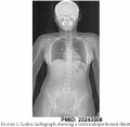
Lodox radiograph showing a ventriculoperitoneal shunt. | Lodox radiograph showing a ventriculoperitoneal shunt. | ||
Ventriculoperitoneal (VP) shunting is a surgical procedure that commenced in the 1960's to treat hydrocephalus and relieve increased pressure inside the skull due to excess cerebrospinal fluid (CSF) on the brain. | |||
:'''Links:''' [[Abnormal Development - Congenital Hydrocephalus|Congenital Hydrocephalus]] | A catheter is placed inside of the brain ventricle, the catheter is then tunneled under the skin from the scalp down into the abdominal cavity, where the other end opens into the peritoneal cavity. | ||
The catheter contains a valve (located in the skin behind the ear) that opens when pressure builds up around the brain. | |||
Complication can include: infections (8-10%), catheter blockage, over-drainage and movement of the catheter. | |||
:'''Links:''' [[Abnormal Development - Congenital Hydrocephalus|ICD11 - LA04 Congenital hydrocephalus]] | [[Abnormal Development - Congenital Hydrocephalus|Congenital Hydrocephalus]] | |||
===Reference=== | ===Reference=== | ||
<pubmed>23243508</pubmed> | <pubmed>23243508</pubmed> | ||
| Line 17: | Line 25: | ||
{{Footer}} | {{Footer}} | ||
[[Category:Abnormal Development]][[Category:Neural]] | |||
Latest revision as of 11:01, 29 May 2017
Ventriculoperitoneal Shunt
Lodox radiograph showing a ventriculoperitoneal shunt.
Ventriculoperitoneal (VP) shunting is a surgical procedure that commenced in the 1960's to treat hydrocephalus and relieve increased pressure inside the skull due to excess cerebrospinal fluid (CSF) on the brain.
A catheter is placed inside of the brain ventricle, the catheter is then tunneled under the skin from the scalp down into the abdominal cavity, where the other end opens into the peritoneal cavity.
The catheter contains a valve (located in the skin behind the ear) that opens when pressure builds up around the brain.
Complication can include: infections (8-10%), catheter blockage, over-drainage and movement of the catheter.
Reference
<pubmed>23243508</pubmed>
S. P. Whiley, et al. Emerg Med Int. 2012;2012:108129. https://www.ncbi.nlm.nih.gov/pmc/articles/PMC3517877/
Copyright
© 2012 S. P. Whiley et al. This is an open access article distributed under the Creative Commons Attribution License, which permits unrestricted use, distribution, and reproduction in any medium, provided the original work is properly cited.
Cite this page: Hill, M.A. (2024, June 26) Embryology Ventriculoperitoneal shunt 01.jpg. Retrieved from https://embryology.med.unsw.edu.au/embryology/index.php/File:Ventriculoperitoneal_shunt_01.jpg
- © Dr Mark Hill 2024, UNSW Embryology ISBN: 978 0 7334 2609 4 - UNSW CRICOS Provider Code No. 00098G
File history
Yi efo/eka'e gwa ebo wo le nyangagi wuncin ye kamina wunga tinya nan
| Gwalagizhi | Nyangagi | Dimensions | User | Comment | |
|---|---|---|---|---|---|
| current | 10:51, 29 May 2017 |  | 584 × 1,000 (56 KB) | Z8600021 (talk | contribs) | |
| 10:48, 29 May 2017 |  | 990 × 966 (73 KB) | Z8600021 (talk | contribs) | Lodox radiograph showing a ventriculoperitoneal shunt. ===Reference=== <pubmed>23243508</pubmed> S. P. Whiley, et al. Emerg Med Int. 2012;2012:108129. https://www.ncbi.nlm.nih.gov/pmc/articles/PMC3517877/ |
You cannot overwrite this file.
File usage
The following 2 pages use this file: