File:Streeter1957 fig08.jpg: Difference between revisions
(Z8600021 uploaded a new version of File:Streeter1957 fig08.jpg) |
mNo edit summary |
||
| (7 intermediate revisions by the same user not shown) | |||
| Line 1: | Line 1: | ||
==Fig. 8. Drawings of | ==Fig. 8. Drawings of sagittal sections of the hypophysis in embryos of horizons xix to xxiii== | ||
Two or more sections were combined in preparing the drawings | Two or more sections were combined in preparing the drawings. | ||
Several sections were utilized in embryos no. {{CE6202}}, xx, no. {{CE1458}}, xxii, and no. {{CE5422}}, xxiii, to represent the lateral processes of the pars intermedia, which grow dorsnlward around the sides of the neural lobe. | |||
<gallery> | |||
File:Streeter1957 fig08-19.jpg|[[Carnegie stage 19|stage 19]] (Embryo {{CE1390}}) | |||
File:Streeter1957 fig08-20.jpg|[[Carnegie stage 20|stage 20]] (Embryo {{CE6202}}) | |||
File:Streeter1957 fig08-21.jpg|[[Carnegie stage 21|stage 21]] (Embryo {{CE1358F}}) | |||
File:Streeter1957 fig08-22.jpg|[[Carnegie stage 22|stage 22]] (Embryo {{CE1458}}) | |||
File:Streeter1957 fig08-23.jpg|[[Carnegie stage 23|stage 23]] (Embryo {{CE5422}}) | |||
</gallery> | |||
{{Streeter1957 figures}} | {{Streeter1957 figures}} | ||
[[Category:Week 8]][[Category:Pituitary]] | [[Category:Week 8]][[Category:Pituitary]][[Category:Carnegie Embryo 1390]] | ||
[[Category:Carnegie Embryo 6202]][[Category:Carnegie Embryo 1458]][[Category:Carnegie Embryo 342]] | |||
[[Category:Carnegie Embryo 5422]][[Category:Carnegie Embryo 1358F]] | |||
[[Category:Carnegie Stage 19]][[Category:Carnegie Stage 20]][[Category:Carnegie Stage 21]] | |||
[[Category:Carnegie Stage22]][[Category:Carnegie Stage 23]] | |||
Latest revision as of 10:16, 23 May 2017
Fig. 8. Drawings of sagittal sections of the hypophysis in embryos of horizons xix to xxiii
Two or more sections were combined in preparing the drawings.
Several sections were utilized in embryos no. 6202, xx, no. 1458, xxii, and no. 5422, xxiii, to represent the lateral processes of the pars intermedia, which grow dorsnlward around the sides of the neural lobe.


| Historic Disclaimer - information about historic embryology pages |
|---|
 |
- Links: 1 Graph Embryos 11-23 | 4 Eye and optic nerve 19-23 | Plate 1 - Cornea | Plate 2 - Hypophysis
Reference
Streeter GL. Developmental Horizons In Human Embryos Description Or Age Groups XIX, XX, XXI, XXII, And XXIII, Being The Fifth Issue Of A Survey Of The Carnegie Collection. (1957) Carnegie Instn. Wash. Publ. 611, Contrib. Embryol., 36: 167-196.
Cite this page: Hill, M.A. (2024, June 16) Embryology Streeter1957 fig08.jpg. Retrieved from https://embryology.med.unsw.edu.au/embryology/index.php/File:Streeter1957_fig08.jpg
- © Dr Mark Hill 2024, UNSW Embryology ISBN: 978 0 7334 2609 4 - UNSW CRICOS Provider Code No. 00098G
File history
Click on a date/time to view the file as it appeared at that time.
| Date/Time | Thumbnail | Dimensions | User | Comment | |
|---|---|---|---|---|---|
| current | 15:03, 8 November 2016 | 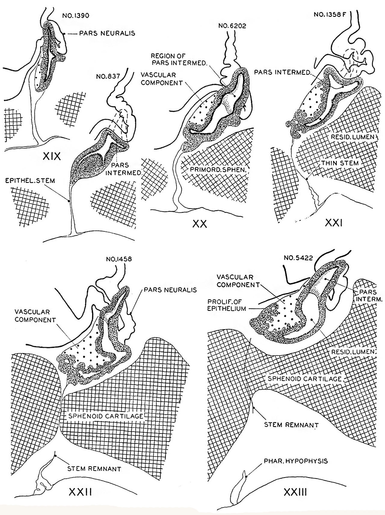 | 1,280 × 1,712 (507 KB) | Z8600021 (talk | contribs) | |
| 14:58, 8 November 2016 | 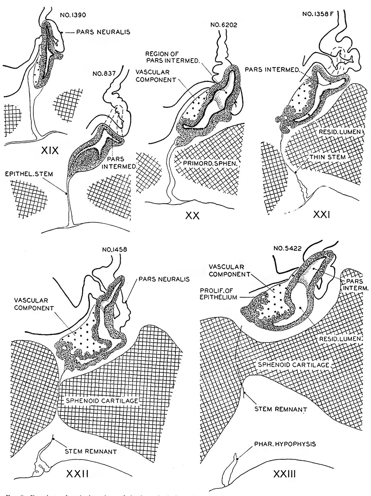 | 1,280 × 1,712 (507 KB) | Z8600021 (talk | contribs) | ||
| 14:57, 8 November 2016 | 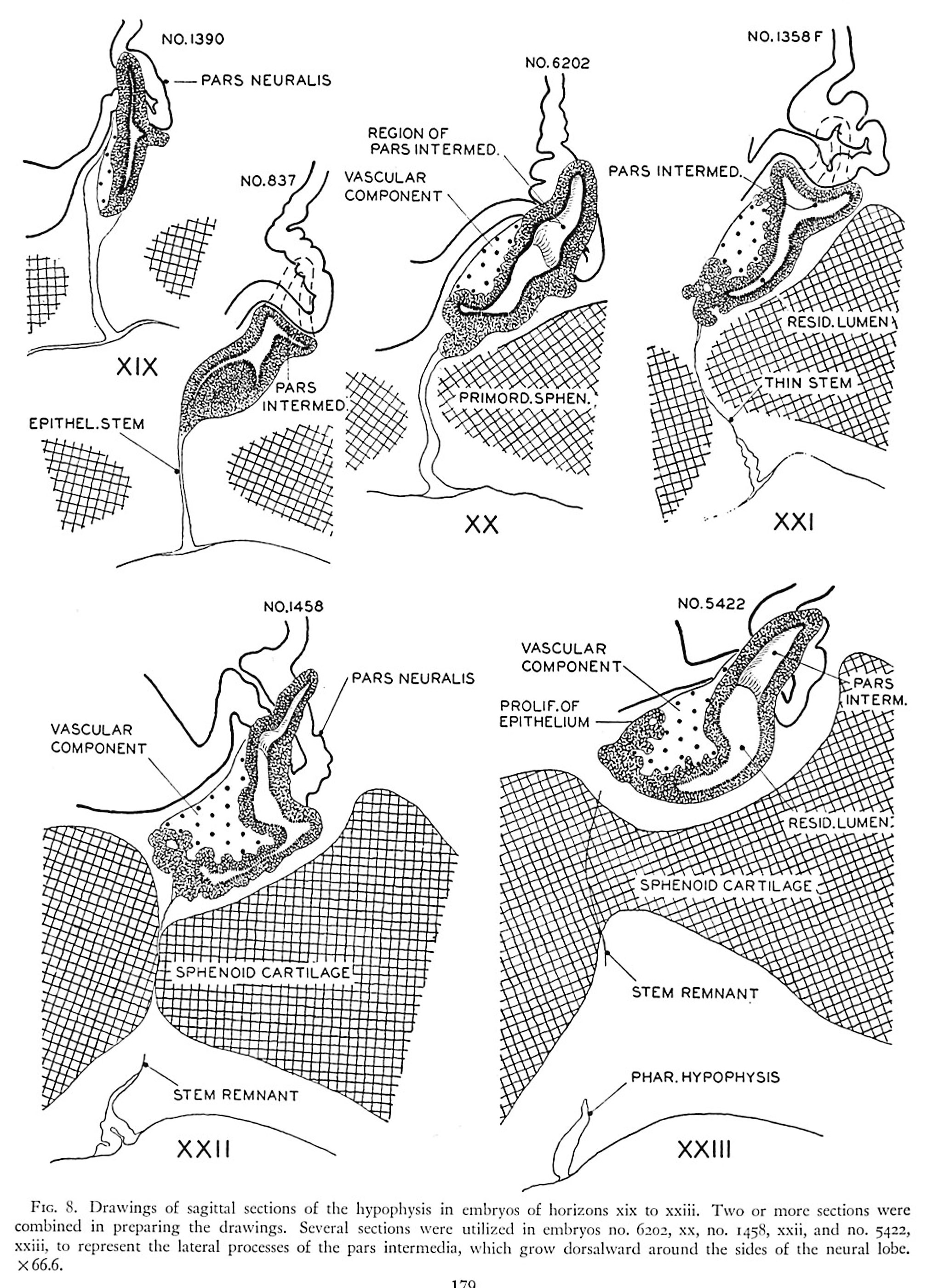 | 2,016 × 2,800 (921 KB) | Z8600021 (talk | contribs) | FIG. 8. Drawings of sngittal sections of the hypophysis in embryos of horizons xix to xxiii. Two or more sections were combined in preparing the drawings. Several sections were utilized in embryos no. 6202, xx, no. 1458, xxii, and no. 342- xxiii, to re... |
You cannot overwrite this file.
File usage
The following 2 pages use this file: