File:Stage11 K17941-01.jpg: Difference between revisions
(==Human Embryo Stage 11== Embryo (Kyoto 17941) mainly showing features of the early developing heart, pharynx and neural groove. The open neural groove is shown at the top of the images. The foregut pharynx can be seen lying between the paired dorsal...) |
mNo edit summary |
||
| (One intermediate revision by the same user not shown) | |||
| Line 1: | Line 1: | ||
==Human Embryo Stage 11== | ==Human Embryo Stage 11== | ||
Embryo ( | Embryo (transverse section) mainly showing features of the early developing heart, pharynx and neural groove. The open neural groove is shown at the top of the images. The foregut pharynx can be seen lying between the paired dorsal aortas (behind) and the heart (in front). The heart tube can be seen to lie in the pericardial cavity, ventral in the embryo, at the bottom of the set histology images. | ||
Embryo (Kyoto 17941) Scale bar 0.5 mm. | |||
:Links: [[Carnegie stage 11]] | |||
{{Kyoto collection}} | {{Kyoto collection}} | ||
{{Footer}} | {{Footer}} | ||
[[Category:Carnegie Stage 11]] | |||
Latest revision as of 12:56, 19 August 2016
Human Embryo Stage 11
Embryo (transverse section) mainly showing features of the early developing heart, pharynx and neural groove. The open neural groove is shown at the top of the images. The foregut pharynx can be seen lying between the paired dorsal aortas (behind) and the heart (in front). The heart tube can be seen to lie in the pericardial cavity, ventral in the embryo, at the bottom of the set histology images.
Embryo (Kyoto 17941) Scale bar 0.5 mm.
- Links: Carnegie stage 11
Image source: The Kyoto Collection images are reproduced with the permission of Prof. Kohei Shiota and Prof. Shigehito Yamada, Anatomy and Developmental Biology, Kyoto University Graduate School of Medicine, Kyoto, Japan for educational purposes only and cannot be reproduced electronically or in writing without permission.
Cite this page: Hill, M.A. (2024, June 27) Embryology Stage11 K17941-01.jpg. Retrieved from https://embryology.med.unsw.edu.au/embryology/index.php/File:Stage11_K17941-01.jpg
- © Dr Mark Hill 2024, UNSW Embryology ISBN: 978 0 7334 2609 4 - UNSW CRICOS Provider Code No. 00098G
File history
Yi efo/eka'e gwa ebo wo le nyangagi wuncin ye kamina wunga tinya nan
| Gwalagizhi | Nyangagi | Dimensions | User | Comment | |
|---|---|---|---|---|---|
| current | 12:54, 19 August 2016 | 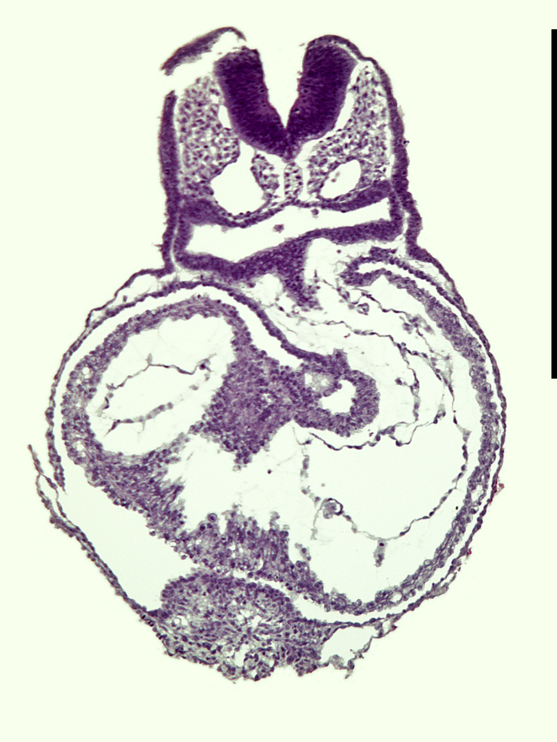 | 800 × 1,066 (625 KB) | Z8600021 (talk | contribs) | ==Human Embryo Stage 11== Embryo (Kyoto 17941) mainly showing features of the early developing heart, pharynx and neural groove. The open neural groove is shown at the top of the images. The foregut pharynx can be seen lying between the paired dorsal... |
You cannot overwrite this file.
File usage
The following page uses this file: