File:Human placenta SERPINE2 expression 01.jpg: Difference between revisions
(==Human placenta SERPINE2 expression== (B) Moderate staining of extravillous trophoblasts (arrow) and decidual cells (arrowhead) in the chorionic plate, and very low staining in the chorionic mesoderm and fibrinoid deposits. Scale bars represent 200...) |
mNo edit summary |
||
| (2 intermediate revisions by the same user not shown) | |||
| Line 1: | Line 1: | ||
==Human | ==Human Placenta SERPINE2 Expression== | ||
(B) Moderate staining of extravillous trophoblasts (arrow) and decidual cells (arrowhead) in the chorionic plate, and very low staining in the chorionic mesoderm and fibrinoid deposits. | (B) Moderate staining of extravillous trophoblasts (arrow) and decidual cells (arrowhead) in the chorionic plate, and very low staining in the chorionic mesoderm and fibrinoid deposits. | ||
| Line 5: | Line 5: | ||
Scale bars represent 200 μm | Scale bars represent 200 μm | ||
===Reference=== | ===Reference=== | ||
<pubmed>21806836</pubmed>| [http://www.rbej.com/content/9/1/106 | |||
Chern et al. Reproductive Biology and Endocrinology 2011 9:106 doi:10.1186/1477-7827-9-106 | Chern et al. Reproductive Biology and Endocrinology 2011 9:106 doi:10.1186/1477-7827-9-106 | ||
====Copyright==== | |||
1477-7827-9-106-1-l.jpg | © 2011 Chern et al; licensee BioMed Central Ltd. | ||
This is an Open Access article distributed under the terms of the Creative Commons Attribution License (http://creativecommons.org/licenses/by/2.0), which permits unrestricted use, distribution, and reproduction in any medium, provided the original work is properly cited. | |||
Panel B cropped from Figure 1. Adjusted in size, contrast and labelling. 1477-7827-9-106-1-l.jpg | |||
{{Footer}} | |||
[[Category:Human]][[Category:Placenta]][[Category:Histology]] | |||
Latest revision as of 09:00, 6 Haziran 2016
Human Placenta SERPINE2 Expression
(B) Moderate staining of extravillous trophoblasts (arrow) and decidual cells (arrowhead) in the chorionic plate, and very low staining in the chorionic mesoderm and fibrinoid deposits.
Scale bars represent 200 μm
Reference
<pubmed>21806836</pubmed>| [http://www.rbej.com/content/9/1/106
Chern et al. Reproductive Biology and Endocrinology 2011 9:106 doi:10.1186/1477-7827-9-106
Copyright
© 2011 Chern et al; licensee BioMed Central Ltd. This is an Open Access article distributed under the terms of the Creative Commons Attribution License (http://creativecommons.org/licenses/by/2.0), which permits unrestricted use, distribution, and reproduction in any medium, provided the original work is properly cited.
Panel B cropped from Figure 1. Adjusted in size, contrast and labelling. 1477-7827-9-106-1-l.jpg
Cite this page: Hill, M.A. (2026, October 29) Embryology Human placenta SERPINE2 expression 01.jpg. Retrieved from https://embryology.med.unsw.edu.au/embryology/index.php/File:Human_placenta_SERPINE2_expression_01.jpg
- © Dr Mark Hill 2026, UNSW Embryology ISBN: 978 0 7334 2609 4 - UNSW CRICOS Provider Code No. 00098G
File history
Yi efo/eka'e gwa ebo wo le nyangagi wuncin ye kamina wunga tinya nan
| Gwalagizhi | Nyangagi | Dimensions | User | Comment | |
|---|---|---|---|---|---|
| current | 18:26, 9 Haziran 2014 | 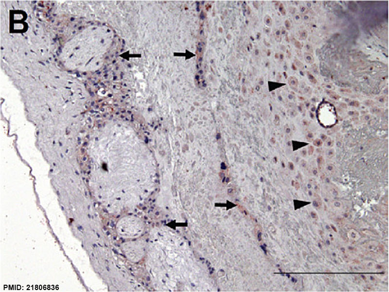 | 800 × 601 (134 KB) | Z8600021 (talk | contribs) | ==Human placenta SERPINE2 expression== (B) Moderate staining of extravillous trophoblasts (arrow) and decidual cells (arrowhead) in the chorionic plate, and very low staining in the chorionic mesoderm and fibrinoid deposits. Scale bars represent 200... |
You cannot overwrite this file.
File usage
The following 2 pages use this file:


