Category:Meiosis: Difference between revisions
From Embryology
No edit summary |
No edit summary |
||
| Line 7: | Line 7: | ||
* meiosis II - (MII) The second part of [[M#meiosis|meiosis]]. In male human [[S#spermatogenesis|spermatogenesis]], producing of four haploid cells (23 chromosomes, 1N) from the two [[H#haploid|haploid]] cells (23 chromosomes, 1N), each of the chromosomes consisting of two sister chromatids produced in [[M#meiosis I|meiosis I]]. In female human [[O#oogenesis|oogenesis]], only a single haploid cell (23 chromosomes, 1N) is produced. '''Phases:''' Prophase II - Metaphase II - Anaphase II - Telophase II | * meiosis II - (MII) The second part of [[M#meiosis|meiosis]]. In male human [[S#spermatogenesis|spermatogenesis]], producing of four haploid cells (23 chromosomes, 1N) from the two [[H#haploid|haploid]] cells (23 chromosomes, 1N), each of the chromosomes consisting of two sister chromatids produced in [[M#meiosis I|meiosis I]]. In female human [[O#oogenesis|oogenesis]], only a single haploid cell (23 chromosomes, 1N) is produced. '''Phases:''' Prophase II - Metaphase II - Anaphase II - Telophase II | ||
'''Links:''' [[Spermatozoa Development]] | [[Ovary Development]] | |||
'''Original web pages:''' [http://embryology.med.unsw.edu.au/Notes/week1.htm Week 1 Notes] | |||
'''Original web pages:''' [http://embryology.med.unsw.edu.au/Notes/week1.htm Week 1 Notes] | |||
Revision as of 10:30, 12 May 2010
This page lists UNSW Embryology content related to meiosis, a form of cell division only occurring in haploid gamete development.
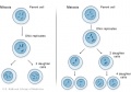
Meiosis is the cell division that occurs only in production of germ cells where there is a reduction in the number of chromosomes (diploid to haploid) which is the basis of sexual reproduction. Note all other non-germ cells divide by mitosis.
- meiosis I - (MI) The first part of meiosis resulting in separation of homologous chromosomes, in humans producing two haploid cells (N chromosomes, 23), a reductional division. Phases: Prophase I - Metaphase I - Anaphase I - Telophase I
- meiosis II - (MII) The second part of meiosis. In male human spermatogenesis, producing of four haploid cells (23 chromosomes, 1N) from the two haploid cells (23 chromosomes, 1N), each of the chromosomes consisting of two sister chromatids produced in meiosis I. In female human oogenesis, only a single haploid cell (23 chromosomes, 1N) is produced. Phases: Prophase II - Metaphase II - Anaphase II - Telophase II
Links: Spermatozoa Development | Ovary Development
Original web pages: Week 1 Notes
Pages in category 'Meiosis'
The following 39 pages are in this category, out of 39 total.
B
P
Media in category 'Meiosis'
The following 51 files are in this category, out of 51 total.
 Adult hermaphrodite gonad arm.jpg 800 × 377; 66 KB
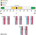
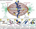
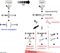 Age-related chromosome segregation errors during MI model.jpg 900 × 794; 112 KB
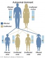 Autosomal dominant inheritance.jpg 307 × 396; 64 KB
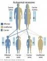 Autosomal recessive inheritance.jpg 307 × 396; 65 KB
 Centrosome cartoon.jpg 732 × 496; 67 KB
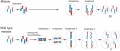
 Chromosome connections in meiosis.jpg 600 × 397; 35 KB
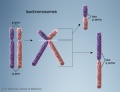
 Chromosome- balanced translocation.jpg 414 × 280; 79 KB
 Chromosome- deletion.jpg 396 × 249; 60 KB
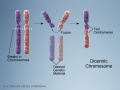 Chromosome- dicentric.jpg 432 × 325; 88 KB
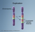 Chromosome- duplication.jpg 286 × 266; 56 KB
 Chromosome- inversion.jpg 467 × 504; 124 KB
 Chromosome- isochromosomes.jpg 469 × 361; 96 KB
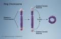 Chromosome- ring chromosome.jpg 448 × 288; 74 KB
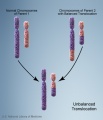 Chromosome- unbalanced translocation.jpg 396 × 460; 98 KB
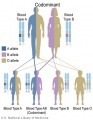 Codominant inheritance.jpg 307 × 396; 65 KB
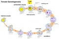 Female gametogenesis.jpg 1,000 × 666; 94 KB
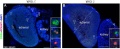
 Fetal adrenal ectopic germ cells 01.jpg 1,092 × 1,280; 358 KB
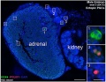
 Fetal adrenal ectopic germ cells 02.jpg 1,086 × 446; 124 KB
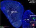
 Fetal adrenal ectopic germ cells 03.jpg 899 × 700; 159 KB
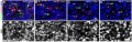
 Fetal adrenal ectopic germ cells 04.jpg 899 × 700; 147 KB
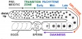
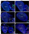
 Fetal ovary meiosis 01.jpg 1,280 × 410; 132 KB
 Fetal ovary meiosis 02.jpg 496 × 600; 77 KB
 Fetal ovary meiosis 03.jpg 652 × 400; 64 KB
 Human-oocyte to blastocyst.jpg 600 × 402; 49 KB
 Karyotype of parthenogenetic blastocysts.jpg 970 × 793; 121 KB
 Kinetochore in mitosis and meiosis 01.jpg 648 × 719; 80 KB
 Male gametogenesis.jpg 1,000 × 666; 121 KB
 Meiosis sister kinetochore geometry.jpg 600 × 464; 34 KB
 Meiosis spindle movement model.jpg 800 × 619; 80 KB
 Meiotic chromosome crossovers 01.jpg 800 × 764; 46 KB
 Meiotic prophase I stages.jpg 1,000 × 341; 66 KB
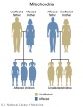 Mitochondrial inheritance.jpg 307 × 396; 53 KB
 Mitosis and meiosis.jpg 1,000 × 423; 58 KB
 Mitosis meiosis1.jpg 500 × 350; 73 KB
 Mouse embryo meiotic to mitotic spindle 01.jpg 800 × 684; 86 KB
 Mouse meiosis pachytene 01.jpg 1,280 × 317; 50 KB
 Mouse oocyte microtubule-associated protein 01.jpg 869 × 1,000; 81 KB
 Oocyte Meiosis 01.mp4 ; 1.22 MB
 Oogenesis and meiosis cartoon.jpg 1,197 × 480; 70 KB
 Rugh 047.jpg 1,156 × 1,000; 186 KB
 Rugh 053.jpg 848 × 800; 128 KB
 Spermatocyte prophase 1 stages 01.jpg 1,280 × 266; 81 KB
 Spindle assembly motors 01.jpg 1,200 × 978; 314 KB
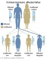 X-Linked dominant (affected father).jpg 307 × 396; 68 KB
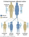 X-Linked dominant (affected mother).jpg 307 × 396; 68 KB
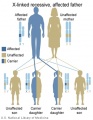 X-Linked recessive (affected father).jpg 307 × 396; 69 KB
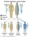 X-Linked recessive (carrier mother).jpg 307 × 396; 69 KB


.jpg)
.jpg)
.jpg)
.jpg)