Category:Somite: Difference between revisions
From Embryology
(Created page with 'This page lists UNSW Embryology content related to somite development. See also mesoderm development.') |
(No difference)
|
Revision as of 02:17, 4 May 2010
This page lists UNSW Embryology content related to somite development. See also mesoderm development.
Subcategories
This category has the following 2 subcategories, out of 2 total.
Pages in category 'Somite'
The following 42 pages are in this category, out of 42 total.
C
M
P
- Paper - Data on the number of somites compared with age in the white rat
- Paper - The development of the anterior post-otic somites in the rabbit
- Paper - The history of the first somite in human embryos
- Paper - The somites of the chick
- Template:Paraxial mesoderm
- Template:Primitive segment
- Template:Primitive segments
R
S
- Template:Sclerotome
- Template:Scoliosis
- Template:Skeletal muscle
- Template:Somite
- Template:Somite cartoon
- Somite Musculoskeletal Movie
- Template:Somite parts table
- Template:SomiteNoTable
- Template:Somites
- Somitogenesis
- Template:Somitogenesis
- Somitogenesis Molecular Movie
- Template:Species somite number table
Media in category 'Somite'
The following 44 files are in this category, out of 44 total.
 Bailey103.jpg 739 × 738; 89 KB
 Bailey104.jpg 878 × 638; 107 KB
 Bailey105.jpg 545 × 567; 61 KB
 Bailey123.jpg 885 × 618; 78 KB
 BaxterBoyd1939-fig04.jpg 642 × 615; 109 KB
 BaxterBoyd1939-fig05.jpg 1,000 × 864; 263 KB
 BaxterBoyd1939-fig06.jpg 489 × 917; 141 KB
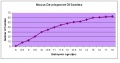 Chart of mouse development 2.JPG 663 × 347; 31 KB
 Chicken - somite.jpg 1,200 × 375; 269 KB
 Chicken body elongation model.jpg 770 × 900; 101 KB
 Chicken-somitogenesis.jpg 600 × 420; 68 KB
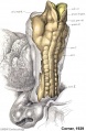
 Corner1929 fig10-11.jpg 1,200 × 1,438; 730 KB
 Hongjun Shi Research photo02.jpg 899 × 677; 58 KB
 Keibel Mall 2 522.jpg 1,280 × 918; 132 KB
 Mesoderm cartoon 05.jpg 80 × 135; 3 KB
 Mesoderm cartoon 06.jpg 216 × 181; 10 KB
 Mesoderm cartoon 07.jpg 138 × 129; 6 KB
 Mesoderm cartoon 08.jpg 176 × 127; 7 KB
 Mesoderm cartoon 09.jpg 270 × 209; 20 KB
 Mesoderm development and Pax 01.jpg 1,211 × 1,000; 171 KB
 Mesoderm development and Pax 02.jpg 984 × 542; 111 KB
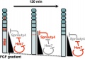 Model for Sprouty4 and FGF in mesoderm segmentation.jpg 481 × 332; 28 KB
 Mouse cleaved intracellular portion of Notch E8.5.jpg 532 × 408; 38 KB
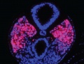
 Mouse somitogenesis gene expression E8.5-9.5.jpg 1,000 × 971; 130 KB
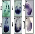
 Mouse somitogenesis genes.jpg 828 × 800; 119 KB
 Rugh 099.jpg 766 × 1,000; 105 KB
 Somite boundary formation model 01.jpg 416 × 1,000; 81 KB
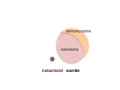
 Somite cartoon1.png 400 × 300; 9 KB
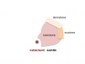
 Somite cartoon2.png 400 × 300; 16 KB
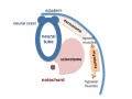
 Somite cartoon3.png 400 × 300; 12 KB
 Somite cartoon4.png 400 × 300; 14 KB
 Somite cartoon5.png 400 × 300; 27 KB
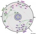
 Somite patterning.jpg 600 × 366; 58 KB
 Somitogenesis 001.mov ; 359 KB
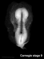
 Stage 10 historic-Corner1929-1.jpg 654 × 1,000; 145 KB
 Stage 10 historic-Corner1929-1a.jpg 523 × 800; 87 KB
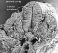
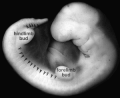
 Stage 9 SEM1.jpg 347 × 450; 42 KB
 Stage11 sem10.jpg 1,000 × 898; 162 KB
 Stage11 sem100.jpg 1,000 × 898; 109 KB
 Stage11 sem100c.jpg 400 × 359; 30 KB
 Stage14 somites limbbuds.png 300 × 245; 24 KB
 Stage9sm.jpg 287 × 378; 6 KB
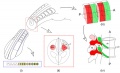
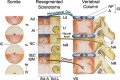 Vertebra development cartoon.jpg 800 × 533; 93 KB
 Wen1928-Fig10.jpg 495 × 1,200; 108 KB


