File:Mouse lung development 01a.jpg: Difference between revisions
mNo edit summary |
|||
| (One intermediate revision by one other user not shown) | |||
| Line 1: | Line 1: | ||
==Histological analyses of Mouse Lungs at Various Embryonic Stages== | ==Histological analyses of Mouse Lungs at Various Embryonic Stages== | ||
Lung sections stained with H and E, and taken at various gestational stages as indicated. | |||
* The first two columns are at lower magnification (10X) and the last two columns at higher magnification (40X). | |||
* The first and third columns are representative sections from wild-type (+/+) embryos and the second and fourth columns from knockout (-/-) embryos. | |||
* No differences were visible at E14.5 or E16.5 (panel A, a-h). Significant differences were visible from E17.5 onwards (panel A, i-x). | |||
* Distal tubules showed dilation and mesenchyme thinning, with progression of septation in wild-type lungs (panel A, i, m and q), whereas the knockout lungs showed less saccular structures and more mesenchyme (panel A, j, n and r). | |||
* At higher magnifications of wild-type lungs (panel A, k, o and s), developing pre-alveoli and thinning mesenchyme (arrowheads) were seen but sections from knockout embryos showed a delay in this sequence of development (panel A, l, p and t). | |||
* At birth, lungs from knockout pups showed deficient septation and thick-walled mesenchyme (panel A, x, arrowheads). | |||
'''Labels:''' | |||
* '''de''' - distal epithelium | |||
* '''m''' - mesenchyme | |||
* '''PA''' - pulmonary artery | |||
* '''a''' - pre-alveoli | |||
* '''b''' - bronchi | |||
Original file name: 1471-213X-4-1-3.jpg http://www.biomedcentral.com/1471-213X/4/1/figure/F3 (Panel A cropped from original full image, resized to fit screen 800px) | Original file name: 1471-213X-4-1-3.jpg http://www.biomedcentral.com/1471-213X/4/1/figure/F3 (Panel A cropped from original full image, resized to fit screen 800px) | ||
| Line 13: | Line 27: | ||
Yu et al. BMC Developmental Biology 2004 4:1 doi:10.1186/1471-213X-4-1 | Yu et al. BMC Developmental Biology 2004 4:1 doi:10.1186/1471-213X-4-1 | ||
====Copyright==== | |||
© 2004 Yu et al; licensee BioMed Central Ltd. This is an Open Access article: verbatim copying and redistribution of this article are permitted in all media for any purpose, provided this notice is preserved along with the article's original URL. | |||
{{Footer}} | |||
[[Category:Mouse]] [[Category:Respiratory]] [[Category:Histology]] [[Category:Abnormal Development]] | [[Category:Mouse]] [[Category:Respiratory]] [[Category:Histology]] [[Category:Abnormal Development]] | ||
[[Category:Mouse E14.5]] [[Category:Mouse E16.5]] [[Category:Mouse E17.5]] [[Category:Mouse E18.5]] [[Category:Mouse E20.5]] | [[Category:Mouse E14.5]] [[Category:Mouse E16.5]] [[Category:Mouse E17.5]] [[Category:Mouse E18.5]] [[Category:Mouse E20.5]] | ||
[[Category:Knockout Mouse]] | |||
Latest revision as of 13:01, 28 September 2015
Histological analyses of Mouse Lungs at Various Embryonic Stages
Lung sections stained with H and E, and taken at various gestational stages as indicated.
- The first two columns are at lower magnification (10X) and the last two columns at higher magnification (40X).
- The first and third columns are representative sections from wild-type (+/+) embryos and the second and fourth columns from knockout (-/-) embryos.
- No differences were visible at E14.5 or E16.5 (panel A, a-h). Significant differences were visible from E17.5 onwards (panel A, i-x).
- Distal tubules showed dilation and mesenchyme thinning, with progression of septation in wild-type lungs (panel A, i, m and q), whereas the knockout lungs showed less saccular structures and more mesenchyme (panel A, j, n and r).
- At higher magnifications of wild-type lungs (panel A, k, o and s), developing pre-alveoli and thinning mesenchyme (arrowheads) were seen but sections from knockout embryos showed a delay in this sequence of development (panel A, l, p and t).
- At birth, lungs from knockout pups showed deficient septation and thick-walled mesenchyme (panel A, x, arrowheads).
Labels:
- de - distal epithelium
- m - mesenchyme
- PA - pulmonary artery
- a - pre-alveoli
- b - bronchi
Original file name: 1471-213X-4-1-3.jpg http://www.biomedcentral.com/1471-213X/4/1/figure/F3 (Panel A cropped from original full image, resized to fit screen 800px)
Mouse lung development 01.jpg
Reference
<pubmed>15005800</pubmed>| BMC Developmental Biology
Yu et al. BMC Developmental Biology 2004 4:1 doi:10.1186/1471-213X-4-1
Copyright
© 2004 Yu et al; licensee BioMed Central Ltd. This is an Open Access article: verbatim copying and redistribution of this article are permitted in all media for any purpose, provided this notice is preserved along with the article's original URL.
Cite this page: Hill, M.A. (2024, June 21) Embryology Mouse lung development 01a.jpg. Retrieved from https://embryology.med.unsw.edu.au/embryology/index.php/File:Mouse_lung_development_01a.jpg
- © Dr Mark Hill 2024, UNSW Embryology ISBN: 978 0 7334 2609 4 - UNSW CRICOS Provider Code No. 00098G
File history
Yi efo/eka'e gwa ebo wo le nyangagi wuncin ye kamina wunga tinya nan
| Gwalagizhi | Nyangagi | Dimensions | User | Comment | |
|---|---|---|---|---|---|
| current | 14:52, 25 August 2011 | 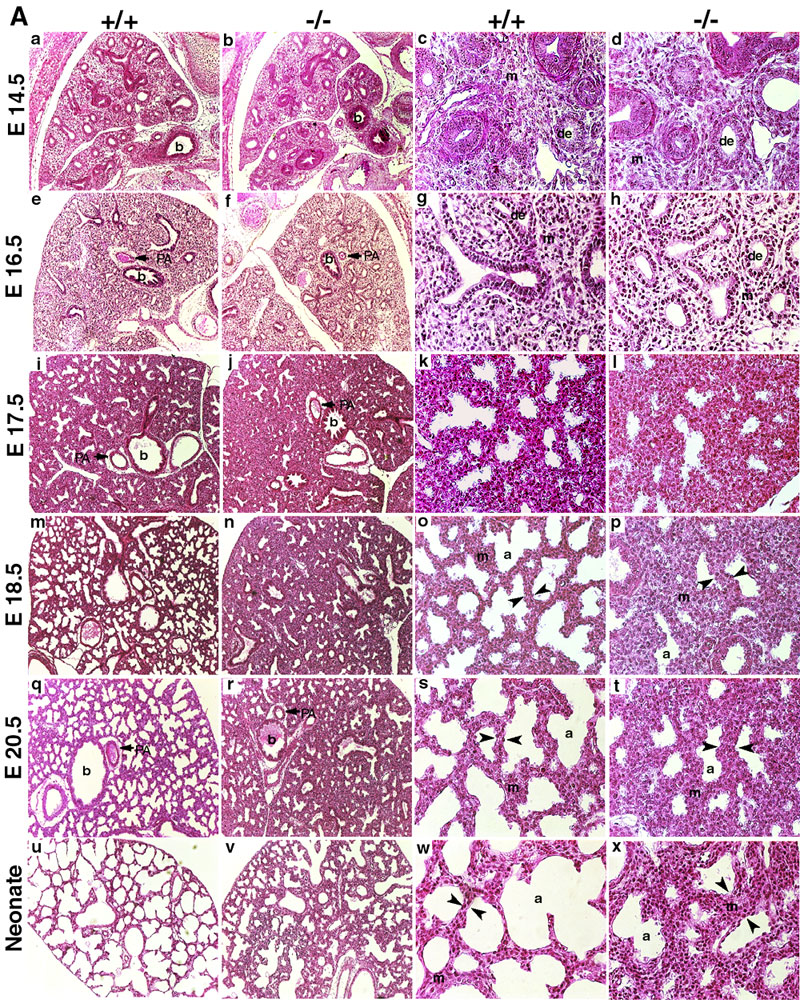 | 800 × 1,003 (495 KB) | S8600021 (talk | contribs) | ==Histological analyses of Mouse Lungs at Various Embryonic Stages== indicates lung sections stained with H and E, and taken at various gestational stages as indicated. The first two columns are at lower magnification (10X) and the last two columns at hi |
You cannot overwrite this file.
File usage
There are no pages that use this file.