File:Fawcett1913 fig01.jpg: Difference between revisions
(Z8600021 uploaded a new version of "File:Fawcett1913 fig01.jpg") |
mNo edit summary |
||
| Line 1: | Line 1: | ||
==Fig.1. Drawing of a model of the right shoulder girdle of the 17mm. (Robinson) embryo== | |||
Viewed from behind. The sternal segment (S.S.) has been cut coronally to expose the interior, and the acromial segment has been cut horizontally for the same purpose. The black area in each is bone, the dotted area surrounding the bone is precartilage, and the area surrounding this is perichondrium. Above the junction of the two segments, two circles and a black dot are seen; the circles represent supraclavicular nerves, the black dot represents the cephalic vein. The scapula has been purposely shortened. | |||
{| | |||
| | |||
* '''Ac. pr.''' - acromion process | |||
* '''A. S.''' - acromial segment of clavicle | |||
* '''C. Al.''' - clei-do-mastoid | |||
* '''C. Cl. I.''' - coraco-clavicular ligament | |||
* '''C. pr.''' - coracoid process | |||
| | |||
* '''T.''' - trapezius muscle | |||
* '''P.M.''' - pectoralis major | |||
* '''St. MI.''' - sterno-miastoid | |||
* '''S. S.''' - sternal segment of clavicle. | |||
|} | |||
{{Fawcett1913 figures}} | {{Fawcett1913 figures}} | ||
Latest revision as of 14:47, 27 December 2014
Fig.1. Drawing of a model of the right shoulder girdle of the 17mm. (Robinson) embryo
Viewed from behind. The sternal segment (S.S.) has been cut coronally to expose the interior, and the acromial segment has been cut horizontally for the same purpose. The black area in each is bone, the dotted area surrounding the bone is precartilage, and the area surrounding this is perichondrium. Above the junction of the two segments, two circles and a black dot are seen; the circles represent supraclavicular nerves, the black dot represents the cephalic vein. The scapula has been purposely shortened.
|
|
Clavicle Links: Fig 1 | Fig 2 | Fig 3 | Fig 4 | Fig 5 | Fig 6 | Fig 7 | Fig 8 | 1913 Clavicle
- Edward Fawcett Links: 1906 Palate | 1910 Head | 1910 Sphenoid | 1911 Maxilla, vomer, and paraseptal cartilages | 1913 Clavicle | 1930 Mandible | Fawcett image | Edward Fawcett
Cite this page: Hill, M.A. (2024, June 20) Embryology Fawcett1913 fig01.jpg. Retrieved from https://embryology.med.unsw.edu.au/embryology/index.php/File:Fawcett1913_fig01.jpg
- © Dr Mark Hill 2024, UNSW Embryology ISBN: 978 0 7334 2609 4 - UNSW CRICOS Provider Code No. 00098G
File history
Yi efo/eka'e gwa ebo wo le nyangagi wuncin ye kamina wunga tinya nan
| Gwalagizhi | Nyangagi | Dimensions | User | Comment | |
|---|---|---|---|---|---|
| current | 14:36, 27 December 2014 | 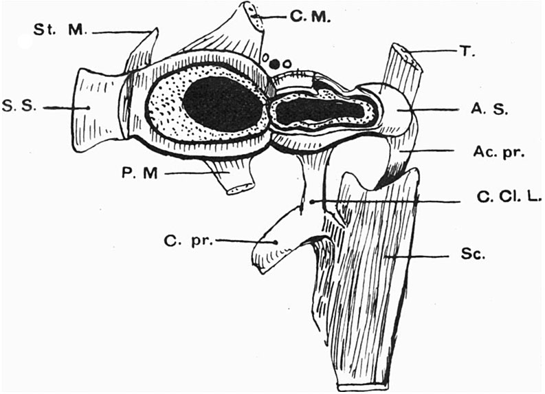 | 793 × 573 (67 KB) | Z8600021 (talk | contribs) | |
| 14:35, 27 December 2014 | 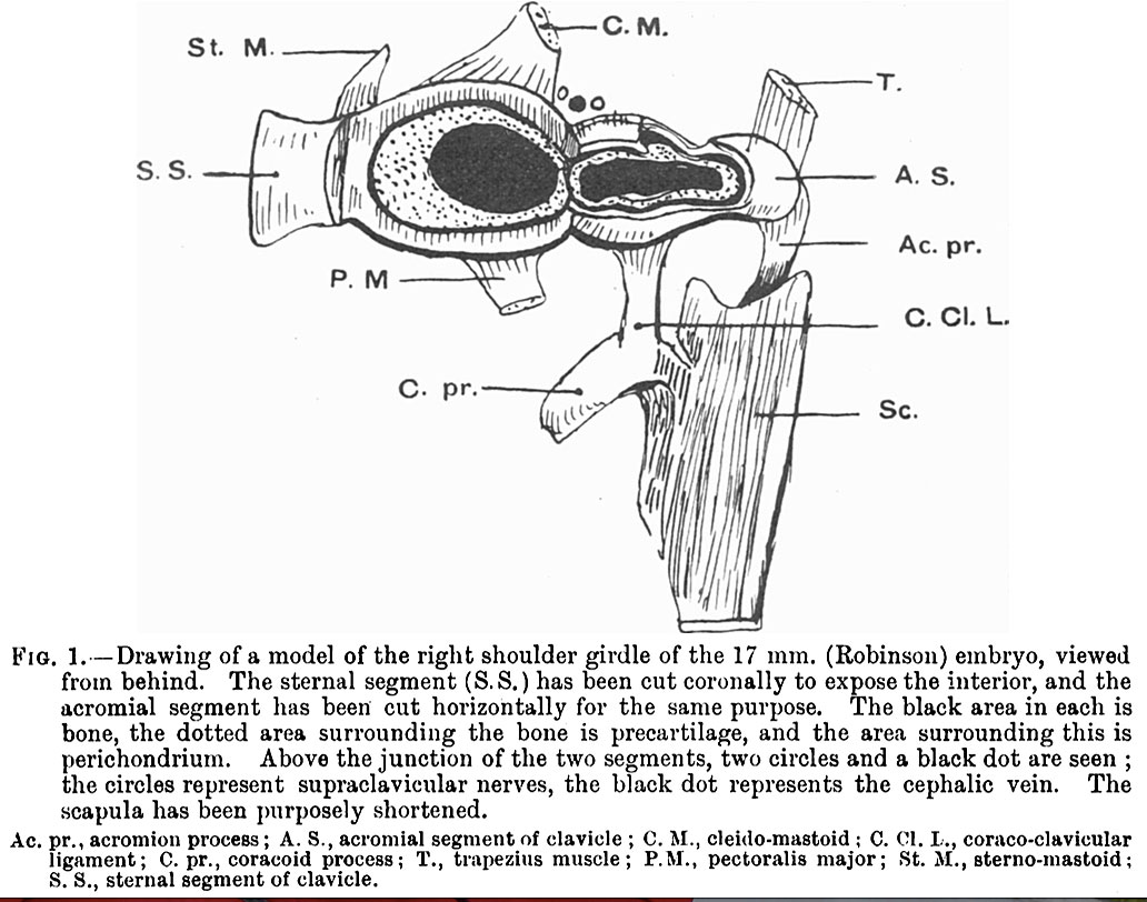 | 1,032 × 812 (163 KB) | Z8600021 (talk | contribs) | {{Fawcett1913 figures}} |
You cannot overwrite this file.
File usage
The following page uses this file: