File:Mouse CT E9.5-E12 head.jpg: Difference between revisions
(==Mouse Embryo Computed Tomography== A - Ontogenetic series of μCT scans showing the range of shape and size variation from E9.5-12. Original File name: Figure 1.A 1471-213X-10-18-1-l.jpg http://www.biomedcentral.com/1471-213X/10/18/figure/F1 Modifica) |
mNo edit summary |
||
| (5 intermediate revisions by 2 users not shown) | |||
| Line 1: | Line 1: | ||
==Mouse Embryo Computed Tomography== | ==Mouse Embryo Computed Tomography== | ||
A - Ontogenetic series of μCT scans showing the range of shape and size variation from E9.5-12. | Heads of embryos E9.5 to E12 shown to scales (ventral view). This image was extracted from 1A of the original paper figure. See also animation based on these images [Mouse Stages Face microCT Movie]]. | ||
'''A''' - Ontogenetic series of μCT scans showing the range of shape and size variation from E9.5-12. | |||
:'''Links:''' [[Mouse Stages Face microCT Movie]] | [[Mouse Development]] | |||
===Reference=== | |||
<pubmed>20163731</pubmed>|[http://www.biomedcentral.com/1471-213X/10/18 BMC Developmental Biology] | |||
====Copyright==== | |||
© 2010 Schmidt et al; licensee BioMed Central Ltd. | |||
This is an Open Access article distributed under the terms of the Creative Commons Attribution License (http://creativecommons.org/licenses/by/2.0), which permits unrestricted use, distribution, and reproduction in any medium, provided the original work is properly cited. | |||
Original File name: Figure 1.A 1471-213X-10-18-1-l.jpg http://www.biomedcentral.com/1471-213X/10/18/figure/F1 | Original File name: Figure 1.A 1471-213X-10-18-1-l.jpg http://www.biomedcentral.com/1471-213X/10/18/figure/F1 | ||
Modification: 1471-213X-10-18-1.PDF Part A resized and cropped to 1000px 72dpi. | Modification: 1471-213X-10-18-1.PDF Part A resized and cropped to 1000px 72dpi. | ||
[[Category:Mouse]] [[Category:Computed Tomography]] [[Category:Mouse E9.5]] [[Category:Mouse E10]] [[Category:Mouse E10.5]] [[Category:Mouse E11]] [[Category:Mouse E11.5]] [[Category:Mouse E12]] | |||
Latest revision as of 13:16, 18 August 2014
Mouse Embryo Computed Tomography
Heads of embryos E9.5 to E12 shown to scales (ventral view). This image was extracted from 1A of the original paper figure. See also animation based on these images [Mouse Stages Face microCT Movie]].
A - Ontogenetic series of μCT scans showing the range of shape and size variation from E9.5-12.
Reference
<pubmed>20163731</pubmed>|BMC Developmental Biology
Copyright
© 2010 Schmidt et al; licensee BioMed Central Ltd. This is an Open Access article distributed under the terms of the Creative Commons Attribution License (http://creativecommons.org/licenses/by/2.0), which permits unrestricted use, distribution, and reproduction in any medium, provided the original work is properly cited.
Original File name: Figure 1.A 1471-213X-10-18-1-l.jpg http://www.biomedcentral.com/1471-213X/10/18/figure/F1
Modification: 1471-213X-10-18-1.PDF Part A resized and cropped to 1000px 72dpi.
File history
Yi efo/eka'e gwa ebo wo le nyangagi wuncin ye kamina wunga tinya nan
| Gwalagizhi | Nyangagi | Dimensions | User | Comment | |
|---|---|---|---|---|---|
| current | 11:44, 17 August 2010 | 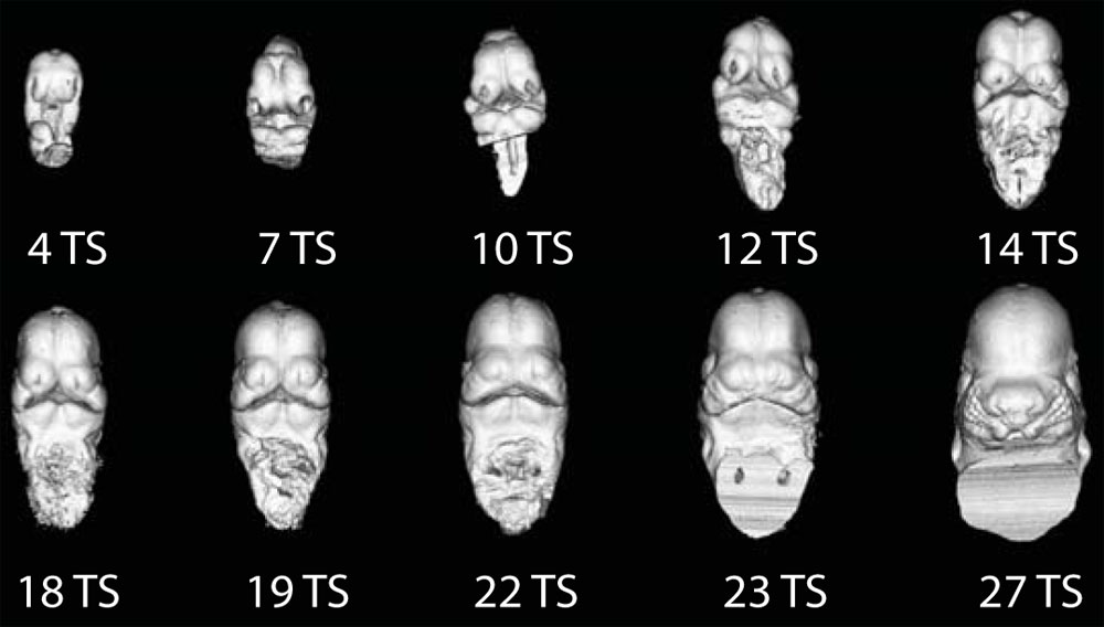 | 1,000 × 568 (56 KB) | S8600021 (talk | contribs) | ==Mouse Embryo Computed Tomography== A - Ontogenetic series of μCT scans showing the range of shape and size variation from E9.5-12. Original File name: Figure 1.A 1471-213X-10-18-1-l.jpg http://www.biomedcentral.com/1471-213X/10/18/figure/F1 Modifica |
You cannot overwrite this file.
File usage
The following 4 pages use this file: